Mucosal incision assisted unroofing technique for endoscopic full thickness resection of a gastric subepithelial lesion
Taylor Bowler, Rahul Karna, Timothy Davie, Carlos Iwamoto, Mohammad Bilal

Abstract
Genes, proteins, chemicals, diseases, species, mutations and cell lines named across the full text — each resolved to its canonical identifier and authoritative record.
Click any figure to enlarge with its caption.
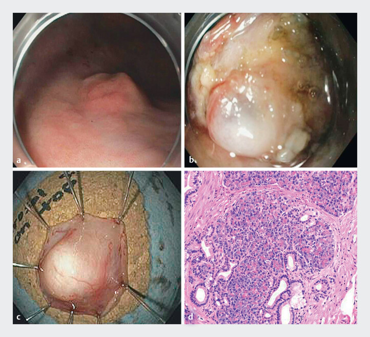 Fig. 1
Peer Reviews
No public reviews on file for this paper yet. If you reviewed it on a platform where reviews are public (OpenReview, ICLR, NeurIPS, ICML), you can paste yours below so the community can read it here.
Videos
No videos yet. Explain this paper in a talk, walkthrough, or lecture? Add one.
Taxonomy
TopicsGastrointestinal Tumor Research and Treatment · Gastric Cancer Management and Outcomes · Gastrointestinal disorders and treatments
Endoscopic full thickness resection (EFTR) using a full thickness resection device (FTRD; OVESCO Endoscopy AG, Tuebingen, Germany) is a safe and effective technique for the management of subepithelial lesions (SELs; 1 ). This technique utilizes a grasping forceps to facilitate retraction into the FTRD prior to clip deployment followed by resection using snare with electrocautery. However, this technique can be challenging with SELs since the grasping forceps often capture the mucosa leading to slippage of the underlying SEL. This limitation can be overcome with mucosal incision and unroofing to expose the underlying lesion and allow for EFTR using the FTRD. This video case report outlines the technique of EFTR using the FTRD after mucosal unroofing of a gastric SEL ( Video 1 ).
Video image: the subepithelial lesion after the successful unroofing technique.Video 1
A 65-year-old male with history of tobacco use was referred for the management of a gastric SEL. Endoscopic ultrasound demonstrated a 11 mm × 5 mm hypoechoic lesion originating from the submucosa (layer III) of the gastric body. Prior fine needle biopsies were non-diagnostic. Endoscopic examination with white light imaging showed a 11 mm subepithelial lesion with a negative pillow sign ( Fig. 1 a ). As demonstrated in the video, initial mucosal incision was performed with an endoscopic submucosal dissection knife to unroof the underlying lesion ( Fig. 1 b ). Exposure of the lesion beneath the mucosal surface allowed for direct grasping of the lesion with the grasping forceps, retraction into the FTRD cap, and subsequent EFTR using the FTRD. EFTR was demonstrated by the fatty patch within the clip and the specimen was successfully retrieved ( Fig. 1 c ). Lesion histopathology showed pancreatic heterotopia involving the submucosa with focal mucosal erosion ( Fig. 1 d ). There were no adverse events with the procedure.
a SELs in the gastric body. b SELs after the mucosal unroofing technique. c Successful retrieval of the lesion after EFTR. d Lesion histopathology demonstrating pancreatic heterotropia.
This case highlights that mucosal unroofing can allow for successful EFTR for the resection of SELs. Future studies are needed to explore the utility of this technique compared to traditional endoscopic resection methods for the management of gastric SELs.
Endoscopy_UCTN_Code_TTT_1AO_2AG_3AD
The reference list from the paper itself. Each links out to its DOI / PubMed record.