Assessment of chemical methods in the extraction of spore surface layers in Clostridioides difficile spores
Javier Sanchez, Alba Romero-Rodriguez, Scarlett Troncoso-Cotal, Morgan S. Osborne, Theresa Ariri, Joseph A. Sorg, Daniel Paredes-Sabja

TL;DR
This study compares different chemical methods for extracting surface layers of Clostridioides difficile spores to improve consistency in research.
Contribution
The study provides a standardized comparison of EBB, USD, and Laemmli methods for spore surface extraction in C. difficile.
Findings
EBB and USD treatments completely remove the spore coat and exosporium while leaving decoated spores intact.
EBB and USD allow for the detection of the spore core germination protease, GPR, after lysozyme treatment.
Lysozyme can degrade the cortex in decoated spores regardless of the extraction method used.
Abstract
Clostridioides difficile spores are essential for initiation, recurrence, and transmission of C. difficile infections (CDI). These outermost layers of the spore, the exosporium and spore coat, are responsible for initial interactions with the host and spore resistance properties, respectively. Several spore coat/exosporium extraction methods have been utilized to study the spore surface, with differing procedures making comparison across studies difficult. Here, we tested how commonly used exosporium and spore coat extraction methods, termed EBB, USD, and Laemmli, remove the spore coat and exosporium layers of C. difficile spores. We assessed the impact of these extraction methods on the spore through transmission electron microscopy, phase contrast microscopy, western blotting, and lysozyme-triggered cortex degradation. Transmission electron microscopy shows that treatment with EBB and…
Genes, proteins, chemicals, diseases, species, mutations and cell lines named across the full text — each resolved to its canonical identifier and authoritative record.
Click any figure to enlarge with its caption.
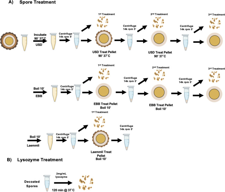 Fig 1
Fig 1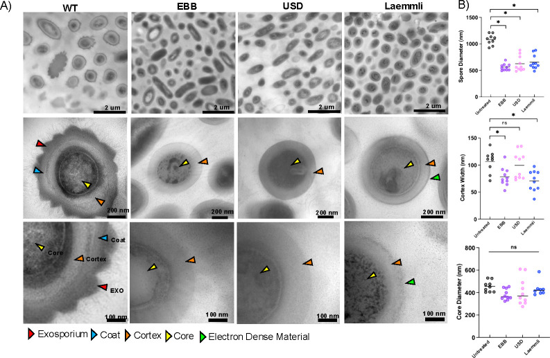 Fig 2
Fig 2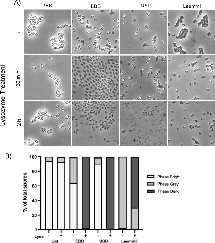 Fig 3
Fig 3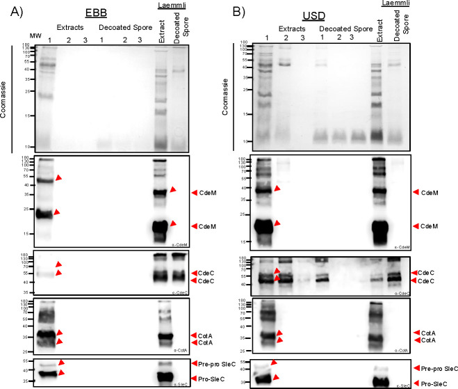 Fig 4
Fig 4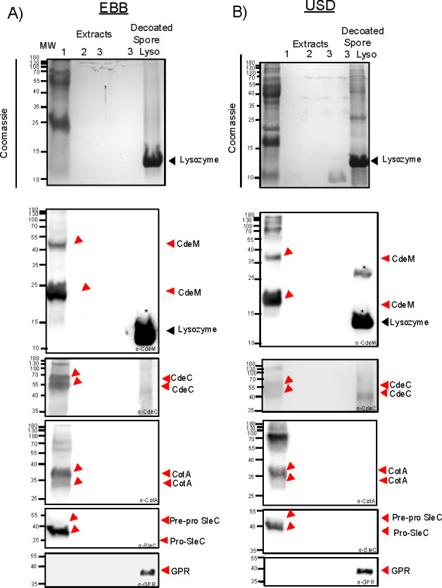 Fig 5
Fig 5- —National Institute of Allergy and Infectious Diseaseshttp://dx.doi.org/10.13039/100000060
- —National Institute of Allergy and Infectious Diseaseshttp://dx.doi.org/10.13039/100000060
- —National Institute of Allergy and Infectious Diseaseshttp://dx.doi.org/10.13039/100000060
Peer Reviews
No public reviews on file for this paper yet. If you reviewed it on a platform where reviews are public (OpenReview, ICLR, NeurIPS, ICML), you can paste yours below so the community can read it here.
Videos
No videos yet. Explain this paper in a talk, walkthrough, or lecture? Add one.
Taxonomy
TopicsClostridium difficile and Clostridium perfringens research · Microscopic Colitis · Viral gastroenteritis research and epidemiology
INTRODUCTION
Clostridioides difficile is a Gram-positive anaerobe that has become a leading cause of antibiotic-associated diarrhea in developed countries (1, 2). Treatment against C. difficile infections (CDI) resolves 95% of primary cases; however, ~15%–30% of recovered CDI patients develop subsequent recurrent episodes of CDI, which result in mortality rates increasing up to 30% (3). Although two major clostridial toxins, TcdA and TcdB, are necessary for disease manifestation (4), the production of C. difficile spores during infection is essential for disease recurrence (5, 6).
C. difficile spores are metabolically dormant and naturally resistant to antibiotics, heat, ethanol, and various cleaning agents (6, 7). The spores are made up of a series of concentric layers that assemble under the control of RNA polymerase sporulation-specific sigma factors, SigF, SigE, SigK, and SigG (6, 7). Importantly, during late stages of sporulation, C. difficile forms two distinctive spores that differ in the thickness of the exosporium morphotype, while the underlying spore coat remains identical (6, 8, 9). The outermost exosporium layer of C. difficile spores plays important roles in infection, recurrence, and interaction with host molecules (10–14).
Both the spore coat and exosporium of C. difficile spores are proteinaceous layers made of >50 and ~10–20 proteins, respectively (15, 16). Both contribute to the spores' resistance against various environmental insults (7). Numerous studies on the spore coat and exosporium layer of C. difficile spores have varied in the methods to extract the spore coat and exosporium layers (12, 15–23). Because these methods dissociate the entire proteinaceous spore surface layers, they are thought to extract the spore coat and exosporium layers, at least in C. difficile (12, 15–23). Although different in their components, most of these chemical approaches use (i) a chaotropic agent, urea or urea + thiourea, to disrupt hydrogen bonds and result in the dissolution of hydrophobic residues within the protein (24); (ii) SDS to denature and solubilize proteins; and (iii) a reducing agent, which can be 2-mercaptoethanol or dithiothreitol. Currently, three methods have been commonly utilized to remove the spore surface layers in C. difficile spores. Several studies utilize 2× Laemmli buffer (1% SDS, 5% 2-mercaptoethanol) followed by boiling for 10 min at 100°C (12, 15, 17), whereas other proteomic studies have used a lysis buffer with 8 M urea and DTT as a reducing agent (16, 21, 22). In C. difficile spore research, of 8 M urea, 2 M thiourea, 5% (wt/vol) SDS, 2% 2-mercaptoethanol, denominated EBB buffer, followed by incubation for 10 min at 95°C, has also been used extensively for the extraction and solubilization of spore proteins mainly in sporulating cultures (18–20). In addition, urea-SDS-DTT, denoted as USD (8 M urea, 50 mM DTT, 1% wt/vol SDS), has been used in the extraction of exosporium and spore coat in C. difficile spores (25). Despite widespread use of these different chemical extraction methods, it is unclear whether these chemical methods, commonly employed in C. difficile spore research, efficiently remove the spore coat and exosporium layer.
Efficient fractionation of the spore surface layers is essential to understand the localization of spore surface constituents in each of these layers. In this regard, we demonstrated that enzymatic digestion with proteinase K or trypsin, or a mechanical treatment with sonication, efficiently removed only the exosporium layer of C. difficile spores (15). This allowed the identification of the exosporium proteome in spores of strain 630 (15); however, a similar validation with chemical extraction methods has not been conducted. Therefore, to improve our toolbox of methods to study C. difficile spore architecture and composition, in this work, we tested commonly used chemical extraction methods, EBB, USD, and Laemmli in their ability to remove the spore coat and exosporium layers and their impact on spore structure. By utilizing antibodies against exosporium markers (i.e., CdeC and CdeM) (12, 17, 26), spore coat specific marker CotA, the spore cortex lytic enzyme SleC, and germination protease GPR as a core specific marker (27–29) we evaluated how these commonly utilized chemical methods to extract these layers through immunoblotting, transmission electron microscopy (TEM), and lysozyme permeability. For comparison, we defined “extraction efficiency” as a measure of the ability to extract protein as seen through SDS-PAGE and immunoblotting, complete removal of exosporium and spore coat as seen through TEM, removal of spore surface markers (CdeM, CdeC, CotA, SleC, and GPR) from decoated spore pellets, and lastly, removal of spore coat following lysozyme treatment.
MATERIALS AND METHODS
Bacterial strains and growth conditions
Bacterial strains used in this study are listed in the main Table 1. C. difficile R20291 strain was grown at 37°C under anaerobic conditions (Coy Laboratory anaerobic chamber, 4% H_2,_ 5% CO_2_, 85% N_2_) on brain heart infusion supplemented with 0.5% yeast extract, and 0.1% L-cysteine (BHIS) broth or 1.5% agar. Escherichia coli DH5α strains were grown on Luria Bertani supplemented with 50 ug/mL chloramphenicol, 10 ug/mL tetracycline, 50 ug/mL kanamycin, or 100 ug/mL ampicillin where indicated.
TABLE 1: Bacterial strains and plasmids used
Plasmid construction
The plasmids pARR19, pMS85, and pHN55 were made from pET22b as follows and listed in the main Table 1. pET22b was digested with NotI/XhoI and purified by gel electrophoresis, extracted, then assembled through restriction cloning with cdeM or cdeC, or gpr fragment via Gibson assembly. The oligonucleotide primers 3931 (5′_CdeM_pET22b) and 3932 (3′_CdeM_pET22b) were used to amplify cdeM, primers FP-CD1067-NdeI and RP-CD1067-XhoI were used to amplify cdeC, and FP-5′pet22b_gpr and RP RP 3′pet22b_gpr were used to amplify gpr from C. difficile R20291. After Gibson assembly and transformation into E. coli DH5α, colonies were re-streaked and tested via PCR for correct assembly, followed by whole plasmid sequencing using Oxford Nanopore Technology by Plasmidsaurus Inc. (Eugene, OR).
Spore preparation and purification
C. difficile strain R20291 was grown in brain heart infusion broth (BHIS) (Difco) supplemented with 0.5% yeast extract and 0.1% cysteine anaerobically at 37°C. Overnight cultures were grown in BHIS broth with 0.1% sodium taurocholate and 0.2% d-fructose. Cultures were diluted to an OD_600_ of 0.5, and 250 uL was plated on 70:30 sporulation media under anaerobic conditions for 5 days at 37°C (12). Sporulating cultures were collected from plates, resuspended in ice-cold MilliQ dH_2_O water, and left at 4°C overnight to allow for lysis of vegetative cells. After incubation, samples were centrifuged, and pellets were washed with ice-cold MilliQ dH_2_O water five times, followed by repeated centrifugation and resuspension to remove vegetative cell debris. Spores were separated using a 60% sucrose gradient and centrifuged for 20 min at 18,400 × g. Spores were then washed with ice-cold water to remove residual sucrose. To assess purity, spores were imaged on a 1% agarose pad and visualized through phase contrast microscopy. Spores were considered pure once the sample was free of vegetative cells and debris. A total of 5 × 10^9^ spores/mL of purified spores were quantified using a Neubauer chamber and stored at −80°C until use.
Spore extraction methods
A total of 1 mL of 5 × 10^9^ spores/mL was pelleted down at 18,400 × g for 5 min and resuspended in 500 uL of buffer. Spores with EBB (EBB 8 M urea, 2 M thiourea, 4% wt/vol SDS, 2% vol/vol β-mercaptoethanol) were boiled for 10 min and spun down at 18,400 × g for 5 min (23). Spores treated with USD (8 M urea, 1% wt/vol SDS, 50 mM DTT, 50 mM Tris-HCl, pH 8) were incubated for 90 min at 37°C and pelleted down (25). All samples were centrifuged using an Eppendorf 5425R centrifuge in 1.5 mL centrifuge tubes. Supernatant containing spore extract (exosporium, spore coat, cortex fraction) was saved, and the spore pellet (cortex, core fraction) was washed three times with MilliQ dH_2_O water to remove residual spore extract and buffer. Spores were then retreated with EBB for a total of three extractions, with supernatant saved between each step. Laemmli-treated spores were boiled for 10 min in 2× Laemmli buffer with 5% (vol/vol) β-mercaptoethanol. Supernatant was saved and decanted spore pellet was washed three times with MilliQ dH_2_O. Decoated spores from USD and EBB treated three times were incubated with 2 mg/mL lysozyme at 37°C for 2 h to degrade the cortex and core for immunoblotting (29).
Germination assay and lysozyme resistance
C. difficile spores were treated with lysozyme to analyze the effects of the extraction methods on the integrity of the spore coat (12). Spores were treated once with USD, EBB, or Laemmli and decoated (1 × 10^7^ spores). Decoated spores were treated with 250 ug/mL lysozyme in 25 mM phosphate buffer (pH 7.4) at 37°C for 30 min and 2 h (12). Samples were washed with MilliQ dH_2_O and analyzed under phase contrast microscopy. Approximately 500 spores were analyzed for germination and binned for phase bright, phase gray, or phase dark at the 2 h time point. The data represents the results of three independent experiments and spore sample preparations.
Transmission electron microscopy and analysis
For transmission electron microscopy, we followed prior protocols with modifications (31). Briefly, C. difficile spores were fixed overnight with 3% glutaraldehyde, 0.1 M cacodylate buffer (pH 7.2) at 4°C. Fixed spores were centrifuged at 18,400 × g for 5 min, supernatant discarded, and stained with 1% osmium tetroxide in 0.05 M HEPES buffer (pH 7.4) overnight at 4°C. Treated samples were washed 5× with MilliQ dH_2_O water. The spores were dehydrated stepwise in 30%, 50%, 70%, and 90% acetone for 15 min, respectively, followed by dehydration with 100% acetone three times for 30 min at each step. A small amount of acetone is left covering the sample to prevent rehydration. Spore samples were embedded in modified Spurr’s resin (Quetol ERL 4221 resin; EMS; RT 14300) in a Pelco Biowave processor (Ted Pella, Inc.). Initially, 1:1 acetone-resin for 10 min at 200 W—with no vacuum, 1:1 acetone-resin for 5 min at 200 W—vacuum 20”Hg, followed by 100% resin four times at 200 W for 5 min—vacuum 20”Hg. The resin was removed, and the sample pellet was transferred to a BEEM conical-tip capsule and filled with 100% fresh modified Spurr’s resin. The sample was left to reach the bottom of the capsule and subsequently left to polymerize for 48 h in a 65°C oven, followed by 24 h at room temperature. Ultrathin sections ~100 nm (silver-gold color) were obtained using a Leica UC7 Ultramicrotome and placed on glow-discharged carbon-coated 300-mesh Cu grids. Grids were double lead-stained with 2% uranyl acetate for 5 min and washed with filter-sterilized (0.2 uM filter) MilliQ dH_2_O water, followed by 5 min staining with Reynold’s lead citrate and subsequent washing as described. Grids were stored in a desiccator containing phosphorus pentoxide until ready for imaging. All ultrathin TEM sections were imaged on a JEOL 1200 EX TEM (JEOL, Ltd.) at 100 kV, and images were recorded on an SIA-15C charge-coupled device (Scientific Instruments and Applications) camera at the resolution of 2,721 by 3,233 pixels using MaxImDL software (Diffraction Limited). All equipment used is located at the Texas A&M University Microscopy and Imaging Center Core Facility (RRID: SCR_022128).
ImageJ was used to measure all samples in nanometers. Ten spores were analyzed for wild-type spores and EBB, USD, and Laemmli decoated spores for a total of 40 spores. Initially, the spore and core diameters were measured from cross sections of circular spores three times, and the average was calculated. The spore diameter was measured using a straight line through the spore center from one end of the outermost layer to the other. Cortex width was measured once from the inner membrane to the cortex. Statistical analysis was done using one-way analysis of variance (ANOVA) with Tukey test for multiple comparison, and ROUT was used to remove outliers. Statistical cutoff for significance with a P value <0.05 was used.
Antibody preparation
E. coli BL21 (DE3) pRIL was used to overexpress CdeC and CdeM proteins. CdeC and CdeM were, respectively, overexpressed using the pETM11 vector with a C-terminal 6xHIS tag (26). Plasmids were transformed into BL21 (DE3) pRIL cells and inoculated in 5 mL of Luria Bertani (LB) broth supplemented with 50 ug/mL chloramphenicol, 10 ug/mL tetracycline, and 50 ug/mL kanamycin in a shaker overnight at 37°C. Overnight culture was used to inoculate a 300 mL flask with the necessary antibiotics at a 1:100 ratio and grown at 37°C until OD_600_ reached 0.7–0.8. Fresh LB with the appropriate antibiotics was added until the flask reached 1 L volume. Once OD_600_ was reached, culture was induced with 0.5 mM isopropyl-β-D-1-thiogalactopyranoside (IPTG) for 18 h at 21°C. Cultures were pelleted down at 3,968 × g for 10 min and stored at −80°C. Pellet was resuspended in 5 mL of soluble buffer (50 mM NaH_2_PO_4_, 300 mM NaCl, 20 mM imidazole, 1 mM phenylmethanesulfonyl fluoride [PMSF], pH 8) and sonicated 15 s on, 15 s off ice for a total of six cycles at 50% amplitude. Lysate was pelleted down at 3,968 × g at 4°C for 30 min and subsequently filtered with a 0.22 uM filter. Filtered lysate was further purified to obtain soluble protein.
Purification of soluble recombinant CdeC and CdeM proteins was done using the Akta Start, Cytiva FPLC protein purification system. Briefly, filtered soluble protein extract was loaded on HisTrap FF crude column (GE Healthcare), washed with 15 column volumes (CV) of wash buffer (50 mM NaH_2_PO_4_, 300 mM NaCl, and 20 mM imidazole, pH 8). Proteins were eluted with 10 CV of elution buffer (50 mM NaH_2_PO_4_, 300 mM NaCl, and 250 mM imidazole, pH 8). Samples were eluted as a single peak, and subsequent fractions were analyzed by 15% SDS-PAGE and stained with Coomassie G250 to determine the purity of fractions. Purified soluble protein was quantified using the Pierce BCA Protein Assay Kit (ThermoFisher Scientific). The purified soluble CdeM and CdeC proteins were subsequently used for antibody production. SDS-PAGE and western blot of purified soluble protein are provided in Fig. S2.
Five BALB/c mice, 7–9 weeks old, were used for immunization with purified soluble CdeC and CdeM proteins, respectively. A pre-immunized bleed was taken at day −1 from isoflurane-anesthetized mice. Blood was incubated for 30 min at room temperature to coagulate and then centrifuged at 2,348 × g for 10 min at 4°C. Serum was collected and stored at −20°C. At day 0, 20 ug of recombinant protein was emulsified with Freund’s Complete Adjuvant (FCA) in a 1:1 ratio. Mice were anesthetized and injected with 200 uL of protein emulsified in FCA subcutaneously in the nape. Mice were inoculated as described at days 14 and 28; however, using Freund’s Incomplete Adjuvant. At day 42, mice were anesthetized with 5% isoflurane and maintained at 1.5% and a cardiac puncture was performed to collect blood. Serum was collected as previously described. The animal protocol was approved by the Comité de Bioética of the Facultad de Ciencias Biológicas at the Universidad Andrés Bello under the approval act code 0035/2018.
CdeC, CdeM, and GPR expression and purification of inclusion bodies
Plasmids pMS85 and pARR19 were transformed independently into E. coli Rosetta BL21 (DE3) and incubated overnight at 37°C on LB-agar supplemented with chloramphenicol (CM, 20 µg/mL) and ampicillin (AMP, 100 µg/mL). A single colony was inoculated into 7 mL of LB broth supplemented with chloramphenicol, ampicillin, and 0.5% glucose. Cultures were grown overnight at 37°C, shaking. 100 mL of LB supplemented with CM, AMP, and 0.5% glucose was inoculated with 1 mL of the overnight culture. Cultures were grown to an OD_600_ between 0.7 and 0.9, shaking. Cultures were induced with 0.5 mM IPTG for 4 h at 37°C, shaking. Cells were pelleted and stored at −80°C until purification of the inclusion bodies.
Inclusion bodies were purified as follows. Cultures were thawed and resuspended in 15 mL of 10 mM EDTA, 0.1% Tween-20, 1 mM PMSF, and 10 mg/mL lysozyme in PBS and incubated for 1 h at 37°C. Samples were sonicated at 20% amplitude for 15 s on and 3 min off six times. Pellets were centrifuged at 1,610 × g at 4°C for 10 min. Supernatant was discarded, and pellets were resuspended in 10 mL of cold PBS with 2% Triton X-100 and 1 mM PMSF. Pellets were centrifuged at 1,610 × g for 10 min at 4°C. Pellets were washed five more times. Pellets were resuspended in 2 mL of PBS supplemented with 2% Triton X-100 and 1 mM PMSF. Pellets were sonicated at 20% amplitude, 30 s on and 30 s off for 5 min. A total of 200 µL of sample was pipetted onto 600 µL of 45% histodenz and centrifuged at 5,900 × g for 20 min at 4°C. This was repeated until all debris was removed. Purity of the inclusion bodies was checked under the microscope using phase contrast. Pellets were washed in cold PBS five times and stored at 4°C. A 3 mg sample in PBS supplemented with 10% glycerol was sent to Pacific Immunology for antibody production in rabbits.
Plasmid pHN55 was GPR, introduced into E. coli Rosetta BL21 (DE3) on LB-agar supplemented with chloramphenicol and ampicillin. Transformed cells were scraped into 1 mL of LB, diluted back to an OD_600_ of 0.01, and used to inoculate 2XTY medium supplemented with ampicillin and chloramphenicol. The culture was incubated at 37°C with shaking at 190 rpm. Once OD_600_ of the culture reached 0.6 and 0.8, the culture was induced with 1 mM IPTG and incubated, with shaking at 180 rpm, for 16 h at 16°C.
After induction, the culture was centrifuged for 30 min at 6,370 × g and 4°C. Supernatant was discarded, and pellets were stored overnight at −80°C. One liter of frozen cells was thawed and suspended in 25 mL of LIB buffer (300 mM NaCl, 50 Mm Tris-HCl, 10% glycerol without imidazole at pH 7.5). After suspension, lysozyme and DNase I were added to each 25 mL of cells and rocked for 30 min at 4°C. Cells were sonicated on ice for 20 min at 27% amplitude and centrifuged at 30,285 × g for 30 min at 4°C. The clarified supernatant was added to Ni-NTA agarose beads and incubated overnight with shaking at 4°C. The beads were washed twice for 15 min with LIB buffer supplemented with 15 mM imidazole (pH 7.5) with shaking. The beads were washed once in 1 mL of LIB buffer with 75 mM imidazole (pH 7.5) with shaking for 15 min at 4°C. Beads were eluted with the same buffer supplemented with 500 mM imidazole. Samples were concentrated using a 30 kDa molecular weight cut-off centrifugal device. An 8 mg/mL solution of expressed protein was aliquoted, flash frozen in a dry ice-ethanol bath, and stored at −80°C. A total of 3 mg of purified protein was sent to Pacific Immunology for generating a polyclonal antibody in a rabbit. Serum was aliquoted and stored at −80°C.
Western blotting
Protein samples were resuspended in 2× SDS-PAGE loading buffer (BioRad) with 5% β-mercaptoethanol and boiled for 10 min. Samples were run in a 15% acrylamide SDS-PAGE gel. Proteins were transferred to nitrocellulose membrane and blocked at 4°C overnight in 4% bovine serum albumin (BSA) in Tris-buffer saline (TBS) (pH 7.4) with 0.1% TWEEN20 (TBS-T). Western blots probed with primary 1:3,000 anti-CdeC, 1:3,000 anti-CdeM, 1:1,000 anti-CotA, 1:10,000 anti-SleC (30, 31), and 1:10,000 anti-GPR produced in rabbit in 1% BSA in TBS-T for 1 h at room temperature. SleC was a gift from Dr. Joseph Sorg at Texas A&M University (32). The membrane was washed three times for 5 min with TBS-T and incubated in secondary antibody 1:10,000 dilution goat anti-rabbit HRP (Sigma) in 1% BSA in TBS-T. The membrane was washed three times as described and imaged with Licor C-DiGit Blot Scanner.
RESULTS AND DISCUSSIONS
Experimental design of spore coat and exosporium extraction
Two chemical methods to extract spore coat and exosporium proteins have been extensively utilized in C. difficile spore research (12, 15–23). However, the extraction efficiency and effect on the oligomerization state of spore coat and exosporium proteins using EBB and USD remain unclear. To investigate and compare extraction treatments, we developed an experimental design that involved multiple rounds of extractions, analysis of extracts, as well as decanted spores (Fig. 1). As shown in Fig. 1, spores were initially treated with either EBB, USD, or Laemmli. For EBB treatment, spores were boiled for 10 min, centrifuged, and the supernatant containing spore coat and exosporium extracts was saved (first extraction). A second and third extraction were subsequently performed on the remaining pellets to assess remaining proteins at each stage. For USD treatments, spores were incubated for 90 min at 37°C, whereas for EBB and Laemmli treatments, spores were boiled for 10 min at 100°C (Fig. 1). An aliquot was saved from each step for immunoblotting to determine the presence of residual protein markers on the spore surface. The remaining decoated pellet was washed to remove residual buffer, and a small aliquot was saved between each treatment (first, second, third decoated spore). To analyze proteins within the core and cortex, spores treated three times with EBB or USD were incubated with 2 mg/mL of lysozyme for 2 h at 37°C. This design provides a method to interrogate proteins within the spore core and cortex peptidoglycan (PG) layer that could not able to be extracted with EBB, USD, or Laemmli alone. For all treatments, exosporium/spore coat/cortex extract and lysozyme-treated decoated spores (cortex/core) fractions were run through SDS PAGE and immunoblotted against specific spore markers (CdeM, CdeC, CotA, SleC, and GPR) (Fig. 1). Moreover, decoated spores were also subjected to TEM and lysozyme-triggered germination for spore core analyses.
Schematic of the extraction of C. difficile spores. (A) Purified R20291 C. difficile spores (5 × 10⁹ spores/mL) were pelleted and resuspended in EBB, USD, or Laemmli. EBB and Laemmli-treated spores were boiled for 10 min, while USD-treated spores were incubated for 90 min at 37°C. After treatment, spores were centrifuged for 5 min at 18,400 rcf. The first extraction supernatant containing exosporium/spore coat/cortex extracts was collected for SDS-PAGE and western blotting. The remaining decanted spore pellet was washed three times with sterile MilliQ water and centrifuged at 18,400 rcf for 5 min in between washes. The decanted spores were treated to two additional rounds of treatment (second and third) following the same procedure described above. At each step, a portion of the extract and spore pellet was saved for analysis. (B) Decoated spores of the third treatment of USD and EBB and the first Laemmli treatment were incubated with 250 ug/mL lysozyme for 2 h to assess the integrity of the spore coat following treatment.
Transmission electron microscopy of treated spores and measurements
To examine the effect of extraction on the spore surface ultrastructure, spores treated with a single extraction with EBB, USD, or Laemmli and subsequently decoated were imaged through TEM. As expected, untreated R20291 spores have electron-dense outer layer, the exosporium, the classical electron-dense bumps shown in thick-exosporium spores, and hair-like projections, followed by the underlying spore coat, cortex, and core (Fig. 2A). Electron micrographs of EBB- or USD-treated spores show that both treatments were able to completely remove the spore coat and exosporium layers, leaving the spore-PG cortex exposed (Fig. 2A). Electron micrographs of Laemmli-treated spores reveal a thin layer of electron-dense material surrounding the spore cortex, suggesting that some residual spore coat material remains attached to the cortex surface (Fig. 2A). Altogether, these results provide ultrastructural evidence that EBB and USD completely remove the spore surface layer after a single round of extraction, whereas Laemmli treatment leaves residual inner spore coat material.
*TEM analysis of treated C. difficile spores. (A) Representative images of wild-type spores and spores treated with EBB, USD, or Laemmli buffers. Scale bars indicate 2 uM, 200 nm, and 100 nm, respectively. Arrowheads point to the edge of exosporium (EXO, red), the coat (blue), cortex (orange), spore core (yellow), and residual electron-dense material (green). Measurements of spore diameter, cortex width, and core diameter were collected using ImageJ (B). Ten wild-type spores and 10 of each treated spore were measured. Statistical significance was performed using a one-way ANOVA with Tukey’s multiple comparisons test. Significance is indicated as ns (not significant), P < 0.05.
To quantitatively assess the impact of these treatments on the ultrastructure of the spore layers, the spore diameter, cortex width, and core diameter were measured. The spore diameter significantly (P < 0.05) decreased by ~40% in spores treated with EBB, USD, and Laemmli when compared to untreated spores (Fig. 2B). C. difficile R20291 spores had an average diameter of ~1,000 nm whereas on average Laemmli-, USD-, and EBB-treated spores were ~600 nm diameter, attributed to the loss of the spore coat and exosporium layers (Fig. 2B). Comparison of the cortex width revealed several differences across treatments (Fig. 2B). A significant decrease was observed in cortex width between untreated spores and EBB (P < 0.05) and Laemmli (P < 0.05) treated spores, respectively. On average, the cortex of untreated spores was ~106 nm compared to ~70–78 nm for EBB and Laemmli-treated spores and 70 nm for Laemmli-treated spores. No significant difference in cortex width between untreated spores (~106 nm) and USD-treated spores (~100 nm) was observed. Interestingly, despite the differences in cortex PG thickness, a slight but not significant decrease in spore core diameter was observed upon comparing USD, EBB, and Laemmli-treated spores to untreated spores (Fig. 2B). The decreased cortex width of EBB and Laemmli-treated spores could be due to the boiling step during extraction, which leads to significant shrinkage in cortex width; this step is absent in USD-treated spores (Fig. 1). Boiling of spores after decoating releases dipicolinic acid, resulting in hydration and potentially release of proteins that might affect the cortex width (17, 33). No significant differences were observed in spore core diameter between untreated wild-type spores and EBB, USD, or Laemmli treatments (Fig. 2B). Collectively, these results indicate that while all three treatments (i.e., EBB, USD, Laemmli) remove the spore coat and exosporium layer, those with a boiling step (i.e., Laemmli and EBB) affect the cortex width.
Effect of EBB, USD, and Laemmli decoating in lysozyme-triggered cortex degradation
The spore coat layer is known to act as an impermeable barrier to molecules >5 kDa, such as enzymes, including proteinase K and lysozyme (25, 34, 35). Moreover, the spore peptidoglycan cortex in decoated spores is susceptible to lysozyme degradation, artificially triggering spore germination and outgrowth (25). We reasoned that, depending on the efficiency with which EBB, USD, and Laemmli remove the coats, spores will have different levels of susceptibility to lysozyme-mediated cortex degradation. In the initial treatments, we observed differential impacts in the refractivity of C. difficile spores with spore cortex degradation seen in phase dark spores at 30 min incubation in treated samples while untreated spores remained phase bright (Fig. 3A). While most of the EBB (64%) and USD-treated spores (89%) remained phase bright after treatment (Fig. 3A), the majority (~99%) of Laemmli-treated spores turned phase gray (Fig. 3B). Next, decoated spores were then treated with lysozyme (250 ug/mL) at 37°C for 30 min and 2 h and observed under phase contrast microscopy (Fig. 3). Untreated control spores, ~94% appeared phase bright before treatment and 93% remained phase bright after lysozyme treatment, indicating lysozyme was unable to degrade the PG cortex due to the spore coat remaining intact (Fig. 3B). In contrast, ~99% of EBB and USD treated spores became phase dark after lysozyme treatment, indicating effective coat removal allowing lysozyme triggered germination supported by TEM data (Fig. 3A and B). Interestingly, USD-treated spores appeared to lyse, leaving debris after 2 h incubation with lysozyme. In the case of Laemmli-decoated spores, subsequent lysozyme treatment resulted in 30% phase gray and 70% phase dark spores, indicating that Laemmli did not completely remove the spore coat from all the spores. Collectively, these observations demonstrate that EBB and USD treatment provides a homogenous population of decoated spores that can be utilized in downstream applications (e.g., studies of the PG cortex and/or spore core proteins).
Effects of extraction treatment on spore coat integrity. (A) Purified R20291 WT spores were treated with either EBB, USD, or Laemmli as previously described in Fig. 1. Spores were treated with 250 ug/mL lysozyme and incubated for 30 min and 2 h at 37°C. Lysozyme-treated spores were imaged under phase contrast to assess the effect of treatment on the integrity of the spore coat and permeability to lysozyme. (B) Spores for the 2 h incubation were quantified using the ImageJ cell counter function and categorized as phase bright, phase gray, or phase dark. Percentages in each category were graphed.
Impact of EBB and USD treatments on spore coat and exosporium removal
Having demonstrated that all three chemical treatments remove most of the spore surface layers, we thought to correlate the ultrastructure of spores treated with EBB, USD, or Laemmli with the efficiency of extraction of spore protein markers by analyzing their protein profile through SDS-PAGE. For this, spore extracts and decoated spores were analyzed by SDS-PAGE electrophoresis ([Fig. 1, 4A, and B](#F1 F4)). Notably, EBB spore extracts from a single EBB treatment were able to extract the majority of spore proteins ~90% within a single extraction (first) ([Fig. 1 and 4A](#F1 F4); Fig. S1). Indeed, comparison of the remaining pellet of EBB- and USD/Laemmli-treated spores shows that EBB-treated spores have less residual spore coat/exosporium material attached ([Fig. 1 and 4A](#F1 F4)). A second (2nd) and third (3rd) EBB treatment led to complete extraction of the spore coat and exosporium extracts and spore-pellets containing negligible remnants of spore coat and exosporium material ([Fig. 1 and 4A](#F1 F4); Fig. S1). In contrast, although a first USD treatment yielded substantial protein extracts removing ~74%, several high and low-molecular-weight protein species remained in USD-spore pellets. A second (2nd) USD treatment led to significant spore coat and exosporium extracts being removed, ~18% with no additional detectable protein in a third extraction (Fig. 4B; Fig. S1). Notably, spore pellets from a second and third treatment with USD still retained detectable levels of unknown low molecular mass protein species (~10 to 15 kDa) (Fig. 4B). Collectively, these results indicate that EBB is more effective than USD and Laemmli in the extraction of spore coat and exosporium proteins from C. difficile spores.
Extraction of C. difficile spore proteins using EBB, USD, or Laemmli. (A) Purified R20291 WT spores were treated with consecutive extractions with EBB or USD and a single extraction with Laemmli buffer. Spores were treated as described in the Materials and Methods and Fig. 1. First (1st), second (2nd), and third (3rd) consecutive extractions and their respective decoated spores were resolved by 15% SDS-PAGE, and gels were stained with Coomassie or subjected to immunoblotting with anti-CdeM, anti-CdeC, anti-CotA, and anti-SleC antibodies. (B) anti-CdeM antibodies, arrows indicate immunoreactive bands at ~70 kDa, 55 kDa, and 27 kDa, respectively. anti-CdeC antibodies, arrows indicate immunoreactive bands indicating extraction of CdeC with bands at ~130, 55, and 37 kDa, respectively. anti-CotA antibodies, arrows indicate extraction with bands ~ 36 kDa. anti-SleC antibodies indicate immunoreactive bands at 55 kDa pre-pro SleC and 37 kDa pro-SleC. Higher molecular weight bands likely represent oligomeric forms of the proteins.
Impact of EBB and USD treatments on spore surface markers
While the above protein profiles correspond to the electrophoretic separation of the total spore coat, exosporium, and cortex extracts, the extraction efficiency of each of these layers remains unclear. Therefore, we utilized antibodies against five proteins as markers. We considered a buffer to be efficient if it was able to remove the specific marker with a single extraction. For exosporium markers, we selected the cysteine-rich and morphogenetic proteins, CdeC and CdeM (12, 15, 17), where CdeM is located on the outermost layer of the exosporium, whereas CdeC is found within the exosporium with presumable interactions with the spore coat (15, 17). The essential morphogenic spore coat protein CotA was selected as a spore coat-specific marker (29, 36). For the spore cortex marker, we selected the cortex-lytic enzyme SleC, known to be located in the cortex layer and essential for cortex degradation (37). Lastly, the germination protease GPR protein, necessary for the breakdown of small acid-soluble proteins within the core, was used as a core marker (19, 38).
For this, aliquots of the first EBB extraction were blotted against anti-CdeM shows immunoreactive bands at ~23 and ~42 kDa likely corresponding to monomer and dimer forms of the protein (Fig. 4A). Higher oligomeric forms are also seen within the membrane (Fig. 4A). In the case of subsequent EBB extractions (second and third), there were no CdeM immunoreactive bands within the extracts or within the decoated spore pellets, indicating that the exosporium layer and CdeM protein were completely removed with a single treatment (Fig. 4A). The absence of exosporium was supported as seen through TEM (Fig. 2A). Extract of Laemmli-treated spores (first) containing spore coat and exosporium extracts shows the presence of ~38 and ~20 kDa immunoreactive species (Fig. 4A), while Laemmli spore-pellet completely lacked CdeM immunoreactive bands; it retained electron dense material in the spore surface as seen through TEM, likely not CdeM ([Fig. 2A and 4A](#F2 F4)). Of note, extraction with EBB led to differences in migration of the respective bands, with EBB CdeM immunoreactive bands migrating slightly higher than those of the Laemmli extracted. Immunoblotting using anti-CdeM antibodies of first USD spore extracts displays immunoreactive bands at ~38 and ~20 kDa and no subsequent CdeM immunoreactive band was observed in subsequent extractions second or third (Fig. 4B). Moreover, no remnants of CdeM were detected in the USD-treated spore pellet (Fig. 4B). These results suggest that all three treatments efficiently remove the exosporium layer but differ in their levels of dissociation of the various CdeM oligomeric species present in the spore coat/exosporium extracts with USD and Laemmli dissociating CdeM as seen through the similar sizes of the immunoreactive bands, whereas EBB leads to higher molecular weight immunoreactive bands of CdeM within the membrane.
Next, to explore whether CdeC, which is an exosporium protein suggested to be at the interface of the spore coat and the exosporium electron dense layer, was removed efficiently with a single treatment, as CdeM, immunoblots were analyzed using anti-CdeC antibodies (12, 17). Results suggest that a single extraction of EBB was sufficient to extract all immunodetectable CdeC of ~44 and 55 kDa, with no CdeC remnants in spore pellets (Fig. 4A and B). However, in the case of USD-extracts, residual immunoreactive bands were detected in USD-decoated spores, which were fully removed after a second extraction with USD (Fig. 4B). These results suggest that EBB can completely extract CdeC with a single treatment compared to USD and Laemmli, which retain CdeC material within the decoated spore following a single extraction. These results together suggest that EBB can extract and dissociate the exosporium and spore coat proteins as seen through CdeM and CdeC, compared to USD and Laemmli buffer, with the latter two requiring multiple rounds for complete removal of these proteins.
We also assessed the extraction of CotA as a spore coat protein marker (36, 39). EBB extracts showed that a single extraction was sufficient to extract CotA with detectable immunoreactive bands at ~37 and ~34 kDa (Fig. 4A). No immunoreactive bands were observed after a second or third extraction in the exosporium/coat/cortex extracts or in the decoated spores (cortex/core). Interestingly, CotA was fully extracted with a single treatment with USD or Laemmli (Fig. 4B). No CotA immunoreactive bands were seen in USD extracts following a second and third extraction, suggesting that most of the protein is removed with a single extraction (Fig. 4B).
A spore cortex protein, SleC, was used as a marker for the extraction of cortex proteins (31). During sporulation, SleC is processed and appears as two immunoreactive bands in spore coat/exosporium extracts, with full-length being ~55 kDa and pro-SleC having a molecular weight of ~37 kDa (37). An initial EBB extraction displayed immunoreactive bands for both versions of SleC at the expected ~55 and ~37 kDa, while EBB-treated spore pellets did not have detectable SleC within the decoated spore (cortex/core fraction) (Fig. 4A), indicating that SleC was completely extracted with a single treatment. In the case of USD, an initial USD treatment extracted the majority of SleC with a single treatment similar to that seen in EBB and Laemmli treatments (Fig. 4B). In all three extraction methods, SleC was completely removed with a single extraction with no detectable SleC in subsequent extractions or in decoated spore pellets. These observations indicate that a single treatment with either treatment is sufficient to remove all of SleC from the spore cortex. Collectively, altogether, these observations suggest that EBB and USD are efficient in extracting exosporium, spore coat, and cortex markers and likely other proteins within these layers.
Protein profile of EBB- and USD-treated spores following lysozyme treatment
The results showing that EBB and USD remove most of the spore coat and that these decoated spores are readily digested with lysozyme led us to speculate whether these steps could be utilized to lyse the spore core for electrophoresis of its contents. As observed in [Fig. 4 and 5](#F4 F5), three total extractions are sufficient to remove all residual spore surface markers. We repeated the three extractions with EBB and USD, observing similar levels of extraction of total protein and exosporium and coat markers CdeM, CdeC, CotA, and SleC in the first extraction (Fig. 5A and B; Fig. S1). Next, decoated spores were subjected to incubation with 2 mg/mL lysozyme for 2 h, followed by electrophoresis and immunoblotting against all four exosporium, coat, and cortex markers, as well as the spore core-specific marker, a germination protease (GPR). Significant protein contents were observable upon treatment of decoated spores with lysozyme in the SDS-PAGE. Immunoblotting against all four coat and exosporium markers yielded immunoreactive bands for CdeM and CdeC only in USD-, but not in EBB-decoated spores (Fig. 5A and B). It is noteworthy to clarify that the immunoreactive band observed in EBB and USD-decoated-lysozyme-treated spores blotted with anti-CdeM is due to non-specific binding to lysozyme used during treatment. Notably, incubation of decoated spores with lysozyme was essential for detecting the spore core protein, GPR, as an immunoreactive band at the expected size of ~37 kDa (Fig. 5A and B). Altogether, these results demonstrate that for spore core protein analyses, EBB allows the extraction of spore core proteins, including GPR, in the absence of spore coat and exosporium.
*SDS-PAGE and immunoblot analysis of lysozyme-treated decoated C. difficile spores. Spore extracts from three subsequent rounds of extractions of EBB and USD treatments were resolved by 15% SDS-PAGE. In addition, fully decoated spores, following three rounds of EBB (A) and USD (B) , were incubated in 2 mg/mL lysozyme for 2 h at 37°C to assess protein accessibility. Gels were stained with Coomassie and immunoblotted using anti-CdeM, anti-CdeC, anti-CotA, anti-SleC, and anti-GPR. Higher molecular weight bands represent oligomeric forms of the proteins. The ~14 kDa molecular weight band corresponding to lysozyme is indicated. Non-specific binding of antibodies is indicated by an asterisk .
Conclusions
This work provides evidence that EBB-decoating treatment is more efficient than USD and Laemmli in removing the spore coat and exosporium layers. EBB also resulted in spore core protein extraction with no residual spore coat and exosporium. These techniques can be adapted to studies seeking fractionation of the different layers of the spore (i.e., exosporium, spore coat, spore peptidoglycan cortex, and spore core), immunoblotting, and functional assays within C. difficile spores.
The reference list from the paper itself. Each links out to its DOI / PubMed record.
- 1Evans CT, Safdar N. 2015. Current trends in the epidemiology and outcomes of Clostridium difficile infection. Clin Infect Dis 60:S 66–S 71. doi:10.1093/cid/civ 14025922403 · doi ↗ · pubmed ↗
- 2Ramsay I, Brown NM, Enoch DA. 2018. Recent progress for the effective prevention and treatment of recurrent Clostridium difficile infection. Infect Dis (Auckl) 11:1178633718758023. doi:10.1177/117863371875802329535530 PMC 5844436 · doi ↗ · pubmed ↗
- 3Stevens VW, Nelson RE, Schwab-Daugherty EM, Khader K, Jones MM, Brown KA, Greene T, Croft LD, Neuhauser M, Glassman P, Goetz MB, Samore MH, Rubin MA. 2017. Comparative effectiveness of vancomycin and metronidazole for the prevention of recurrence and death in patients with Clostridium difficile infection. JAMA Intern Med 177:546–553. doi:10.1001/jamainternmed.2016.904528166328 · doi ↗ · pubmed ↗
- 4Rupnik M, Wilcox MH, Gerding DN. 2009. Clostridium difficile infection: new developments in epidemiology and pathogenesis. Nat Rev Microbiol 7:526–536. doi:10.1038/nrmicro 216419528959 · doi ↗ · pubmed ↗
- 5Deakin LJ, Clare S, Fagan RP, Dawson LF, Pickard DJ, West MR, Wren BW, Fairweather NF, Dougan G, Lawley TD. 2012. The Clostridium difficile spo 0A gene is a persistence and transmission factor. Infect Immun 80:2704–2711. doi:10.1128/IAI.00147-1222615253 PMC 3434595 · doi ↗ · pubmed ↗
- 6Paredes-Sabja D, Cid-Rojas F, Pizarro-Guajardo M. 2022. Assembly of the exosporium layer in Clostridioides difficile spores. Curr Opin Microbiol 67:102137. doi:10.1016/j.mib.2022.01.00835182899 · doi ↗ · pubmed ↗
- 7Paredes-Sabja D, Shen A, Sorg JA. 2014. Clostridium difficile spore biology: sporulation, germination, and spore structural proteins. Trends Microbiol 22:406–416. doi:10.1016/j.tim.2014.04.00324814671 PMC 4098856 · doi ↗ · pubmed ↗
- 8Pizarro-Guajardo M, Calderón-Romero P, Castro-Córdova P, Mora-Uribe P, Paredes-Sabja D. 2016. Ultrastructural variability of the exosporium layer of Clostridium difficile spores. Appl Environ Microbiol 82:2202–2209. doi:10.1128/AEM.03410-1526850296 PMC 4807528 · doi ↗ · pubmed ↗