Howell-Jolly-like inclusions in granulocytes of a liver transplant recipient
Verónica Roldán Galiacho, Sara Hormaza de Jauregui, Lourdes Elicegui Fernández

Abstract
Genes, proteins, chemicals, diseases, species, mutations and cell lines named across the full text — each resolved to its canonical identifier and authoritative record.
Click any figure to enlarge with its caption.
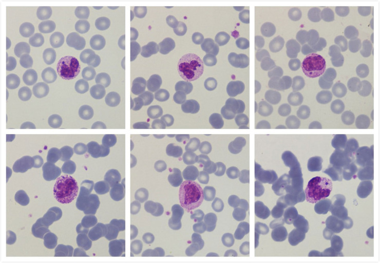 Figure 1
Peer Reviews
No public reviews on file for this paper yet. If you reviewed it on a platform where reviews are public (OpenReview, ICLR, NeurIPS, ICML), you can paste yours below so the community can read it here.
Videos
No videos yet. Explain this paper in a talk, walkthrough, or lecture? Add one.
Taxonomy
TopicsMycobacterium research and diagnosis · Autoimmune and Inflammatory Disorders · Chronic Lymphocytic Leukemia Research
A 38-year-old woman with history of a liver transplant performed four months earlier, presented with fever and multiple lymphadenopathies. She was taking mycophenolate, tacrolimus and prednisone for chronic rejection, lamivudine because of hepatitis B virus serology, and valganciclovir due to recent reactivation of cytomegalovirus.
On admission the complete blood count findings included: hemoglobin 9.7 g/dL, platelets 260 × 10^9^/L, leukocytes 1.5 × 10^9^/L with 0.2 × 10^9^/L neutrophils and elevated C-reactive protein (120 mg/L).
Peripheral blood examination showed hyposegmentation in neutrophils with Howell-Jolly body-like inclusions (Figure 1).Figure 1. Peripheral blood smear showing atypical inclusions (“Howell-Jollylike-bodies”) in granulocytes(Optical microscopy images using May-Grünwald Giemsa stain - x1000 magnification).Figure 1:
Blood cultures for bacteria and fungus did not support growth of any organism and serologic tests were negative. Additionally, lymph node aspiration cytology did not reveal tumoral cells however, a polymerase chain reaction-based assay to detect Mycobacterium tuberculosis in the ganglion was positive. With the diagnosis of ganglionic tuberculosis, the patient received treatment with isoniazid, pyrazinamide, myambutol and levofloxacin. After one year of treatment, the leukocyte count is normal and the adenopathies have disappeared in a full body scan.
Howell-Jolly body-like inclusions in granulocytes are small dense basophilic inclusions similar to Howell-Jolly in erythrocytes. Their appearance in neutrophils may indicate a nuclear fragmentation induced by antiviral treatment with nucleoside analogs, which act on viral DNA. They arise secondary to stressed granulopoiesis often induced by immunosuppressive states including congenital conditions or acquired due to drugs for HIV infection or chemotherapy [1,2]. They are also been described in patients with Mycobacterium avium infection and more rarely in myelodysplastic syndromes [3]. These inclusions must be differentiated from other neutrophil inclusions such as those observed in intracellular bacterial infections, those found in genetic conditions such as Chédiak-Higashi syndrome, or Döhle bodies [1].
Conflicts of interest
The authors of this paper have no conflicts of interest, including specific financial interests, relationships, and/or affiliations relevant to the subject matter or materials included.
The reference list from the paper itself. Each links out to its DOI / PubMed record.
- 1Morales-Indiano C.Arenillas Rocha L.Mas Bosch V.Florensa Brichs L.Howell-jolly body-like inclusions in immunocompromised patients with antiviral treatment Ann Hematol 9312201420912092 Dec 2484478210.1007/s 00277-014-2109-x · doi ↗ · pubmed ↗
- 2Omman R.Kwong C.Shepherd D.Molnar J.A.Velankar M.M.Mirza K.M.Revisiting Howell-Jolly body-like cytoplasmic inclusions in neutrophils: a report of two cases and confirmation of nuclear origin J Hematol 642017101104 Oct 3230040210.14740/jh 334w PMC 7155841 · doi ↗ · pubmed ↗
- 3Mattana Dionisio L.Koehler J.de Faria Moss M.Howell-jolly body-like inclusions in neutrophils of a patient with a myelodysplastic syndrome Br J Haematol 19252021799 Mar 3321636610.1111/bjh.17209 · doi ↗ · pubmed ↗