A Diagnostic Triad in the Vesicular Stage of Incontinentia Pigmenti
Thien Nguyen, Tuan Anh Vu

Abstract
Genes, proteins, chemicals, diseases, species, mutations and cell lines named across the full text — each resolved to its canonical identifier and authoritative record.
Click any figure to enlarge with its caption.
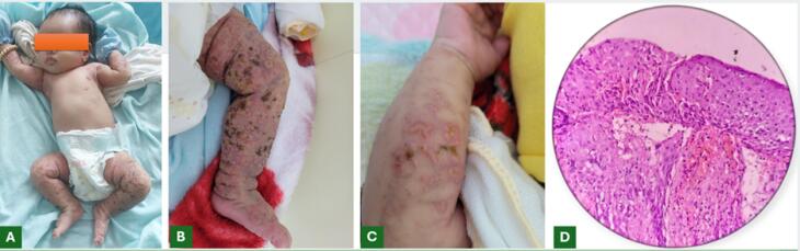 Figure 1
Peer Reviews
No public reviews on file for this paper yet. If you reviewed it on a platform where reviews are public (OpenReview, ICLR, NeurIPS, ICML), you can paste yours below so the community can read it here.
Videos
No videos yet. Explain this paper in a talk, walkthrough, or lecture? Add one.
Taxonomy
TopicsGenetic and rare skin diseases. · Hedgehog Signaling Pathway Studies · RNA regulation and disease
A 2-month-old female infant presented with cutaneous symptoms since birth, characterized by vesicles and tense bullae on an erythematous base. The lesions seem to exhibit a Blaschko-linear distribution predominantly involving the extremities (Figure 1A, B and C). The infant was hemodynamically stable, alert, feeding well, afebrile, and without irritability. Clinical examination revealed no extracutaneous abnormalities. Family history revealed similar cutaneous symptoms during infancy in the patient’s mother, maternal aunt, and maternal grandmother. Complete blood count demonstrated leukocytosis with eosinophilia dominance. Histopathology of lesions revealed prominent eosinophilic spongiosis (Figure 1D).
The vesicular stage is the first stage of incontinentia pigmenti (IP), a disease with four distinct stages, each corresponding to the individual’s growth. The estimated incidence of IP is approximately 0.7 cases per 100 000 births.^1^ As an X-linked dominant genetic disorder, IP manifests predominantly in females because affected males typically cannot survive until birth. The characteristic clinical symptoms of vesicular stage IP include vesicles and tense bullae on an erythematous base, distributed along Blaschko lines.^2^ Diagnosing IP is generally straightforward but may pose challenges for less experienced physicians. When clinical assessment is inconclusive, supportive diagnostic tools include complete blood count demonstrating eosinophil-predominant leukocytosis and histopathological evidence of eosinophilic spongiosis. Family history evaluation is also crucial, particularly investigating dermatological conditions in female relatives on the maternal side of the patient’s family and documentation of miscarriages or absence of male offspring in the maternal lineage. In most cases, the diagnosis of vesicular stage IP still relies primarily on cutaneous manifestations.^3^ This principle is reflected in the 2014 diagnostic criteria,^4^ which require at least one major criterion, characteristic stage 1 skin findings, and at least one minor criterion, frequently histopathological evidence of eosinophilic spongiosis, particularly when extracutaneous involvement is relatively subtle or goes unnoticed in otherwise healthy infants like our case.
In summary, the triad of characteristic skin lesions, peripheral eosinophilia, and histopathological eosinophilic spongiosis can be considered the cornerstone for diagnosing the vesicular stage of IP.
The reference list from the paper itself. Each links out to its DOI / PubMed record.
- 1Cammarata-Scalisi F Fusco F Ursini MV Incontinentia pigmenti Actas Dermosifiliogr (Engl Ed)20191104273810.1016/j.adengl.2019.03.00930660327 · doi ↗ · pubmed ↗
- 2Çetinarslan T Fölster-Holst R Van Gysel D Buchner M Happle R Incontinentia pigmenti stage 1 is not simply vesiculo-bullous but vesiculo-pustular Pediatr Dermatol 2024411182310.1111/pde.1546538284782 · doi ↗ · pubmed ↗
- 3Pal T Agrawal S Grover C Incontinentia pigmenti Indian Pediatr 202461879910.1007/s 13312-024-3268-z 39001787 · doi ↗ · pubmed ↗
- 4Andrade A Gonçalves CF Fernandes PV Jacinto T Teixeira F Costa E Incontinentia pigmenti - one case, two diagnoses J Clin Images Med Case Rep 202452283110.52768/2766-7820/2831 · doi ↗