Infected left atrial myxoma with Streptococcus gordonii: case report and literature review
Liqin Ruan, Shixiong Chen, Jing Zhang, Guiping Peng

TL;DR
A rare case of infected heart tumor caused by Streptococcus gordonii is reported, highlighting the importance of thorough diagnostic methods to avoid misdiagnosis.
Contribution
Presents a novel case of infected left atrial myxoma confirmed using metagenomic sequencing and highlights diagnostic strategies for similar cases.
Findings
Infectious left atrial myxoma can present with nonspecific symptoms, leading to delayed diagnosis.
Streptococcus gordonii was identified as the causative agent using postoperative blood culture and metagenomic sequencing.
Combining mNGS, PCR, and TEE is recommended for diagnosing febrile cardiac myxoma with suspected infection.
Abstract
Cardiac myxoma is a relatively common type of benign heart tumor, but infectious myxoma is rare. The symptoms of non-infected cardiac myxoma and infected cardiac myxoma are similar and mostly nonspecific, which can easily lead to delayed diagnosis, missed diagnosis, and delayed treatment. A 57-year-old male patient presented with nonspecific systemic symptoms such as anorexia, fever, and cough, and was initially considered to have gastrointestinal disease or pulmonary infection. Preoperative bacterial culture was negative, and imaging features were consistent with cardiac myxoma. A small amount of vegetation was found attached to the surface of the tumor. Postoperative blood culture, surgical specimen culture, and postoperative blood metagenomic next-generation sequencing (mNGS) examination all showed positive results for Streptococcus gordonii, confirming the diagnosis of infectious…
Genes, proteins, chemicals, diseases, species, mutations and cell lines named across the full text — each resolved to its canonical identifier and authoritative record.
Click any figure to enlarge with its caption.
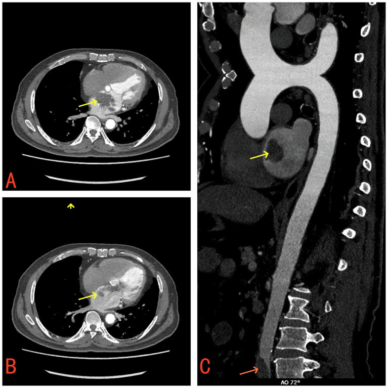 Figure 1
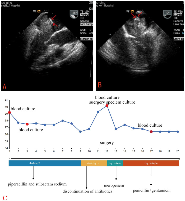 Figure 2
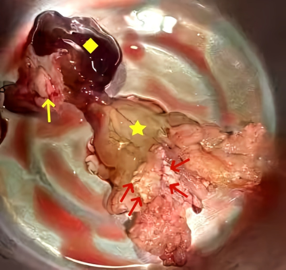 Figure 3
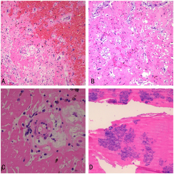 Figure 4
| Serial | Year | Author | Sex | Year | Microorganism | Diagnostic means | Location of myxoma | Surgery | Risk factor | Complication | Outcome | Symptom | Time of antibiotic | References |
|---|---|---|---|---|---|---|---|---|---|---|---|---|---|---|
| 1 | 2022 | José M | Male | 63 | Fusobacterium nucleatum | TTE, TEE | LA | yes | diabetes mellitus | splenic abscesses | survival | fever, sweating, epigastric discomfort, weight loss | 6 weeks | ( |
| 2 | 2018 | Gerald Paul Fitzgerald | male | 23 | Streptococcus | TTE, TEE | LA | yes | none | none | survival | Low fever, anorexia, weight loss, | 6 weeks | ( |
| 3 | 2022 | Coutinho | Female | 33 | Haemophilus spp | TTE | MV | yes | none | splenic abscesses, | survival | fever, sweating, shortness of breath, weight loss | 4 weeks | ( |
| 4 | 2023 | Shi A Kim, | woman | 39 | Haemophilus parainfluenzae | TTE, TEE | LA | yes | none | none | survival | fever, chills, myalgia, headache, | 3 weeks | ( |
| 5 | 2019 | Neil Patel | Male | 43 |
| TTE, TEE | LV | yes | intravenous Heroin | cerebral infarction | survival | shortness of breath, weakness | 4 weeks | ( |
| 6 | 2019 | Matthew J. Peters | male | 34 | Streptococcus parasanguinis | TEE | LA | yes | none | middle cerebral | survival | a feeling of pressure on the chest, symptoms in the left arm | 6 weeks | ( |
| 7 | 2022 | Masi Javeed | male | 70 | Escherichia coli | TTE, TEE | RA | yes | post bilateral femoral artery stents, diabetes mellitus | none | survival | Shortness of breath | 6 weeks | ( |
| 8 | 2021 | Jayaweera | male | 46 | Kodamaea ohmeri | TEE | TV | yes | a dental procedure | none | survival | fever, malaise, | 6 weeks | ( |
| 9 | 2023 | Kawabori | female | 60 | Streptococcus vestibularis | TEE | LA | yes | none | cerebral infarction | survival | fever | 6weeks | ( |
| 10 | 2019 | Masashi Kawabori | male | 55 | Staphylococcus epidermidis | TTE | LA | yes | none | splenic infarct | survival | malaise, fever, weight loss | 6 weeks | ( |
| 11 | 2021 | Takashi Yamamoto | female | 72 | Streptococcus mitis | TEE | RA | yes | a dental procedure | septic pulmonary | survival | bilateral purpura, leg edema, general fatigue, weight loss | 4 weeks | ( |
Peer Reviews
No public reviews on file for this paper yet. If you reviewed it on a platform where reviews are public (OpenReview, ICLR, NeurIPS, ICML), you can paste yours below so the community can read it here.
Videos
No videos yet. Explain this paper in a talk, walkthrough, or lecture? Add one.
Taxonomy
TopicsCardiac tumors and thrombi · Infective Endocarditis Diagnosis and Management · Advanced Surface Polishing Techniques
Background
Cardiac myxomas are uncommon neoplasms. The classic clinical presentation of cardiac myxomas often encompasses a triad of symptoms: systemic, obstructive, and embolic manifestations (1, 2). Infected cardiac myxomas represent an exceedingly rare clinical scenario, with only isolated cases documented in the medical literature (3, 4).
Case report
A 57-year-old male patient presented to the hospital emergency department with a one-month history of intermittent fever, fatigue of unknown origin, anorexia, occasional cough, expectoration of white sputum, and frequent urination. Initially admitted to the gastroenterology department of our hospital, the patient was suspected of having a digestive tract disease, lung infection, or urinary tract infection. The patient has a history of newly diagnosed type 2 diabetes mellitus and denies any history of dental procedures. On physical examination, his blood pressure was 118/73 mmHg, heart rate was 110 beats per minute, body temperature was 38.2°C, and respiratory rate was 18 breaths per minute. Notably, the first heart sound was intensified, and an occasional soft early diastolic murmur was auscultated. Lung examination revealed coarse breath sounds bilaterally. The abdominal examination was unremarkable, but mild bilateral edema was observed in the extremities. The remaining physical examination findings were normal.
Laboratory tests revealed a raised white blood cell count of 12.58×10^9/L, with 83% polymorphonuclear cells. The hemoglobin concentration was 10.2 g/dL, and the hematocrit was 30.8%. The fasting blood glucose level was 9.73 mmol/L. Serum albumin and C-reactive protein (CRP) levels were 31.7 g/L and 16.10 mg/dL, respectively. Urine analysis showed glucose 2+ and protein 1 +. Chest computed tomography (CT) and electrocardiograms were normal. Aortic and coronary CT angiography (CTA) indicated a space-occupying lesion in the left atrium and a filling defect at the intersection of the abdominal aorta and bilateral common iliac arteries, suggestive of thrombosis (Figure 1). Transthoracic echocardiography (TTE) and TEE revealed an elongated, irregular mass originating from the fossa ovalis in the left atrium, with a base of approximately 9 mm and dimensions of approximately 7 cm × 3 cm. The tumor prolapsed into the left ventricle during diastole, causing mitral valve obstruction. TEE revealed punctate non-shadowing echogenic foci without comet tail artifact on the surface of the tumor (Figure 2; Supplementary Video 1). These findings were consistent with atrial myxoma. Additionally, the echocardiographic findings included left atrial dilation, mild to moderate tricuspid regurgitation, tachycardia, and an estimated ejection fraction of 55% in the left ventricle.
(A) Contrast-enhanced CT demonstrates a mass in the left atrium (yellow arrow). (B) The left atrial mass prolapses through the mitral valve into the left ventricle (yellow arrow). (C) A filling defect suggestive of thrombus observed at the bifurcation of the abdominal aorta and bilateral common iliac arteries (red arrow).
TEE revealed an elongated, irregular mass originating from the fossa ovalis in the left atrium, with a base of approximately 9 mm and dimensions of approximately 7 cm × 3 cm and punctate echogenic foci observed on the surface of the tumor (red arrow). (A) Long-axis view of the left ventricle (B) Four-chamber view (C) Clinical Timeline.
Empirical antibiotic therapy with piperacillin and sulbactam sodium was initiated due to the patient’s presentation of fever, cough, frequent urination, along with elevated inflammatory markers, suggesting a likely respiratory or urinary tract infection. On the third day after admission, blood cultures showed the growth of Gram-positive bacilli, which was regarded as a contaminant. A repeat blood culture was negative for bacterial growth. The patient’s body temperature gradually returned to normal. Given the likelihood that the fever was caused by the cardiac myxoma, antibiotics were discontinued after the patient was transferred to the cardiothoracic surgery department. However, following the discontinuation of antibiotics, the patient’s body temperature rose again to 39°C. The patient underwent left atrial tumor resection under cardiopulmonary bypass on the third day after the transfer. Intraoperative exploration revealed a pedunculated mass located in the fossa ovalis in the left atrium. The tumor was a lobulated, dark-red, gelatinous mass attached to the interatrial septum, with a tail-like portion exhibiting a yellow, villous mass. Multiple small vegetations were visible on the surface of the tumor (Figure 3). The tumor and a portion of the interatrial septum were resected.
Gross examination revealed a pedunculated, lobulated, dark-red, gelatinous mass (yellow square) was observed attached to the interatrial septum (yellow arrow), with a tail-like portion exhibiting a yellow, villous tumor (five-pointed star) with an irregular surface and vegetations (red arrows).
Postoperatively, the patient was managed with intravenous meropenem (1 g every 8 hours) due to the suspicion of infective endocarditis (IE). On the second postoperative day, cultures of both blood and the vegetation of the surgery specimen yielded Streptococcus gordonii, which was sensitive to penicillin, vancomycin, ampicillin, ceftriaxone, cefotaxime, cefoxitin, and erythromycin. Infectious disease experts recommended treatment with penicillin (3.2 million units every 6 hours) and gentamicin. (80 mg every 12 hours). Postoperative mNGS of the patient’s blood detected both Streptococcus gordonii and Epstein-Barr virus. A transthoracic echocardiogram (TTE) performed on the fourth postoperative day showed no residual mass.
Histopathological examination of the surgical specimen showed spindle-shaped cells, and stellate cells scattered on the background of a myxomatous matrix. The tumor surface was covered with fibrin and bacterial masses (Figure 4). These findings were compatible with a diagnosis of infected cardiac myxoma.
Histopathological examination (A) the myxoma with hemorrhage (×100) (B) spindle-shaped cells, and stellate cells scattered on the background of a myxomatous matrix (eosin stain, ×100) (C) the myxoma (×400) (D) Bacterial colonies and fibrin on the surface of the myxoma (×400).
Postoperative blood cultures were negative. The patient was prescribed a 1-month course of penicillin and gentamicin. The patient had an uneventful postoperative recovery. Additionally, considering the presence of an abdominal aortic thrombus, continued anticoagulation therapy with aspirin will be provided after discharge.
Discussion
Cardiac myxomas are uncommon neoplasms. Comprehensive reviews of infected cardiac myxomas were conducted by Revankar & Clark in 1998 and Shi-Min Yuan in 2015, compiling a total of 40 and 39 cases, respectively (5, 6). These reviews provided detailed characterizations of the clinical profiles associated with this rare condition. Their analyses revealed that while there is no definitive distinction between infected and non-infected cardiac myxomas, infected myxomas tend to be associated with more pronounced fever-related symptoms and a heightened risk of embolic complications. Recent dental procedures, recent infections, history of invasive surgery, and immunocompromised status are risk factors for infection of cardiac myxoma. We conducted a comprehensive literature search for all cases reported between 2015 and 2025. Using the MeSH terms “infection”, “myxoma”, “endocarditis”, “blood culture” and “bacteremia”, we performed a full-text search of the PubMed database and identified 11 relevant references (3, 7–16). Our findings are consistent with the previously reported cases in symptoms, risk factors, and complication (Table 1).
Fever is observed in approximately 20% of patients with non-infected cardiac myxomas (17), whereas the proportion rises significantly to 92%-97.3% in those with infected myxomas (5, 6). Although infective cases commonly present with elevated white blood cell counts (76.9%), anemia (92.9%), and increased erythrocyte sedimentation rate (ESR) and CRP (6). Non-infected cardiac myxomas with prominent systemic symptoms may also exhibit abnormal inflammatory markers (18). Consequently, fever and abnormal inflammatory markers alone cannot reliably distinguish between infected and non-infected myxomas.
In this case, the patient’s prior use of antibiotics before blood culture resulted in a negative blood culture, further complicating the diagnostic process. However, the observation that the patient’s fever was suppressed by antibiotics and recurred upon their discontinuation strongly suggested the possibility of a concomitant infection. Additionally, mNGS and PCR techniques offer significant advantages in diagnosing culture-negative IE, effectively improving the detection rate of pathogens (19).
While TEE is nearly 100% sensitive for detecting cardiac myxomas, differentiating the imaging characteristics of infected and non-infected cardiac myxomas remains challenging. In this case, a retrospective review of TEE revealed intrasolid punctate nonshadowing echogenic foci without comet tail artifact on the surface of the tumor, which were interpreted as ultrasonic manifestations of tiny, nodular vegetations that were not detected by TTE. A finger-like projecting structure seen attached to the mass is considered to be TEE manifestation of a vegetation on the tumor (20). This highlights the indispensable role of TEE in identifying infections in cardiac myxomas, particularly its enhanced capability in detecting tiny vegetations.
Systemic embolism is a common complication in patients with left atrial myxoma, with potential embolism sites including the brain, coronary arteries, aorta, kidneys, spleen, extremities, and pulmonary arteries (6). The incidence of embolism is significantly higher in infected myxomas compared to non-infected myxomas (5, 21, 22). Although this patient did not exhibit overt embolic symptoms, he was found to have an abdominal aortic thrombosis, necessitating anticoagulation and antiplatelet therapy postoperatively. Therefore, preoperative assessments should include Doppler ultrasonography of the extremity vessels, visceral vasculature CTA and cranial CT to avoid missing cases of vascular thrombus.
Streptococci are the second most common cause of IE after staphylococci, with streptococci viridans accounting for approximately 30% of all streptococcal-related endocarditis cases (23). However, Streptococcus gordonii, a member of the Streptococcus sanguinis group, is rarely reported as a cause of IE (24–28). A study in Denmark showed a higher proportion of endocarditis among patients with sepsis caused by this bacterium, which may be related to geographical distribution or differences in bacterial flora (23). Major risk factors include oral trauma, dental procedures, and immunocompromise (24). It is likely that Streptococcus gordonii escapes from the oral cavity and enters sterile body sites, causing a variety of severe infections when the immune system is compromised, including IE (24), empyema in the lungs (29), septic arthritis (28), and pyogenic spondylitis (30) or spondylodiscitis (31). To date, there have been no reported cases of infected cardiac myxoma caused by Streptococcus gordonii. The virulence factors PadA and Hsa proteins of Streptococcus gordonii enable the bacterium to effectively bind to the tumor cells of cardiac myxomas and platelets, forming complex biofilms composed of bacterial-platelet-fibrin complexes. Additionally, the serine-rich glycoprotein GspB in the cell wall of S. gordonii further promotes platelet aggregation, which can lead to thrombus formation.
Epstein–Barr virus (EBV) was detected in the postoperative mNGS. However, preoperative blood tests for EBV were negative, and the number of EBV sequences and the coverage were both low in the mNGS results. This suggests that there is no active EBV infection. Moreover, there was no pathological evidence of EBV infection in the cardiac myxoma. Therefore, we conclude that the detection of EBV in the mNGS was likely a coincidental finding and not related to the pathogenesis of the infected cardiac myxoma in this case.
Currently, there is no consensus regarding the optimal timing of surgery for patients with infected cardiac myxoma. Most patients are inclined to undergo initial antimicrobial therapy to stabilize their condition (6). For those who experience embolic events and disseminated intravascular coagulation, emergency surgery is more likely to be considered (4). Based on current evidence, a 30-day course of postoperative antibiotic use is considered safe and effective (6).
In conclusion, we report a case of infected cardiac myxoma caused by Streptococcus gordonii. This case underscores several critical clinical lessons: cardiac myxoma patients presenting with fever should be closely monitored for potential concurrent infections. Obtaining blood cultures before initiating antibiotic therapy is crucial for accurate diagnosis. When blood cultures are negative, mNGS and PCR testing can provide valuable insights into the causative pathogens. TEE is indispensable for detecting vegetations on the surface of cardiac myxomas. Early TEE can provide critical information for diagnosis. The use of appropriate antibiotics and surgical resection of the infected myxoma, are key to improving patient outcomes. Avoiding premature discontinuation of antibiotics is also crucial to ensure complete eradication of the infection.
The reference list from the paper itself. Each links out to its DOI / PubMed record.
- 1Gasparovic I Artemiou P Bezak B Michut S Hulman M . Surgery for cardiac myxomas: 12-year experience. Bratisl Lek Listy. (2023) 124:635–8. doi: 10.4149/BLL_2023_098, PMID: 37635659 · doi ↗ · pubmed ↗
- 2Li Y Yang W Liao S Zuo H Liu M . Cardiac myxomas as great imitators: a rare case series and review of the literature. Heart Lung. (2022) 52:182–9. doi: 10.1016/j.hrtlng.2022.01.010, PMID: 35101277 · doi ↗ · pubmed ↗
- 3Jayaweera JAAS Kothalawala M Sooriyar S . Infected tricuspid valve myxoma with Kodamaea ohmeri: Case report. Indian J Med Microbiol. (2021) 39:252–5. doi: 10.1016/j.ijmmb.2020.12.002, PMID: 33966863 · doi ↗ · pubmed ↗
- 4Yoshioka D Takahashi T Ishizaka T Higuchi T . Successful surgical resection of infected left atrial myxoma in a case complicated with disseminated intravascular coagulation and multiple cerebral infarctions: case report. J Cardiothorac Surg. (2011) 6:68. doi: 10.1186/1749-8090-6-68, PMID: 21569401 PMC 3108288 · doi ↗ · pubmed ↗
- 5Revankar SG Clark RA . Infected cardiac myxoma: case report and literature review. Med (Baltimore). (1998) 77:337. doi: 10.1097/00005792-199809000-00003, PMID: 9772922 · doi ↗ · pubmed ↗
- 6Yuan S-M . Infected cardiac myxoma: an updated review. Braz J Cardiovasc Surg. (2015) 30:571–8. doi: 10.5935/1678-9741.20140112, PMID: 26735605 PMC 4690663 · doi ↗ · pubmed ↗
- 7Abuelo JM García Carro J Caneiro J El-Diasty M Fernandez Gonzalez AL . Left atrial myxoma infected with fusobacterium nucleatum. Port J Card Thorac Vasc Surg. (2022) 29:55–7. doi: 10.48729/pjctvs.287, PMID: 36197820 · doi ↗ · pubmed ↗
- 8Fitzgerald GP Coughlan JJ Satti Z Arnous S . Atrial myxoma presenting as infective endocarditis. BMJ Case Rep. (2018) 2018:bcr–2017-223656. doi: 10.1136/bcr-2017-223656, PMID: 29525758 PMC 5847994 · doi ↗ · pubmed ↗