A Rare Case of Actinomycosis in a Patient With Systemic Lupus Erythematosus
Vinaya Gopalaswamy, Sarit S Pattanaik, Manoj K Parida, Saumya R Tripathy, Bidyut K Das

TL;DR
A rare case of skin actinomycosis in a lupus patient shows the need for biopsy, long-term antibiotics, and surgery to manage recurring infections.
Contribution
Highlights diagnostic and treatment challenges of cutaneous actinomycosis in immunosuppressed SLE patients.
Findings
Cutaneous actinomycosis occurred in a 35-year-old SLE patient on immunosuppressive therapy.
Prolonged antibiotic therapy and surgical excision were required to manage recurring lesions.
No recurrence was observed over a two-year follow-up after surgical intervention.
Abstract
Actinomycosis is a rare, chronic infection caused by Actinomyces species, which are normal commensals of human mucosa. It commonly involves the orocervicofacial, thoracic, abdominal, and pelvic regions. It can also affect other sites, such as cutaneous tissue, as seen in our case. We report a case of a 35-year-old woman with systemic lupus erythematosus (SLE) and lupus nephritis on immunosuppressive therapy who developed a large, nodular cutaneous actinomycosis lesion over the right calf. A biopsy confirmed Actinomyces, and she responded to a six-month course of amoxicillin-clavulanate. However, the lesion recurred a year later, requiring surgical excision due to persistent disease despite prolonged antibiotic therapy. Over a two-year follow-up, no further recurrence was noted. This case highlights the diagnostic challenges of cutaneous actinomycosis, particularly in SLE patients, and…
Genes, proteins, chemicals, diseases, species, mutations and cell lines named across the full text — each resolved to its canonical identifier and authoritative record.
Click any figure to enlarge with its caption.
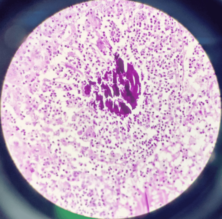 Figure 1
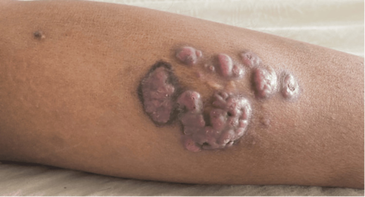 Figure 2
| Patient details | Background disease | Immunosuppression | Phenotype | Precipitating event | Treatment | reference |
| 35/F | SLE with ESRD | Details not specified | Largyneal actinomycosis | Intubation 1 year back | Excision followed by oral penicillin for 3 months | [ |
| 47/M | SLE with ESRD post-transplant | NA | Laryngeal | Intubation recent past | Penicillin for 3 months | [ |
| 49/F | SLE with Class IV LN | Steroids and cyclophosphamide followed by azathioprine | Pelvic actinomycosis | IUCD 2 years back | Excision followed by ampicillin IV for 1 month followed by oral antibiotic | [ |
| 28/F | SLE with ESRD on haemodialysis | Details not specified | Oesophageal actinomycosis | NA | IV penicillin for 1 week followed by ampicillin for 6 months | [ |
| 27/M | SLE with ESRD post-cadaveric transplantation | Prednisolone praquenil prograf | Oesophageal actinomycosis | NA | IV penicillin for 4 weeks followed by oral antibiotic | [ |
| 34/F | SLE | Methotrexate hydroxychloroquine | Cervical | NA | Amoxicillin for 12 months | [ |
Peer Reviews
No public reviews on file for this paper yet. If you reviewed it on a platform where reviews are public (OpenReview, ICLR, NeurIPS, ICML), you can paste yours below so the community can read it here.
Videos
No videos yet. Explain this paper in a talk, walkthrough, or lecture? Add one.
Taxonomy
TopicsActinomycetales infections and treatment · Fungal Infections and Studies · Infective Endocarditis Diagnosis and Management
Introduction
Actinomycosis is a rare, chronic, granulomatous infection caused by gram-positive, filamentous Actinomyces species [1]. These bacteria are normal commensals of the human mucosa and typically become pathogenic following a breach in the mucosal barrier [1]. Actinomyces primarily affect the orocervicofacial, thoracic, abdominal, and pelvic regions [2]. However, it can also involve other sites, such as cutaneous and subcutaneous tissue.
Systemic lupus erythematosus (SLE) is an autoimmune disease characterized by multi-organ involvement and immune system dysregulation. Patients with SLE, especially those undergoing immunosuppressive therapy, are at an increased risk of infections, including bacterial, fungal, and opportunistic infections [3]. Although actinomycosis is not classically considered an opportunistic infection, increasing reports suggest that immunocompromised individuals like patients on steroids [2], may have a predisposition to developing Actinomyces infections. Here, we present a unique case of cutaneous actinomycosis in an SLE patient with lupus nephritis, highlighting diagnostic challenges, treatment responses, and a review of similar reported cases.
Case presentation
This report is of a 35-year-old female diagnosed with SLE for the past 12 years with mucocutaneous and nephritis as the affected domains. She received induction therapy with three doses of pulse methylprednisolone and intravenous (IV) cyclophosphamide (Euro-Lupus regimen) in 2017 for nephritis, followed by maintenance therapy with mycophenolate (2 g/day), prednisolone (2.5 mg/day), and hydroxychloroquine (200 mg/day).
In April 2021, she presented with a large nodular mass lesion over the right calf for two months which was progressively increasing in size and non-painful. The lesion was not fixed to the underlying muscle. There was no discharge or surface changes. She had no history of fever or lymphadenopathy. As the patient was immunosuppressed, the differential diagnosis of hypertrophic variant of lupus vulgaris, actinomycosis, sporotrichosis, and fungal infection were considered. Her routine labs were unremarkable. Ultrasonography showed a hypoechoic, nodular, conglomerated, elongated lesion measuring 11 × 7 cm in the subcutaneous plane in the posterior aspect of the right mid-calf with minimal internal flowing debris with normal underlying muscle. Punch biopsy of the mass showed infiltration of polymorphs and actinomycotic colony formation in the epidermis and dermis with Splendore Hoeppli formation (Figure 1). She was given amoxycillin-clavulanate for six months with good response.
Clumps of basophilic bacteria with branching filaments surrounded by eosinophilic club-shaped ends (Splendore Hoeppli reaction) surrounded by dense acute inflammatory infiltrate with necrotic areas.
In April 2022, she presented with recurrence of a nodular variegated mass with satellite lesions in the same region (Figure 2) and repeat biopsy showed non-specific granulomatous tissue with foci of ill-defined suppurative granuloma and foreign body-type giant cells and positive staining for Actinomyces. Simultaneously, the patient had mucocutaneous and arthritis flares that were managed by increasing the steroid dose. In view of the frequent flares of SLE that required disease-modifying antirheumatic drugs (DMARDs), excision of the lesion was performed. In the follow-up at two years, the patient had not shown any further recurrence.
Clinical image of the lesion during recurrence in April 2022
Discussion
Actinomycosis is an uncommon, endogenous infection affecting all age groups caused by Actinomyces species [1]. Actinomyces is a gram-positive, filamentous, non-acid-fast, anaerobic to microaerophilic bacteria that normally resides on the mucosal surface of the mouth, colon, and genitourinary tract. Classically, it is an indolent, slowly progressive granulomatous disease that mimics malignancy, as it does not respect tissue boundaries [4].
Mucosal disruption can lead to infection, most commonly orocervicofacial, abdominal, thoracic and pelvic region but virtually any site can be involved [1]. Our case was atypical in its location involving skin and subcutaneous tissue of the lower extremities. There are previous reports of lower extremity disease among intravenous drug users due to infection from of an injection needle, following foot trauma from a toothpick puncture. No such antecedent trauma was identified in our patient.
The disease can occur in both immunocompetent and immunocompromised individuals [2]. Infections are common in individuals who do not seek or have access to health care or an intrauterine device (IUD) in place for a prolonged duration or who receive bisphosphonates [5], hence has not been considered an opportunistic infection. Reports have shown individuals with HIV, transplantation, common variable immunodeficiency (CVID), chronic granulomatous disease (CGD) [6], or on treatment with tumor necrosis factor (TNF) inhibitors [7], steroids, bisphosphonates [5], radiotherapy [8], and chemotherapy are susceptible to actinomyces infection.
Of the six documented cases (Table 1) [9-14], four occurred in females, with a collective age range of 27 to 49 years. A prominent feature was the high prevalence of renal disease (five out of six), and had end-stage renal disease (ESRD), including two post-transplant recipients, and most are on some form of immunosuppressive treatment. The clinical presentations of actinomycosis were diverse, involving the oesophagus (two cases), larynx (two cases), neck, and pelvis. An identifiable precipitating factor either prior to intubation [9,10] or an IUD [11] was noted, though a significant latency period of over one year preceded diagnosis [9,11]. This extended timeframe complicates a direct temporal association but remains consistent with the characteristically insidious progression of actinomycotic infections.
Routine blood investigations are nonspecific and may show anemia, mild leukocytosis, and elevated erythrocyte sedimentation rate (ESR) and C-reactive protein (CRP). Imaging in early stages may show nonspecific inflammatory changes with abscess formation but no lymphadenopathy. At later stages, it may demonstrate tissue infiltration mimicking malignancy, with sinus formation, which, although characteristic, is not specific to actinomycosis [2].
Diagnosis is primarily based on histopathology, which identifies sulfur granules and demonstrates gram-positive filamentous bacteria [2]. Sulfur granules, though not specific to actinomycosis, appear as round or oval basophilic masses (colonies of organisms) with eosinophilic terminal clubs. Microbiologic isolation confirms the diagnosis, but the isolation rate is less than 50% [6].
Treatment requires prolonged treatment with high doses of antibiotics (penicillin, amoxicillin, erythromycin, tetracyclines, doxycycline, and clindamycin) [2]. Combined medical-surgical therapy is advocated in selected cases. Our patient had recurrent lesions on her calf, without any obvious trauma or inciting event and didn’t respond to prolonged high doses of therapy requiring complete surgical excision. Although rare, diagnosis can be challenging, especially in atypical sites or without a history of trauma. Correct diagnosis is imperative to determine the appropriate antibiotic duration and the need for surgical excision.
Conclusions
This case highlights a cutaneous presentation of actinomycosis in a patient with SLE on immunosuppressive therapy. The unusual site of infection, absence of trauma, and recurrence despite prolonged antibiotics made diagnosis and management challenging. Histopathology was key to diagnosis, and surgical excision was required due to incomplete response to medical therapy. A combined approach of antibiotics and surgery may be needed in resistant or recurrent cases.
The reference list from the paper itself. Each links out to its DOI / PubMed record.
- 1Actinomyces and related organisms in human infections Clin Microbiol Rev Könönen E Wade WG 4194422820152578851510.1128/CMR.00100-14PMC 4402957 · doi ↗ · pubmed ↗
- 2Actinomycosis BMJ Wong VK Turmezei TD Weston VC 0343201110.1136/bmj.d 609921990282 · doi ↗ · pubmed ↗
- 3Systemic lupus erythematosus and risk of infection Expert Rev Clin Immunol Barber MR Clarke AE 5275381620203247862710.1080/1744666 X.2020.1763793 · doi ↗ · pubmed ↗
- 4Actinomycosis Harrison’s Principles of Internal Medicine. 21st ed Russo TA 13401344 New York Mc Graw-Hill Education 2022 https://accessmedicine.mhmedical.com/content.aspx?bookid=2129§ionid=192023164
- 5Osteopathology associated with bone resorption inhibitors - which role does Actinomyces play? A presentation of 51 cases with systematic review of the literature J Oral Pathol Med Schipmann S Metzler P Rössle M 5875934220132336916610.1111/jop.12038 · doi ↗ · pubmed ↗
- 6Actinomyces in chronic granulomatous disease: an emerging and unanticipated pathogen Clin Infect Dis Reichenbach J Lopatin U Mahlaoui N 170317104920091987420510.1086/647945 PMC 4100544 · doi ↗ · pubmed ↗
- 7Cutaneous actinomycosis associated with anti-TNF-alpha therapy: report of two cases Dermatology Breton AL Lamblin G Pariset C Jullien D 1121142282014 https://doi.org/10.1159/000357522.2457725810.1159/000357522 · doi ↗ · pubmed ↗
- 8Actinomyces in infected osteoradionecrosis--underestimated?Hum Pathol Hansen T Kunkel M Kirkpatrick CJ Weber A 6167372006 https://doi.org/10.1016/j.humpath.2005.09.018.1636041710.1016/j.humpath.2005.09.018 · doi ↗ · pubmed ↗