Parsing Glomerular and Tubular Structure Variability in High-Throughput Kidney Organoid Culture
Kristiina Uusi-Rauva, Anniina Pirttiniemi, Antti Hassinen, Ras Trokovic, Sanna Lehtonen, Jukka Kallijärvi, Markku Lehto, Vineta Fellman, Per-Henrik Groop

TL;DR
This study examines how different culture methods affect kidney organoid development, showing that high-throughput techniques can reduce variability.
Contribution
The study introduces a novel patient-derived iPSC line and evaluates high-throughput methods for kidney organoid culture.
Findings
Culture approach and conditions significantly influence glomerular and tubular structure development in kidney organoids.
High-throughput methods explain 35–77% of variability in organoid structure development.
The protocol is adaptable for other labs to reduce organoid size variability.
Abstract
High variability in stem cell research is a well-known limiting phenomenon, with technical variation across experiments and laboratories often surpassing variation caused by genotypic effects of induced pluripotent stem cell (iPSC) lines. Evaluation of kidney organoid protocols and culture conditions across laboratories remains scarce in the literature. We used the original air-medium interface protocol to evaluate kidney organoid success rate and reproducibility with several human iPSC lines, including a novel patient-derived GRACILE syndrome iPSC line. Organoid morphology was assessed with light microscopy and immunofluorescence-stained maturing glomerular and tubular structures. The protocol was further adapted to four microplate-based high-throughput approaches utilizing spheroid culture steps. Quantitative high-content screening analysis of the nephrin-positive podocytes and…
Genes, proteins, chemicals, diseases, species, mutations and cell lines named across the full text — each resolved to its canonical identifier and authoritative record.
Click any figure to enlarge with its caption.
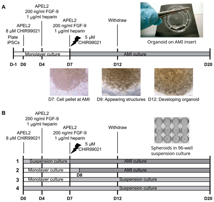 Figure 1
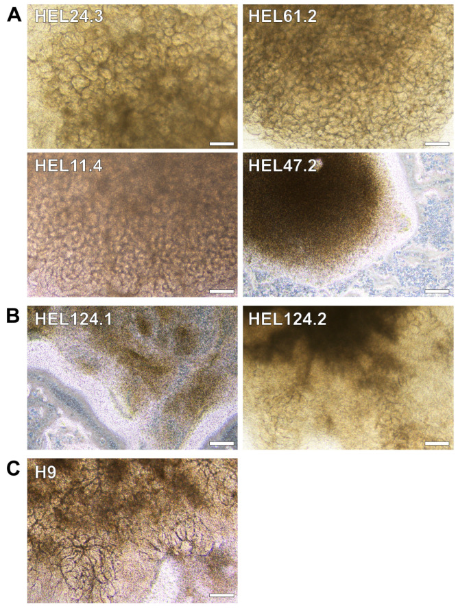 Figure 2
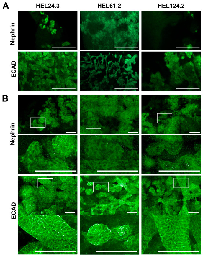 Figure 3
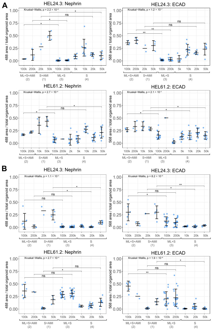 Figure 4
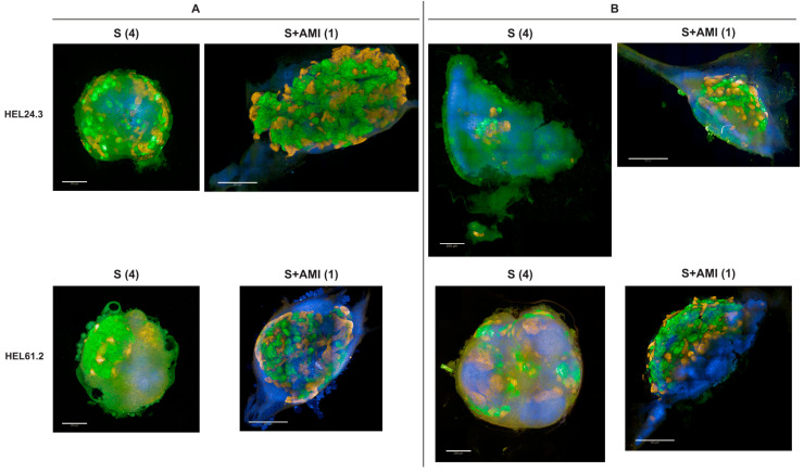 Figure 5
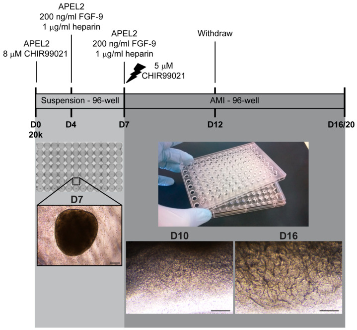 Figure 6
- —Folkhälsan Research Foundation
- —Finnish Academy
- —Wilhelm and Else Stockmann Foundation
- —Novo Nordisk Foundation
- —Foundation for Pediatric Research
- —Swedish Research Council
- —Medical Society of Finland
- —Faculty of Medicine, University of Helsinki
- —Doctoral School in Health Sciences and the Doctoral Programme in Clinical Research, University of Helsinki, Finland
- —Biocenter Finland
- —Helsinki University Library
Peer Reviews
No public reviews on file for this paper yet. If you reviewed it on a platform where reviews are public (OpenReview, ICLR, NeurIPS, ICML), you can paste yours below so the community can read it here.
Videos
No videos yet. Explain this paper in a talk, walkthrough, or lecture? Add one.
Taxonomy
TopicsRenal and related cancers · Pluripotent Stem Cells Research · 3D Printing in Biomedical Research
1. Introduction
Human-induced pluripotent stem cell (hiPSC)-derived kidney organoids represent an attractive micro-scale source of patient-specific kidney tissues, which are otherwise difficult to obtain [1,2]. Although structurally and functionally immature, pluripotent stem cell-derived kidney organoids have been shown to mimic embryonic kidney development remarkably up to the second trimester, resulting in tubular and glomerular structures with foot processes and a primitive endothelial network with architectural resemblance to nephron structures in vivo [3,4,5,6]. Initial reports on the differentiation of human pluripotent stem cells, which include hiPSCs and human embryonic stem cells (hESCs), into kidney cells and kidney organoids [4,7,8,9,10] were soon followed by studies aiming to enhance the level of maturation and complexity of generated structures, as well as efficiency and cost-effectiveness of the production [11,12,13,14,15,16,17,18,19].
Deployment of kidney organoid models, or any stem cell differentiation, can be challenging when the technology is transferred from one laboratory to another, as the choice of protocol, cell lines, and culture conditions may greatly affect the end result of the differentiation [20,21,22]. While it has been reported that laboratory-based variation can surpass genotypic effects due to issues such as organoid cell type heterogeneity, high-throughput applications and third-party testing of the available methodology remain scarce in the literature [12,20,23,24,25,26,27].
In order to evaluate the reproducibility of kidney organoid differentiation, we set out to test success rates of several available iPSC lines that were not preselected based on their capacity to differentiate towards kidney cells. The present study utilized primarily previously reported healthy donor iPSC lines, but the array of cell lines was complemented by novel iPSC lines generated and characterized in the present study. These include patient-derived iPSC lines of the GRACILE syndrome, a rare early-onset and lethal mitochondrial disorder manifested by fetal growth restriction, aminoaciduria, cholestasis, iron overload in the liver, lactic acidosis, and early death, from which the disease acronym was constructed [28,29]. For kidney organoid differentiation, we used one of the first originally described protocols [4,7,10], a 6-well format protocol by Takasato et al. allowing the production of self-organized organoids generated in albumin polyvinyl alcohol essential lipids (APEL) medium at air-medium interface (AMI) (“APEL/air-medium interface protocol”) [4] (Figure 1A).
Subsequently, the original kidney organoid protocol was modified to incorporate spheroid culture steps in four high-throughput approaches utilizing multi-well plates (Figure 1B). The proportion of immunostained nephrin-positive glomerular structures and epithelial cadherin (ECAD)-positive tubular structures were assessed with a quantitative high-content confocal screening system to set up a protocol to fulfil the requirements of high-throughput analyses. A multiple linear model was used to assess the effects of the culture approach, iPSC line, experimental replication, and initial cell number on the glomerular and tubular structure development.
As high variability is a well-known limitation in stem cell research, here we aimed to assess the success rate of kidney organoid structure development and further develop high-throughput culture approaches to enable better distinction of technical and biological sources of variation. Using quantitative high-content screening and multivariate statistics, we show that the culture approach, iPSC line, experimental replication, and initial cell number are significantly associated with nephrin-positive glomerular and ECAD-positive tubular structure development. Our microplate-based high-throughput culture approach demonstrated that spheroid culture can efficiently control organoid size, while air-medium interface culture proved most beneficial for overall renal structure development.
2. Materials and Methods
2.1. Ethics
The present study was performed as a collaboration between Folkhälsan Research Center and University of Helsinki and conducted in accordance with the Declaration of Helsinki. The study was carried out from October 2014 onward and benefited from previously collected human fibroblasts and reprogrammed iPSC lines. All ethical approval statements were obtained from the ethics committee of the Helsinki and Uusimaa Hospital District, Finland (HUS; 77/E7/2007, obtained in 2007, for GRACILE patient fibroblasts; an update of 77/E7/2007, obtained in 2011, for GRACILE iPSCs and differentiation; 423/13/03/00/08, obtained in 2009, for healthy donor fibroblasts/iPSCs and differentiation). Written informed consent for participation was obtained from the donor or the guardians. Healthy donor fibroblasts were collected, and respective iPSC reprogramming experiments were started between January 2010 and May 2012. GRACILE samples were collected, and respective iPSC reprogramming experiments were started between April 2007 and September 2011. The authors had no access to information that could identify individual participants during the study.
2.2. Cell Culture
Human iPSC lines HEL11.4, HEL24.3, and HEL47.2 from healthy donors have been generated and described previously [30,31,32]. hESC line H9 (WA09) was originally from WiCell and Dr. James Thomson, University of Wisconsin and provided by BSCC, Helsinki, Finland. Healthy control iPSC line HEL61 and GRACILE syndrome HEL124 were generated in this study.
Pluripotent stem cells were cultured on Matrigel-coated (BD Biosciences, San Jose, CA, USA) cell culture plates in E8 medium (Life Technologies, Carlsbad, CA, USA) and split using 0.5 mM ethylenediaminetetraacetic acid (EDTA; Thermo Fisher Scientific, Waltham, MA, USA). Rho-associated protein kinase (ROCK) inhibitor (Y-27632; Sigma, St. Louis, MO, USA) was utilized to enhance the cell survival after dissociation and thawing (5.0–10.0 μM). Media were changed every other day. Antibiotics were utilized at routine cell culturing. GRACILE iPSC lines HEL124.1 and HEL124.2 were supplemented with 200 μM uridine. Uridine was also added to the media of all cell cultures in the experiments, where the growth or characteristics of HEL124.1 and HEL124.2 were compared to the healthy donor cell lines. All cells were kept in an incubator at +37 °C and 5% CO_2_. Cell cultures were regularly tested for the mycoplasma infection (LookOut^®^ Mycoplasma qPCR Detection Kit, Sigma).
2.3. Generation and Characterization of Healthy Control iPSC Line HEL61 and GRACILE Syndrome iPSC Line HEL124
iPSC reprogramming was done at Biomedicum Stem Cell Center, University of Helsinki (BSCC; Helsinki, Finland). Dermal fibroblast cultures of a 57-year-old healthy female and a newborn female GRACILE patient (ZCC11-72) were cultured in Dulbecco’s modified Eagle’s medium (DMEM; Life Technologies/Thermo Fisher Scientific) supplemented with 10% fetal bovine serum (FBS; Life Technologies) and 2 mM GlutaMAX (Life Technologies/Thermo Fisher Scientific) (the “fibroblast medium”). The medium of GRACILE fibroblasts was additionally supplemented with 200 μM uridine (Calbiochem, San Diego, CA, USA).
The fibroblasts from the healthy female were seeded four days before reprogramming. On the day of reprogramming (day 0), cells were dissociated into single cells with trypsin and washed with phosphate-buffered saline (PBS). Cells were transfected using a Sendai virus (SeV) kit according to the manufacturer’s instructions (CytoTune-iPS Reprogramming Kit, Life Technologies) and plated on mitomycin C-inactivated mouse embryonic fibroblasts. After five days, cell culture medium was changed to human embryonic stem cell medium (hESC medium; KnockOut DMEM supplemented with 20% KnockOut-Serum Replacement (KOSR), 1% GlutaMAX, 0.1 mM β-mercaptoethanol, 1% nonessential amino acids; all from Life Technologies) containing 6 ng/mL recombinant human basic fibroblast growth factor (FGF-2; Sigma) and supplemented with 0.25 mM sodium butyrate (Sigma). Emerging colonies were picked manually, plated on Matrigel-coated cell culture plates in Essential-8 medium (E8 medium; Life Technologies), and further expanded for the generation of HEL61 iPSC lines. Media were changed every other day. The clone HEL61.2 was included in the analyses of the study.
The fibroblasts (1 × 10^5^ cells) of the GRACILE patient were transduced with SeVdp (a single vector harboring the four reprogramming factors) as previously described [33,34], and plated on Matrigel (BD Biosciences) in the presence of the fibroblast medium supplemented with 200 μM uridine. After five days, cell culture medium was changed to the hESC medium supplemented with 0.25 mM sodium butyrate (Sigma) and 200 μM uridine. When first colonies started to emerge, cell culture medium was changed to E8 medium. iPSC colonies were picked manually and further expanded in the presence of 200 μM uridine. The clones HEL124.1 and HEL124.2 were characterized and the presence of the disease-causing BCS1L mutation was verified by Sanger sequencing.
Chromosomal integrity of HEL124.1 and HEL124.2 was analyzed from extracted DNA at the Finnish Microarray and Sequencing Centre (FMSC, Turku, Finland) by a KaryoLite™ BACs-on-Beads™ method (KaryoLite™ BoBs™, PerkinElmer, Waltham, MA, USA) [35] with two technical replicates per sample. The karyotype of HEL61.2 was analyzed by G-banding.
In the characterization of selected iPSC clones, the expression of embryonic stem cell marker proteins TRA-1-60, OCT3/4, and SSEA3 or SSEA4 was assessed by immunofluorescence microscopy of iPSCs, and the expression of embryonic stem cell marker genes OCT3/4, SOX2, NANOG, and TDGF1 as well as the clearance of the SeV were verified by quantitative reverse transcription PCR (RT-qPCR). Spontaneous differentiation capacity of the generated iPSC lines was tested by embryoid body formation and subsequent immunofluorescence staining of the markers of the three germ layers (α-fetoprotein, β-III tubulin, MAP-2, vimentin, and desmin).
2.4. Reverse Transcription and Quantitative PCR (qPCR)
Total RNA was extracted using RNA Spin II kit (Macherey-Nagel, Düren, Germany) according to the manufacturer’s instructions. cDNA was synthesized from 1 μg total RNA by SuperScript III reverse transcriptase (Invitrogen, Waltham, MA, USA) with oligo dT primer (Invitrogen) in 20 μL volume. One percent of the above cDNA was used for each qPCR reaction in a 20 μL mixture containing 10 μL of SYBR green-Taq mixed solution (Sigma) and 5 μL of 2 μM-primer mix. PCR reactions were carried out in a Corbette thermal cycler (Qiagen, Hilden, Germany) for 40 cycles and each cycle contained +95 °C for 15 s, +60 °C for 30 s and +72 °C for 30 s. Relative gene expression was determined using the ΔΔCt method, with a glyceraldehyde-3-phosphate dehydrogenase (GAPDH) utilized as a housekeeping gene. Expression levels are relative to the RNA sample of hESC line H9 (positive control). Primer sequences (5′→3′) were as follows: GGATCACTAGGTGATATCGAGC (SeV-F), ACCAGACAAGAGTTTAAGAGATATGTATC (SeV-R); TTGGGCTCGAGAAGGATGTG (OCT4-F), TCCTCTCGTTGTGCATAGTCG (OCT4-R), GCCCTGCAGTACAACTCCAT (SOX2-F), TGCCCTGCTGCGAGTAGGA (SOX2-R), CTCAGCCTCCAGCAGATGC (NANOG-F), TAGATTTCATTCTCTGGTTCTGG (NANOG-R), TCAGAGATGACAGCATTTGGC (TDGF1-F), TTCAGGCAGCAGGTTCTGTTTA (TDGF1-R), GGTCATCCATGACAACTTTGG (GAPDH-F), TGAGCTTCCCGTTCAGCTC (GAPDH-R).
2.5. Embryoid Bodies
Cells were dissociated into small aggregates and passaged on ultra-low-attachment 6-well culture plates (Corning, Incorporated, Corning, NY, USA) in hESC medium without FGF-2. The embryoid body culture medium was supplemented overnight with 5 µM ROCK inhibitor (Y-27632, Selleck Chemicals, Houston, TX, USA or Sigma) after the initial cell plating to improve cell viability. In total, 200 μM uridine was added in the medium of HEL124.1 and HEL124.2 GRACILE cell line cultures. Embryoid bodies were grown in suspension for 4–19 days, and the medium was changed regularly. Thereafter, the embryoid bodies were plated on gelatine-coated coverslips and cultured for an additional 6–10 days before processing for immunocytochemistry.
2.6. Immunocytochemistry of iPSCs and Embryoid Bodies
Cultures were fixed at room temperature with 4% paraformaldehyde (PFA) for 15 min and permeabilized using 0.2% Triton X-100 in PBS for 30 min. Non-specific proteins were blocked by Ultra V block (Thermo Fisher Scientific) or 0.5% BSA in PBS. The cells were then incubated with diluted primary antibodies overnight at +4 °C or 1 h at room temperature followed by washes. Secondary antibody incubations were done at room temperature for 40 min in the presence of Hoechst nuclei stain followed by washes. Antibodies are described in Supplementary Table S1.
2.7. Differentiation of iPSCs Towards Hepatocyte Lineage Cells
The protocol utilized for the definitive endodermal differentiation was based on previously published protocol [36]. After modifying the protocol to exclude sodium butyrate and testing cell-line-specific requirements for insulin, healthy donor iPSC line HEL24.3 and both available GRACILE iPSC clones (HEL124.1 and HEL124.2 representing the same patient, ZCC11-72) were successfully differentiated to definitive endoderm. Differentiation of HEL61.2 required insulin supplementation, which was observed to result in a more heterogenous culture in this particular cell line as compared with the differentiated cultures of other utilized cell lines.
2.8. Western Blotting
Samples were lysed in cold 50 mM Tris-HCL pH 7.4, 150 mM NaCl, 1% Triton X-100, and 1 mM ethylene glycol tetra acetic acid (EGTA) lysis buffer supplemented with protease inhibitor cocktail (Roche, Basel, Switzerland). The cells were disrupted on ice with a pestle, and the final lysate was collected after centrifugation at +4 °C for 10 min (13,000 rpm in a microcentrifuge). Protein concentrations of the lysates were analyzed by DC Protein Assay (Bio-Rad, Hercules, CA, USA). Protein lysates (1 or 10 μg) were loaded into single SDS-PAGE gel per comparison (Mini-Protean TGX Any kDa, Bio-Rad), electrophoresed, and semi-dry blotted on 0.2 μm PVDF membrane (Trans Blot Turbo, Bio-Rad). Blots were cut horizontally for simultaneous probing of different proteins (>50 kDa, 37–50 kDa, and <37 kDa). The stainings were carried out according to standard procedure with well-established primary and secondary antibodies (Supplementary Table S2). Stained blots were exposed to HRP substrate (Pierce ECL Western Blotting Substrate or SuperSignal West Femto Maximum Sensitivity Substrate, Thermo Fisher Scientific) and imaged fresh for chemiluminescence (ChemiDoc Imager, Bio-Rad). Chemiluminescence signals were quantified (ChemiDoc Image Lab Software 6.1, Bio-Rad) and normalized against the signals of mitochondrial (porin or SDHA) or cytoplasmic (β-actin) loading controls. The relative expressions of samples representing the same genotype (i.e., control or mutated cell lines) were pooled for visualization of the mean results.
2.9. Differentiation of Pluripotent Stem Cells into Kidney Organoids
Kidney organoids were generated as described [4,15] with the following technical modifications and additional notes (Figure 1A). In APEL/air-medium interface differentiation [4,37], cells were detached with 0.5 mM EDTA (Thermo Fisher Scientific), and small cell aggregates were plated at different densities on Matrigel-coated cell culture dishes in E8 medium (Thermo Fisher Scientific) supplemented with a 2.5–5.0 μM ROCK inhibitor (Y-27632, Sigma). On the next day, the cells were washed, and the differentiation was continued according to the original protocol, except that 5% Protein-Free Hybridoma Medium (PFHM-II; Thermo Fisher Scientific) was separately added to the basal medium available at the time of the study (a recombinant protein-based, animal product-free APEL medium, i.e., APEL2; Stem Cell Technologies, Vancouver, BC, Canada). Of note, none of the tested cell lines were able to differentiate without PFHM. At day 4, the 8 µM glycogen synthase kinase 3 inhibitor (CHIR99021; Tocris Bioscience, Bristol, UK) containing medium was changed to 200 ng/mL animal-free FGF9 (PeproTech, Cranbury, NJ, USA) and 1 µg/mL heparin (Sigma) containing medium according to the original protocol. Approximately 6 × 10^6^ cells were obtained from 12-well (3.8 cm^2^) plates by day 7 of the differentiation. At day 7, cells were detached and aliquoted in microtubes, in which the one-hour shock with 5 μM CHIR99021 (Tocris Bioscience) in APEL2 medium in a cell incubator was carried out, before the cells were pelleted and placed on Transwell membranes (Corning/Sigma).
The following Transwell membranes were utilized: 10 µm thick polyester membrane with 0.4 µm pores and 4 × 10^6^/cm^2^ pore density (#3450), and 10 µm thick polycarbonate membrane with 0.4 µm pores and 1 × 10^8^/cm^2^ pore density (#3412). Kidney organoids were harvested at day 20 unless otherwise stated. Throughout the differentiation, the medium was changed every second day, and the differentiating cells were monitored daily under the light microscope. Uridine was also added to the media of all cell cultures in the experiments where the growth or characteristics of the GRACILE cell lines were compared to the healthy donor cell lines.
The following changes allowing higher throughput by incorporating spheroid culture steps were applied to the APEL/air-medium interface protocol (Figure 1B, approaches 1–4). For spheroid formation, iPSCs were detached with TrypleSelect (Thermo Fisher Scientific), counted, and diluted in different densities (ranging from 2000 to 50,000 cells for approaches 1 and 4 or 100,000 to 200,000 cells for approaches 2 and 3) in 100 µL of medium per 96-well of a CellCarrier Spheroid Ultra-low attachment (ULA) microplate (PerkinElmer) followed by spinning to enhance cell aggregation/spheroid formation. For the spheroid cultures, the CHIR99021 shock at day 7 was carried out in the ULA wells (Figure 1B, approaches 1 and 4), while the shock for the cells detached from the monolayer cultures, for subsequent spheroid formation, was carried out in a microtube containing a master dilution to be aliquoted in each 96-well (Figure 1B, approaches 2 and 3). For the Opera Phenix high-content screening of structures, approaches 1–4 were tested with two selected healthy-donor iPSC lines, HEL24.3 and HEL61.2, both in two separate experiments (experiment A and B), and 24-well AMI inserts were used in approaches 1 and 2 (Supplementary Table S3).
In the differentiation of approach 1 at AMI on 96-well membranes, an array of 96 wells with permeable inserts connected by a rigid, robotics-friendly tray was utilized [HTS Transwell-96 Permeable Support with 10 µm thick polyester membrane containing 1.0 µm pores (#3380)]. The medium was changed regularly according to the original protocol [4,37], except that the medium for the spheroid cultures from day 9 onwards was changed daily. The same or subsequent batches of each iPSC line were utilized throughout the study, unless otherwise stated.
2.10. Immunofluorescence Staining of Kidney Organoids
Samples were fixed with 4% PFA in PBS supplemented with 1 mM CaCl_2_ and 0.5 mM MgCl_2_ for 45 min and washed with PBS. Samples differentiated at AMI in 6-well Transwell inserts (Corning) were detached from the membrane and cut in half for separate staining. The stainings were processed in multiwell plates, except in case of large organoids, which were incubated in small droplets of antibody dilution placed on a clean sealing film. Cells were permeabilized and blocked for unspecific binding with 0.3% Triton X-100 in 10% FBS-PBS, for 4–5 h at room temperature. After washing with 0.1% Triton X-100 and 10% FBS in PBS the samples were incubated in primary antibody dilution, at +4 °C overnight. Fluorescein Lotus tetragonolobus lectin (LTL; Vector Laboratories, Newark, CA, USA, FL-1321, 1:500) and following primary antibodies were utilized: guinea pig anti-mouse nephrin (Progen, Heidelberg, Germany, GP-N2, 1:300) and mouse anti-human epithelial cadherin (ECAD; BD Biosciences 610181, 1:400). After washing, the samples were incubated in a secondary antibody dilution in the dark, for 4–5 h at room temperature, or alternatively, at +4 °C overnight. The following secondary antibodies were utilized: Goat anti-Guinea pig AlexaFluor 488 (Thermo A11073, 1:1000), and goat anti-mouse AlexaFluor 594 (Thermo A11005, 1:1000). Finally, samples were washed, and nuclei were stained (Hoechst 33258, Sigma), followed by rinsing with lab-quality water and mounting with an in-house-made Mowiol mounting medium or Fluoromount Aqueous Mounting Medium (Sigma).
2.11. Microscopy and Morphological Analyses of Kidney Organoids
Whole mount organoids were viewed and imaged for overall appearance using the Nikon Eclipse Ts2 light microscope and DS-Fi3 camera with DS-L4 imaging applications. The immunostained organoids were viewed and imaged using an Axioplan 2 microscope and the AxioCam HRc camera with an AxioVision imaging system (Carl Zeiss Microscopy GmbH, Jena, Germany), and additionally, using Axio Imager M2, AxioCam 503, and ZEN 2.3 software. Kidney organoids were also imaged using a PerkinElmer Opera Phenix spinning disk confocal microscope.
2.12. Opera Phenix High-Content Screening and Image Analysis
High-content imaging of whole mount organoids was performed with a PerkinElmer Opera Phenix spinning disk confocal microscope (High Content Imaging and Analysis Unit, FIMM, HiLIFE, University of Helsinki, Helsinki, Finland) using a 5× widefield (NA 0.16) pre-scan objective and 40× water-immersion objective for the determined organoid area (NA 0.6, working distance 0.62 mm, depth of focus 1.2 µm) using three excitation lasers (405 nm with emission band-pass filter 435/480; ex 488, em 500/550 and ex 561, em 570/630). Nine fields-of-view with 5% overlap were imaged per well using 15 predetermined Z focus planes with laser-based autofocusing. The images were captured with two Andor Zyla sCMOS cameras (16-bit, field of view 650 × 650 μm^2^, effective xy resolution 0.66 µm). Z-stacks of 15 images were analyzed from a maximum projection using the PerkinElmer Harmony 4.9 software package as described in Supplementary File S1.
2.13. Statistical Analysis
Statistical analyses were conducted with R version 4.3.1. The proportional nephrin and ECAD-positive area per total Hoechst-stained area in an organoid were calculated based on the Opera Phenix-acquired images and data files. For the higher throughput approaches 3 and 4, the spheroid organoid diameter was calculated based on the total Hoechst-stained area. Mann–Whitney U test was used for two-group analyses, while the comparison of multiple groups was done using the Kruskal–Wallis test, and Spearman’s rank correlation coefficient was used to assess correlation between nephrin and ECAD positive area per organoid. Multiple linear models were fitted using the proportional nephrin- and ECAD-positive area treated as dependent variables and the higher throughput approaches (1–4), cell line (HEL24.3 and HEL61.2), experimental replicates (experiments A and B), and cell number at spheroid initiation (2000 to 200,000 cells) as explanatory variables. The nephrin and ECAD-proportion values were logit-transformed into a continuous scale, using a pseudo-count (value 0.01) in order to include zero values in the data. The assumptions of a multiple linear model were assessed with data visualization, which were sufficiently met, although mild skewedness was observed in the Q-Q plot of the residuals (Supplementary Figure S1). Multiple linear models were fitted and results reported based on the logit-transformed data.
3. Results
3.1. Generation and Characterization of iPSC Lines
Differentiation capacity was evaluated in our study utilizing six human iPSC lines, namely HEL11.4, HEL24.3, HEL47.2, HEL61.2, HEL124.1, and HEL124.2. The generation and characterization of HEL61.2 (and its clone HEL61.1), representing a 57-year-old, healthy female donor, and HEL124.1 and HEL124.2, the two clones representing a female newborn GRACILE patient (ZCC11-72), are described in the present study. Other iPSC lines have been published earlier [30,31,32].
All the established iPSC clones expressed typical stem cell marker proteins TRA-1-60, OCT3/4, and SSEA3 or SSEA4 (Supplementary Figure S2, data shown for HEL61.2; and Supplementary Figure S2, for HEL124.1 and HEL124.2), as well as endogenous pluripotency-associated genes NANOG, OCT3/4, SOX2, and TDGF1 (Supplementary Figures S2 and S3). Absence of the SeV was confirmed by RT-PCR (Supplementary Figures S2 and S3). All iPSC lines generated in the study were able to differentiate into the three germ layers (Supplementary Figure S2).
3.2. The GRACILE Syndrome Molecular Phenotype Is Replicated in Patient-Derived iPSC-Lines Differentiated into Hepatocyte Cells and Kidney Organoids
In the GRACILE syndrome, the incorporation of Rieske iron-sulfur protein (RISP/UQCRFS1) into the mitochondrial respiratory chain complex III is decreased due to the BCS1L mutation [38,39], but the magnitude is highly tissue-specific. Deficiencies in the mitochondrial oxidative phosphorylation pathway tend to manifest poorly in non-differentiated proliferating cells. Loss of RISP and subsequent CIII deficiency has not been detected in cultured fibroblasts from patients with BCS1L mutations, whereas it is detectable in liver samples [38,40]. Therefore, the molecular phenotype of the novel GRACILE iPSC lines, HEL124.1 and HEL124.2, was assessed with Western blot analysis of mitochondrial respiratory chain proteins in iPSC-derived definitive endodermal cells differentiating towards hepatocyte lineage cells [36] (Supplementary Figure S5A,B) and kidney organoids [4] (Supplementary Figure S5C,D).
The analysis of the definitive endodermal cells with two healthy donor (HEL24.3 and HEL61.2) and both GRACILE iPSC lines (HEL124.1 and HEL124.2) showed adequate endodermal differentiation capacity and confirmed that the expression of RISP and BCS1L were decreased in the GRACILE iPSC-derived hepatocyte lineage cells (Supplementary Figure S5B). Moreover, kidney organoids were generated with the previously published APEL/air-medium interface protocol [4] (Figure 1A) with the two healthy donor iPSC lines and two batches of GRACILE iPSC line HEL124.2. The expression of both BCS1L and RISP were also low in the GRACILE kidney organoids compared to that in the healthy donor organoids (Supplementary Figure S5D). All other analyzed mitochondrial proteins (NDUFA9 for complex I, SDHA for complex II, Core1 for complex III, and COX1 for complex IV) were unaffected (Supplementary Figure S5A–D), indicating that the loss of RISP is replicated in the iPSC-derived hepatocyte and kidney tissue models of the GRACILE syndrome.
3.3. The APEL/Air-Medium Interface Protocol Shows Variability in Kidney Organoid Structure Development Across Cell Lines
Success rates and replicability of the original APEL/air-medium interface kidney organoid protocol [4] (Figure 1A) were further evaluated across seven stem cell lines, which were not preselected based on their capacity to differentiate towards kidney cells. The success rate was determined based on the appearance of intrinsic structures seen under the light microscope and subsequently confirmed by a fluorescence microscopy analysis of markers for maturing kidney cells.
The overall success rate by day 20 was encouraging when tested with six iPSC lines, which have not been used for kidney organoid generation previously, and one hESC line, which has been successfully utilized for kidney organoid generation earlier [41] (Figure 2, Table 1). However, the success rates varied between the cell lines, with some producing organoids from experiment to experiment, while others showed more variable outcomes, and the healthy-donor line HEL47.2 and GRACILE line HEL124.1 failed to produce kidney organoids (Figure 2, Table 1). Interestingly, while GRACILE cell line clone HEL124.2 showed good success rates overall in our study, in four initial experiments the organoids showed tubular degradation before reaching day 20 (Supplementary Figure S6).
Immunostaining with kidney cell markers demonstrated structures of nephrin-positive podocyte cells, ECAD-positive maturing tubular epithelial cells, and LTL-positive proximal tubule cells (representative images shown for healthy-donor cell lines HEL24.3, HEL61.2, and GRACILE cell line HEL124.2 in Figure 3 and Figure S7). Experiment-to-experiment variation was observed in the number of nephrin-positive glomerular structures in particular, yet the overall appearance of tubular structures at the end of the differentiation was primarily cell-line-dependent. Compared with the other tested cell lines, the healthy donor iPSC line HEL61.2 produced organoids with seemingly more complex tubular structures (Figure 3 and Figure S7).
3.4. Optimization of the APEL/Air-Medium Interface Protocol Towards Higher Throughput Shows Association Between Culture Conditions and Kidney Organoid Structure Development
Since the original APEL/air-medium interface protocol is laborious and the organoids need to be manually processed one by one in relevant steps, the protocol was modified towards higher throughput. Altogether, four modified approaches with different culture systems and amounts of cells were tested for their ability to support the differentiation while enabling simultaneous processing of a plateful of samples (24–96 wells) (Figure 1B, Supplementary Table S3). Comparable to the original protocol (Figure 1A), two of the approaches were started with monolayer cultures (Figure 1B, approaches 2 and 3). However, all approaches utilized 96-well-plate spheroid suspension cultures (one spheroid per well) for the generation of small 24- and 96-well-compatible spheroids (Figure 1B, approaches 1–4). The spheroids were either placed on membranes at the air-medium interface for final differentiation (24-well utilized for approaches 1 and 2) or were differentiated further as spheroids (approaches 3 and 4).
The approaches were simultaneously tested with two healthy-donor iPSC lines, HEL24.3 and HEL61.2, and in two separate experiments (experiment A and B, n = 292, Supplementary Table S3). Since spheroid size may affect the number of differentiated structures [15], cultures were started with a variable number of cells, with approaches 1 and 4 spheroids formed using 2000 to 50,000 cells at day 0 and approaches 2 and 3 spheroids formed using 100,000 or 200,000 cells at day 7. To image and quantify the proportional amount of immunostained kidney structures, a pipeline for Opera Phenix high-content screening analysis of the organoids was developed (Supplementary File S1). For the screening, we selected podocyte-specific nephrin as a marker for glomerular structures, and ECAD as a marker for tubular structures, as it should recognize adequate number or maturing proximal/distal tubule epithelial cells considering that the kidney organoids generated here have not reached full maturity [4,37].
Opera Phenix analysis demonstrated that all tested higher throughput approaches allowed the formation of structures positive for the podocyte and tubular markers (Figure 4 and Figure 5). Experiment-to-experiment variation in structure development was observed, with the approaches utilizing spheroid cultures in particular (Figure 4, compare the approaches 3 and 4 between A and B). Moreover, approaches 3 and 4 produced spheroid organoids of uniform size by day 20 of culture with good replicability across cell lines; however, more experiment-to-experiment variation was seen for approach 3 (Supplementary Figure S8A). With structure development, there were challenges in replicability for all included samples (Figure 4, compare A and B for both nephrin and ECAD in HEL61.2 with approach 1 and 10,000 cells). Nevertheless, there was a trend indicating that the utilization of the air-medium interface at the end of the differentiation had overall more potential in producing kidney structures compared with the approaches continuing with spheroid cultures to the end of the differentiation (Figure 4, showing results of two independent experiments, Supplementary Figure S8B showing pooled data from Figure 4; and Figure 5 showing representative Opera Phenix immunofluorescence images for the comparison between the approaches 4 and 1). On the other hand, spheroid approaches 3 and 4 allowed for the generation of a larger number of organoids providing more statistical power (Supplementary Table S3). Accordingly, the most statistically significant correlation between nephrin and ECAD-positive structure development per organoid was seen for spheroid approach 4, although the other approaches supported the overall structure development better (Supplementary Figure S8C).
The effects of the high-throughput approaches and culture conditions on the observed structure variability were further assessed with multiple linear models, using the high-throughput approach, cell line, experimental replication and cell number at spheroid initiation as explanatory variables (Table 2 and Table 3). Since approaches S+AMI (1) and S (4) used initial cell numbers ranging from 2000 to 50,000 for spheroid formation at day 0 (n = 149), they were analyzed as a separate subgroup from approaches ML+S+AMI (2) and ML+S (3), which used an initial cell number ranging from 100,000 to 200,000 for spheroid formation at day 7 (n = 143). For the subgroup of approaches 1 and 4, the cell line showed no significant effect on structure formation, while the cell number at spheroid initiation and the experiment played a significant role. The model for approaches 1 and 4 explained 35% of the variability in the logit-transformed proportion of nephrin-positive area and 48% of the variability in the logit-transformed proportion of ECAD-positive area. On the contrary, for the subgroup of approaches 2 and 3, the cell line and experiment had a significant impact, while cell number at spheroid initiation had no significant impact on structure formation. The model of the subgroup of approaches 2 and 3 explained 76% of the variability in the logit-transformed proportion of the nephrin-positive area and 44% of the variability in the logit-transformed proportion of the ECAD-positive area. Both subgroup analyses indicated that the approaches utilizing an air-medium interface at the end of the protocol, S+AMI (1) and ML+S+AMI (2), increased structure formation compared to the approaches ML+S (3) and S (4), which used spheroid culture to the end. Approach 1 (spheroid suspension culture followed by AMI), the most potential approach in terms of simplicity and ability to support the differentiation, was also successfully adapted to the 96-well level with a robotics-friendly array of 96-well membrane inserts (Figure 6).
4. Discussion
Human iPSC-derived kidney organoids represent an alternative strategy to generating tissue models for the studies of genetic diseases, disease mechanisms, and drug screening [5]. Along with the efforts to enhance the maturation, glomerular vascularization, and the functionality of the kidney cells [42,43,44], it is also crucial to report on line-to-line and experiment-to-experiment variations that may play a significant role, especially in long multistep differentiations. These variations are a known challenge in the iPSC field, but remain scarce in the kidney organoid literature, regarding negative results in particular [20,21,45]. Previous studies have utilized transcriptomics for the testing of a specific protocol (the protocol of Takasato et al.) and have shown variation in the maturation and abundance of kidney and non-kidney cells of the organoids produced from the same clone but at a different time [46].
In addition to the experimental variation, the protocol and cell line of choice may play a significant role in the transferability of the methods across laboratories. Available kidney organoid protocols differ in terms of basal medium, targeted signaling pathways, the concentrations and timings of each chemical induction, culture systems and vessel format, and cell lines used in the establishment of the protocol. Moreover, each patient-derived iPSC line and its subclones need to be extensively tested to select optimal conditions, e.g., for functional analyses, which further increases the methodological complexity in this field. The APEL/air-medium interface protocol [4] by Takasato et al. showed generally positive success rates in our study, although the overall structure development was largely cell-line-dependent. The negative success rates observed in the present study, however, do not exclude the possibility that the tested cell lines may show different outcomes with protocols and induction methods not tested in this study.
Previous studies have compared kidney organoid protocol outcomes, e.g., by reporting transcriptional differences in cell type ratios and maturation state between the organoids derived using the protocols of Takasato et al. and Morizane and Bonventre [23]. Accordingly, we show here that the culture method, initial cell number, and experimental replication are significantly associated with glomerular and tubular structure proportion in immunostained kidney organoids. Moreover, a modified Takasato protocol has been utilized in the validation of the development of nephron-like structures generated in the established adherent high-throughput differentiation [12]. The present study highlights the importance of high-throughput applications of morphological analyses, along with the frequently applied omics approaches, as immunostaining-based analyses remain indispensable in mapping the structural orchestration of organoids [6,19].
Air-medium interface culture was here found to have a generally positive impact on the production of glomerular and tubular structures, and based on the present study and the data of others, it can be adapted to higher throughput with the possibility to control the size of an organoid [47,48,49]. High-throughput applications, e.g., for functional analyses and drug-based screenings, will need a large number of uniformly sized organoids. These requirements may be difficult to obtain with co-cultures of more than one organoid. Published high-throughput kidney organoid protocols have utilized co-cultures or suspension cultures of multiple organoids [12,15], which may not be optimal for reliable screening and statistical analyses, as technical variation can significantly affect organoid size and maturation [50]. Indeed, bioprinting has been successfully utilized in the placement of the cells at air-medium interface to increase throughput of the APEL/air-medium interface protocol and to control the size of the organoids [47]. In the present study, a spheroid suspension culture step was incorporated in the protocol to reduce and control the size of the organoids, which were cultured in individual wells up to 96-well level at air-medium interface. Thereby, our method can enable better statistical management of technical and biological sources of variation, such as the effects of cell lines, initial cell populations, or experimental replication. Although limited by efficiency when compared with bioprinting, our microplate-based approach can serve as an accessible option for kidney organoid screening studies in laboratories where cell printing technology is not available.
The newly characterized iPSC lines in our study included also a novel GRACILE patient-derived line, which enabled the evaluation of this genetic mitochondrial disease cell line for kidney organoid generation. Our study showed that kidney organoids represent a promising tissue model for the study of the GRACILE syndrome, as the molecular Rieske iron-sulfur protein (RISP/UQCRFS1) phenotype of the disease was replicated in iPSC-derived kidney cells, in a cell-autonomous manner. The GRACILE syndrome is caused by a homozygous missense mutation in BCS1L (c.A232G, p.S78G), which encodes a translocase required for the incorporation of RISP into mitochondrial Complex III [29]. Proximal tubulopathy is an early postnatal feature of the disease, causing generalized Fanconi type aminoaciduria with increased excretion of lactate, glucose, pyruvate, and phosphate [28]. Pathomechanisms on a molecular level have previously been studied in human tissue samples and in a knock-in mouse model with the human mutation [38,39,40,51]. The present study is the first where GRACILE patient-derived iPSCs and respective tissue models have been generated, but full functional characterization was not within the scope of this study. Of note, only one out of the two patient-derived clonal GRACILE iPSC lines produced kidney organoids, with some experiment-to-experiment variation in viability. Whether the observed variability in structure formation or viability may be connected to the disease-causing mutation still requires future studies with additional patient cell lines and isogenic controls, as our preliminary attempts to utilize the CRISPR/Cas9 approach in the generation of a genome-edited, isogenic control cell line for GRACILE HEL124.2 were not successful.
Our study highlights the potential of iPSC-derived tissue organoids for the modelling of genetic and mitochondrial diseases, but also underlines the importance of extensive testing of available iPSC lines and methodology before proceeding to large-scale functional analyses and comparison of data obtained by different laboratories. Our high-throughput approach for the production and morphological analysis of kidney organoids may enable more robust distinction between technical and biological sources of cell type variability. Moreover, the method can help combat the issue of size variability and is likely easily adaptable by other laboratories. Overall, the present study promotes validation, transparency, and transferability of kidney organoid technologies across laboratories, hopefully serving as a reference for similar studies in the future.
The reference list from the paper itself. Each links out to its DOI / PubMed record.
- 1Takahashi K. Tanabe K. Ohnuki M. Narita M. Ichisaka T. Tomoda K. Yamanaka S. Induction of Pluripotent Stem Cells from Adult Human Fibroblasts by Defined Factors Cell 200713186187210.1016/j.cell.2007.11.01918035408 · doi ↗ · pubmed ↗
- 2Takahashi K. Yamanaka S. Induction of Pluripotent Stem Cells from Mouse Embryonic and Adult Fibroblast Cultures by Defined Factors Cell 200612666367610.1016/j.cell.2006.07.02416904174 · doi ↗ · pubmed ↗
- 3Little M.H. Combes A.N. Kidney Organoids: Accurate Models or Fortunate Accidents Genes Dev.2019331319134510.1101/gad.329573.11931575677 PMC 6771389 · doi ↗ · pubmed ↗
- 4Takasato M. Er P.X. Chiu H.S. Maier B. Baillie G.J. Ferguson C. Parton R.G. Wolvetang E.J. Roost M.S. Chuva de Sousa Lopes S.M. Kidney Organoids from Human i PS Cells Contain Multiple Lineages and Model Human Nephrogenesis Nature 201552656456810.1038/nature 1569526444236 · doi ↗ · pubmed ↗
- 5Trush O. Takasato M. Kidney Organoid Research: Current Status and Applications Curr. Opin. Genet. Dev.20227510194410.1016/j.gde.2022.10194435785592 · doi ↗ · pubmed ↗
- 6Namestnikov M. Cohen-Zontag O. Omer D. Gnatek Y. Goldberg S. Vincent T. Singh S. Shiber Y. Rafaeli Yehudai T. Volkov H. Human Fetal Kidney Organoids Model Early Human Nephrogenesis and Notch-Driven Cell Fate EMBO J.2025444681471910.1038/s 44318-025-00504-240691416 PMC 12402132 · doi ↗ · pubmed ↗
- 7Freedman B.S. Brooks C.R. Lam A.Q. Fu H. Morizane R. Agrawal V. Saad A.F. Li M.K. Hughes M.R. Werff R.V. Modelling Kidney Disease with CRISPR-Mutant Kidney Organoids Derived from Human Pluripotent Epiblast Spheroids Nat. Commun.20156871510.1038/ncomms 971526493500 PMC 4620584 · doi ↗ · pubmed ↗
- 8Morizane R. Lam A.Q. Freedman B.S. Kishi S. Valerius M.T. Bonventre J.V. Nephron Organoids Derived from Human Pluripotent Stem Cells Model Kidney Development and Injury Nat. Biotechnol.2015331193120010.1038/nbt.339226458176 PMC 4747858 · doi ↗ · pubmed ↗