Native Wound-Repair Proteins Retained in Multilayer Placental CAMPs
Pragya Singh, Shantanu Guha, Odalis Landa, Andrew Ryan King, Diego Valdes Cavazos, Joanna Marquez, Shauna Hill

TL;DR
This study analyzes the protein composition of multilayer placental biomaterials and shows they support wound healing and tissue repair.
Contribution
The study provides the first comprehensive proteomic and functional characterization of multilayer placental CAMPs.
Findings
Proteomic analysis identified 8908 proteins, with 32.5% linked to tissue repair and remodeling.
Multiplex analysis confirmed the presence of biologically relevant soluble factors.
Soluble proteins from CAMPs supported endothelial tube formation, indicating pro-angiogenic potential.
Abstract
The human placenta is a complex organ that supports fetal development and is rich in extracellular matrix proteins and growth factors, making it suitable as a biomaterial in wound care. Placenta-derived amnion-only allografts have traditionally been used in the clinic, but they lack the structural and biochemical complexity of the full three-layer placental membrane, which includes the amnion, intermediate, and chorion layers. Advances in tissue engineering have enabled preservation of multiple layers, giving rise to multilayer placental-based Cellular and Acellular Matrix-like Products (CAMPs) such as Full-Thickness (FT; amnion, intermediate, chorion) and ACA (amnion, intermediate, chorion, amnion). Although these advanced CAMPs are increasingly applied clinically, their molecular composition has not been comprehensively defined. This study presents a global proteomic analysis of FT…
Genes, proteins, chemicals, diseases, species, mutations and cell lines named across the full text — each resolved to its canonical identifier and authoritative record.
Click any figure to enlarge with its caption.
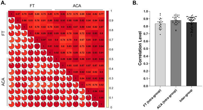 Figure 1
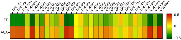 Figure 2
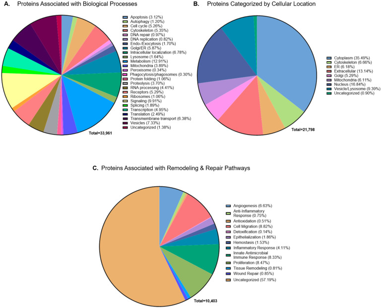 Figure 3
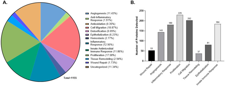 Figure 4
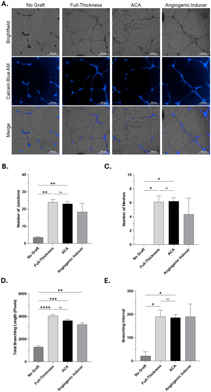 Figure 5
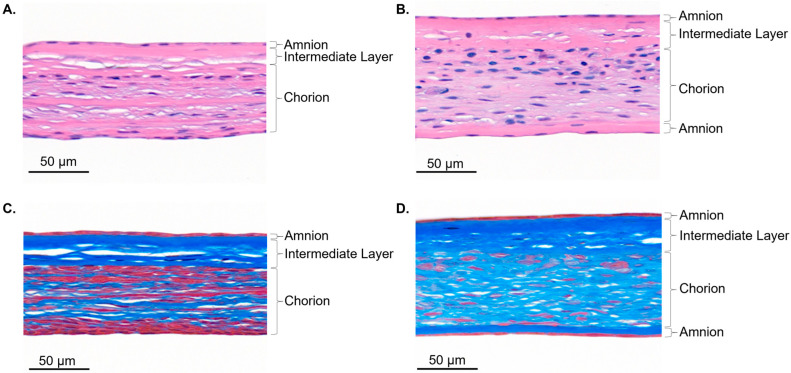 Figure 6
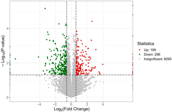 Figure 7
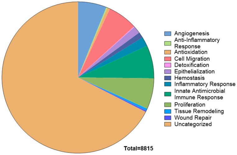 Figure 8
- —Tiger Wound Care Medical, LLC.
Peer Reviews
No public reviews on file for this paper yet. If you reviewed it on a platform where reviews are public (OpenReview, ICLR, NeurIPS, ICML), you can paste yours below so the community can read it here.
Videos
No videos yet. Explain this paper in a talk, walkthrough, or lecture? Add one.
Taxonomy
TopicsNeonatal Respiratory Health Research · Preterm Birth and Chorioamnionitis · Bone fractures and treatments
1. Introduction
The human placenta is a complex organ that has long been utilized in medicine as a biomaterial [1]. Its native function is to act as a protective barrier while facilitating nutrient and gas exchange. Its composition, which includes extracellular matrix (ECM) proteins, growth factors, and other bioactive molecules, offers structural and biological properties that make it valuable as a biomaterial [2,3,4]. One of its most common medical applications is in wound care, where its inherent barrier function is leveraged to serve as a wound covering, protecting the wound, reducing the risk of contamination, and maintaining a local environment that supports tissue repair. In clinical practice, placental membranes are engineered into allografts that retain these properties and are broadly categorized as Cellular and Acellular Matrix-like Products (CAMPs), which are routinely applied as wound coverings in modern medical practice [5,6].
Placental allografts have traditionally been engineered as amnion-only membranes. Amnion is the thinnest layer of the placenta, composed of a collagen-rich matrix with well-documented anti-inflammatory and epithelial-supportive properties [7,8]. Despite its clinical utility, amnion alone lacks the full structural and biochemical complexity of the native placental membrane. In addition to amnion, the placenta contains an intermediate layer and chorion, which contribute distinct biological features. The intermediate layer is a rich source of soluble biomolecules, with over 900 identified to date [9], while the chorion is the thickest layer and is enriched in ECM proteins such as collagen, fibronectin, laminin, and elastin [10,11]. Recent advances in tissue engineering have enabled the retention of all three layers, resulting in allografts that are both structurally more robust and compositionally more diverse. These advances have laid the groundwork for the development of next-generation CAMPs that better preserve the native characteristics of the placenta.
Building on these advances, multilayer CAMPs such as Full-Thickness (FT; amnion, intermediate, chorion) and ACA (amnion, intermediate, chorion, amnion) represent the next stage in placental allograft development. Previous studies have demonstrated that these CAMPs retain higher levels of extracellular matrix proteins and growth factors including ANG-2 (angiopoietin-2), EGF (epidermal growth factor), PDGF-AA (platelet-derived growth factor), and VEGF (vascular endothelial growth factor), as well as signaling factors associated with angiogenesis, tissue remodeling, inflammation, and host defense [12,13,14,15]. By preserving the full membrane structure, these CAMPs may improve graft durability and establish a microenvironment more favorable to tissue repair [16,17].
Previous studies have relied on targeted assays that capture only a limited subset of proteins or focus on individual tissue layers, leaving the broader proteomic landscape of these advanced CAMPs unexplored. This lack of comprehensive characterization restricts our understanding of how retention of the full membrane architecture contributes to graft composition and biological relevance.
To address this gap, the present study provides a molecular analysis of two advanced CAMP technologies, FT and ACA, in their ready-to-use form. We combined untargeted global proteomic profiling, targeted multiplex analysis of soluble proteins, and an in vitro angiogenesis assay to evaluate both molecular composition and associated biological relevance. Together, these findings offer new insights into the structural and compositional integrity of advanced CAMPs and highlight their potential to provide durable wound coverage while supporting a local environment conducive to tissue repair.
2. Results and Discussion
2.1. Proteomic Composition of Multilayer Placental Technologies
Two multilayer CAMP technologies were evaluated for their structural characteristics and overall protein composition in their ready-to-use form. The Full-Thickness (FT) technology retains the three native placental layers amnion, intermediate, and chorion. The four-layer ACA technology contain these same layers together with an additional preserved second amnion layer.
Histological analysis demonstrated the presence and organization of these membrane layers in both technologies (Appendix A Figure A1). Hematoxylin and Eosin (H&E) staining demonstrated preservation of membrane architecture, and Masson’s Trichrome staining revealed dense, collagen-rich extracellular matrix (ECM) across both technologies (Appendix A Figure A1). These findings demonstrate the expected structural composition and support previous observations of increased collagen staining.
Building on this structural evaluation, proteomic profiling was performed on allografts derived from different donors. Previous global proteomic studies have characterized unprocessed placental tissue [18], providing insights into the native protein composition. Extending this type of analysis to processed placental allografts, this study applied a global proteomic approach to multilayer CAMPs in their ready-to-use form.
A total of 8908 proteins were detected as shared between FT and ACA. Protein abundance profiles were consistent across samples, with Pearson correlation coefficients of 0.84 within FT, 0.88 within ACA, and 0.85 between groups (Figure 1A,B). In Figure 1A, a modified heat map is shown to visually demonstrate the correlation between all samples and technologies tested. The plot is divided into two analogous sections across the diagonal axis, representing intra-group (ACA vs. ACA; FT vs. FT) and inter-group (ACA vs. FT) comparisons. The top half displays Pearson correlation coefficients as numerical values, color-coded according to the similarity scale (0 = complete dissimilarity; 1 = exact match). The diagonal axis represents self-comparisons, with coefficients of 1.0 indicating 100% alignment. The bottom half shows corresponding pie charts that represent the percentage of overlap between each sample comparison. In Figure 1B, these correlation coefficients (excluding self-comparisons) are summarized in a bar graph, highlighting the distribution of intra-group (ACA vs. ACA; FT vs. FT) and inter-group (ACA vs. FT) comparisons. These results demonstrate that both CAMPs exhibit a similar proteomic profile, indicating a high degree of shared proteins.
The proteomic profile of ACA is largely shared with that of FT. Across the proteome, the relative levels of proteins are highly similar between the technologies, which is expected given that both technologies retain the same membrane layer types, with the ACA technology preserving an additional amnion layer. This is further illustrated in the volcano plot comparing ACA to FT (Appendix A Figure A2). With FT protein identification and relative expression as reference, approximately 5.6% of proteins in ACA are differentially expressed. These results confirm that FT and ACA share similar proteomic profiles.
Previous studies have shown that FT and ACA retain similar levels of selected growth factors and ECM-associated proteins, with collagen being the primary differentiating factor [13,14]. These findings are further supported by the current dataset, which confirms up to 50%, or 0.5-fold, higher levels of 31 collagen isoforms in ACA compared to FT (Figure 2). The higher relative levels of collagen isoforms are attributed from the preservation of an additional amnion layer, which is naturally rich in collagens [14]. This study identified elevated levels of Collagens I, II, III, IV, V, VI, XVII, XVIII, and XVIV in ACA compared to FT technologies. These results align with previous reports showing that ACA allografts contain significantly higher total collagen than FT allografts [14]. Higher collagen content may enhance the mechanical stability and handling characteristics of ACA in clinical use, given its influence on the structural properties of CAMPs [9,13,19].
2.2. Pathway Enrichment Analysis of the CAMP Proteome
To further characterize the CAMP proteome and assess its biological relevance, pathway enrichment analysis was performed on the 8908 detected proteins in both FT and ACA. Proteins were annotated by Gene Ontology (GO) biological processes and cellular components to better understand their subcellular localization and functional roles. This analysis aimed to identify key cellular pathways and mechanisms supported by the CAMPs technology.
Annotation resulted in 33,961 assignments across GO categories, with many proteins mapped to more than one biological process. Analysis of biological processes revealed that the most represented processes include metabolism, signal transduction, and intracellular transport (Figure 3A). These pathways are fundamental for supporting cell viability, regulating signaling and supporting molecular trafficking.
In addition to biological processes, the detected proteins were also mapped to cellular components to identify subcellular localization. This analysis resulted in 21,798 assignments across compartments, with the majority localized to the cytoplasm, followed by the nucleus and extracellular space (Figure 3B). This distribution indicates that CAMPs retain a broad spectrum of intracellular proteins as well as extracellular components, the latter being particularly relevant as extracellular proteins contribute to matrix remodeling and the regulation of the local cellular environment during tissue repair.
Building on this, the proteome was further evaluated to determine whether the retained proteins are linked to biological processes associated with tissue repair. This analysis generated 10,403 mapping assignments across twelve repair-associated categories, as individual proteins contribute to more than one process. The categories included angiogenesis, inflammation, epithelialization, proliferation, hemostasis, antioxidant activity, detoxification, antimicrobial activity, innate immune response, and general wound healing associated mechanisms were employed [20,21,22,23,24,25,26,27,28,29,30]. For this, a custom classification approach was applied to group proteins into twelve functional categories encompassing key repair-associated processes (Table 1). Proteins from both FT and ACA were represented across all twelve categories (Figure 3C). Using a conservative classification approach that restricted each protein to a single pathway, 32.52% of the shared proteome was associated with repair-associated processes (Appendix A Figure A3).
2.3. Evaluation of Soluble Proteins Under Physiological Conditions
To complement the global proteomic analysis, this study evaluated the availability of soluble proteins from both CAMP technologies following hydration. The objective was to determine which proteins may become accessible in the local environment under physiologically relevant conditions and whether these proteins are associated with biological processes involved in tissue repair. CAMPs were incubated at 37 °C for 72 h, and the resulting conditioned media containing solubilized proteins was analyzed using multiplex immunoassays targeting a broad panel of cytokines and growth factors. In total, 507 soluble analytes were detected across both CAMPs, with similar distribution patterns observed between groups.
To assess the potential biological relevance of these proteins, detected analytes were mapped to a predefined set of biological processes associated with tissue remodeling (Figure 4A). This classification, based on GO annotations, included key pathways such as proliferation (229 proteins), cell migration (202 proteins), and angiogenesis (143 proteins), as well as epithelialization, inflammatory response, and innate immune activity (Figure 4B). Importantly, the proteins detected in this study overlap with those reported in unprocessed placental tissue, further indicating that the molecular features of the native membrane are preserved in these technologies [32].
These findings are consistent with prior studies demonstrating that three-layer placental technologies retain growth factors and ECM-associated proteins detectable under similar in vitro conditions [31,33]. Together with the retained proteome data, these results offer a more complete molecular characterization of placental technologies. The presence of soluble proteins linked to biological processes associated with repair suggests that these CAMPs may help support a local environment favorable for tissue remodeling.
2.4. In Vitro Angiogenesis Assessment of Soluble Proteins
The global and targeted proteomic analysis showed that CAMPs retain proteins associated with wound repair. To demonstrate the functional relevance of available soluble proteins detected in the FT and ACA allografts, an in vitro tube formation assay was conducted to assess whether available proteins can support endothelial cell activity associated with angiogenesis. Angiogenesis represents a central process in tissue repair, as the formation of blood vessels enables delivery of oxygen and nutrients, removal of waste, and establishment of a biologically conducive microenvironment [20].
In this study, primary human umbilical vein endothelial cells (HUVECs) were cultured on Geltrex matrix and incubated with conditioned media collected from FT and ACA allografts after 72 h at 37 °C. Basal media alone served as a negative control, while media supplemented with an angiogenic inducer was used as a positive control. After 16 h of incubation, tube formation was qualitatively assessed using brightfield and fluorescence imaging using Calcein Blue AM. Compared to the negative control, conditioned media from both FT and ACA groups supported the formation of capillary-like structures (Figure 5). These included elongated tubes, branching points, and enclosed meshes similar to those observed in the positive control, indicating that proteins available in the media can contribute to a local environment that supports endothelial network formation.
Quantitative analysis further confirmed these observations, demonstrating higher numbers of junctions (Figure 5B), meshes (Figure 5C), and overall branching (Figure 5D,E) in both FT and ACA groups relative to the negative control, with values comparable to the angiogenic inducer. Notably, no significant differences were observed between FT and ACA allografts, indicating that both technologies provide a comparable capacity to support endothelial network formation.
These findings align with previous literature evaluating placenta-derived materials and their ability to support neovascularization in vitro [34,35,36,37]. Importantly, they provide functional context to the proteomic and multiplex findings presented earlier. While molecular analyses confirmed the presence of growth factors associated with angiogenic pathways, these results demonstrate that the proteins remain in a state that can provide a conducive environment to support tissue repair and remodeling under relevant in vitro conditions.
3. Materials and Methods
3.1. Placental Membrane-Derived CAMP Technologies
The placental tissue, donated from consenting healthy birth mothers over the age of 18 following cesarian and vaginal deliveries, was screened and tested before processing in accordance with U.S. Food and Drug Administration (FDA) regulations and American Association of Tissue Banks (AATB) standards. Placental based allografts were generated as described in [14]. Briefly, FT is a three-layer allograft that consists of amnion, intermediate, and chorion layers, while ACA is a more advanced four-layer allograft that includes FT layers with an additional amnion layer preserved during processing. Placental allografts used in this study include Full-Thickness technology (CompleteFT™, Tiger Wound Care LLC, Conshohocken, PA, USA) and ACA technology (ACApatch™, Tiger Wound Care LLC, Conshohocken, PA, USA).
3.2. Histological Assessment
Histology analysis of FT and ACA technology was conducted utilizing Hematoxylin and Eosin (H&E) and Masson’s trichrome staining. For the staining, samples were fixed overnight utilizing 10% neutral-buffer formalin (VWR, Radnor, PA, USA), 5 µm-thick cross-sections of the allografts were utilized, and stained by the Precision Pathology Laboratory (San Antonio, TX, USA) according to their standardized protocols. H&E staining was utilized for the assessment of the differences in the tissue structures between ACA and Full-Thickness (FT) technology. MTS staining was used for the qualitative ECM collagen content assessment. The representative cross-section images of H&E and Masson’s trichrome staining were taken using Slide Viewer (Version 2.8.0) at a 60× magnification. For histological analyses, eight distinct donors from FT and ten from ACA were evaluated.
3.3. Global Proteomic Profiling
Sample preparation and LC-MS/MS acquisition for global proteomic analysis were conducted by Metware Biotechnology Inc. (Woburn, MA, USA). Full-Thickness (n = 7 donors) and ACA (n = 8 donors) were cryopulverized in liquid nitrogen and processed via ethanol solution precipitation, followed by lysis with 8 M urea, 1 mM PMSF, and 2 mM EDTA lysis buffer. The protein concentration was determined using a BCA assay kit (Thermo Scientific, Waltham, MA, USA). Proteins were subsequently separated based on hydrophobicity using a NanoElute UHPLC (Bruker, Billerica, MA, USA) system with a nanoliter flow rate on a C18 column for LC-MS/MS analysis. The mass spectrometry data were acquired using the ddaPASEF mode of a timsTOF HT mass spectrometer to establish an appropriate acquisition window for the diaPASEF acquisition method. The parameters used in the analysis included a valid gradient of 47 min, positive ion detection mode, parent ion scanning range of 100–1700 m/z, ion mobility 1/K_0 in the range of 0.6–1.6 Vs/cm^2^, ion accumulation and release time of 100 ms, ion utilization rate approximating 100%, capillary voltage of 1600 V, drying gas rate of 3 L/min, and drying temperature of 180 °C. Parameters used in the diaPASEF acquisition mode included a mass range of approximately 400–1200, a mobility range of 0.6–1.6 Vs/cm^2^, a mass width of 25 Da, a mass overlap of 0.1, 24 mass steps per cycle, and two mobility windows, totaling 48 acquisition windows.
3.3.1. Proteomic Analysis
In database searching with DIA-NN (v1.8.1), precursor ion and protein-level identifications were filtered at a 1% false discovery rate (FDR), ensuring high confidence in peptide and protein assignments. For protein quantification, DIA-NN’s MaxLFQ algorithm was applied with normalization across samples to minimize systematic bias. Differential expression analysis was then performed using either t-tests (for two-group comparisons) or ANOVA (for multi-group comparisons) in FT and ACA replicates. For differential expression analyses which include p-value testing, FDR is calculated using the Benjamini–Hochberg (BH) method. Global protein expression profiles for FT and ACA were used to generate a custom heatmap for all collagen variants. Protein expression was determined by averaging protein expression across FT and ACA groups and assessing relative abundance of each variant to the overall average. Variance in protein abundance within each group was assessed by an ANOVA test, and associated p-values were calculated using unpaired two-tailed t-tests with Welch’s correction for the volcano plot.
3.3.2. Bioinformatics and Functional Category Analysis
To characterize the biological relevance of total proteins within the global proteomics dataset, a comprehensive bioinformatics pipeline was applied. Functional enrichment analysis included Gene Ontology (GO) categories, Kyoto Encyclopedia of Genes and Genomes (KEGG) pathways, EuKaryotic Orthologous Groups (KOG) functional classification, subcellular localization, and signal peptide (SignalP) prediction. Functional categorization of proteins was performed using the GO-based clustering tool Categorizer [38], enabling the classification of detected proteins in FT and ACA technology into biologically relevant pathways. Gene Ontology (GO) terms encompassing biological pathways associated with tissue remodeling were curated from QuickGO (European Bioinformatics Institute, Hinxton, United Kingdom) [39] as listed in Table 1. This included over 170 GO terms mapped to the processes: wound repair, proliferation, cell migration, innate immune response, angiogenesis, inflammatory response, tissue remodeling, and epithelialization. These curated GO pathways were matched against the Homo sapiens annotation dataset from the EBI Gene Ontology Annotation (GOA) database [40] then mapped to the detected protein list derived from the global proteomic profiling to assign proteins for functional overlap where annotations intersected.
Downstream enrichment analyses (GO, KEGG, KOG, protein domains) were also evaluated with respect to significance p-values derived from hypergeometric testing against the background of all identified proteins. The raw p-values were further subjected to FDR correction through the BH method to control for multiple comparisons.
3.4. L-Series Human Antibody Array for Soluble Protein Profiling
The L-Series multiplex enzyme-linked immunosorbent assay (ELISA) array (RayBiotech, Inc., Peachtree Corners, GA, USA, Cat. No.: AAH-BLG-1-4) was used to semi-quantitatively assess 507 growth factors, cytokines, and other signaling molecules in the soluble extracts from FT and ACA. Soluble extract preparation used a surface area of 3 cm^2^ per 500 μL ratio of DMEM cell culture media for each placental allograft. Extracts were incubated at 37 °C at 700 RPM on a thermomixer (Eppendorf, Hamburg, Germany, Cat. No. 5382) for 3 days. After incubation, extracts were centrifuged at 10,000 RPM for 5 min to remove debris. The supernatant was sterilized using a 0.22 µm filter (VWR, Radnor, PA, USA, Cat. No. 29442-752). The samples were measured in duplicate and reported as Relative Fluorescence Units (RFUs). For the assay, extracts from four distinct FT donors and five distinct ACA donors were utilized.
The pathway-based bioinformatics approach used in the global proteomic dataset was applied to the L-Series antibody array dataset. Detected soluble analytes (n = 507) were compared against a curated list of GO biological pathways (Table 1). Proteins matching these pathways were further categorized using the AmiGO2 database to identify functional enrichment, as listed in Appendix A Table A1.
3.5. In Vitro Angiogenesis Assay
Angiogenic potential was assessed using the Angiogenesis Starter Kit Assay (Thermo Scientific, Waltham, MA, USA, Cat. No. A14609-01) following manufacturer’s instructions. Primary Human Umbilical Vein Endothelial Cells (HUVECs) (Thermo Scientific, Waltham, MA, USA, Cat. No. C0035C) were cultured in Medium 200 (Thermo Scientific, Waltham, MA, USA, Cat. No. M200500) supplemented with LVES (Thermo Scientific, Waltham, MA, USA, Cat. No A1460801) and maintained at 37 °C with 5% CO^2^. Cells were seeded at a density of 25,000 cells per well on Geltrex-coated 48-well plates (Thermo Scientific, Waltham, MA, USA, Cat. No. A1413202).
Experimental groups were treated with FT or ACA soluble extracts prepared as described in L-series Human Antibody Array with Medium 200 containing 2% heat-inactivated fetal bovine serum (VWR, Radnor, PA, USA, Cat. No.: 45000-734). Positive controls received growth media with LVES (containing angiogenic factors); negative controls received Medium 200 without LVES. Following a 16 h incubation, wells were washed with DPBS (Corning, Corning, NY, USA, Cat. No. 21-030-CV), and stained with Calcein Blue AM (6 µg/mL; Thermo Scientific, Waltham, MA, USA, Cat. No.: C1429) for 30 min at 37 °C. After a final DBPS wash, cells were imaged in brightfield and fluorescence modes using a BioTek Cytation 5 Multimode Reader (Agilent Technologies, Santa Clara, CA, USA) at 10× magnification.
Assays were performed using extracts from FT (n = 4 donors) and ACA (n = 9 donors) allografts, with three technical replicates per condition across two independent experiments.
Quantitative Analysis of Angiogenic Activity
Captured images were analyzed through NIH’s ImageJ (National Institutes of Health, Bethesda, MD, USA) with the Angiogenesis Analyzer plugin [41] to quantify the number of junctions, meshes, and branch lengths. Quantitative analysis was conducted in batches across three technical replicates of brightfield images per condition at 10× magnification after a 24 h treatment, accounting for non-selective bias. The following parameters were used in the Angiogenesis Analyzer plugin: minimum object size = 20 pixels, minimum branch size = 25 pixels, artificial loop size = 850 pixels, isolated element size threshold = 15 pixels, master segment size threshold = 15 pixels, and iteration number = 3 pixels. Statistical significance was determined using a normal Gaussian one-way ANOVA test using multiple comparisons of column means.
3.6. Data Analysis and Handling
For all analyses, a significance threshold of p < 0.05 was used. Statistical processing was conducted using GraphPad Prism (Version 10.5) and Microsoft Excel. Experiment specific statistical analyses are described in the corresponding methods subsections. To ensure reproducibility and independence of the data, all global and targeted proteomic analyses were performed by an independent third party, minimizing the potential for author-related bias. Analyses were conducted on biological replicates to confirm consistency across samples. Quantitative assessment of angiogenesis was conducted on representative experiments using ImageJ Fiji Software (Java Version 21.0.7) plugin Angiogenesis Analyzer (Version 1.0.C). This software applies objective algorithms for tube formation metrics and reduces the risk of subjective interpretation.
4. Conclusions
This study presents a detailed molecular characterization of advanced multilayer CAMPs. Through a combination of global proteomics, targeted multiplex protein detection, and an in vitro angiogenesis assay, we evaluated the protein composition and its relevance to tissue repair processes. The proteomic analysis revealed a highly consistent protein profile across both technologies, supporting the idea that the structural layers preserved in these CAMPs retain a diverse set of biologically relevant proteins. Functional categorization highlighted a substantial portion of these proteins associated with key processes involved in tissue remodeling and repair. Collagen was notably higher in ACA compared to FT, a distinguishing characteristic that aligns with previous studies and reflects the additional preserved amnion layer in the four-layer technology.
In addition to overall protein composition, the availability of soluble proteins was assessed. Soluble proteins detected in conditioned media were mapped to pathways associated with tissue repair, offering additional insight into the molecular profile accessible in ready-to-use CAMPs. An in vitro angiogenesis assay using endothelial cells further demonstrated that both FT and ACA technologies contain soluble proteins that can support endothelial tube formation. These findings demonstrate that the proteins preserve their functional integrity and provide a conducive environment to support cellular activities related to tissue repair.
Together, these findings demonstrate that CAMPs retain proteins linked to angiogenesis, cell proliferation, and immune modulation, which may contribute to a local environment supportive of tissue repair. This work advances the field by moving beyond targeted assays to offer broader proteomic profile of advanced CAMPs in their ready-to-use form. Preservation of these proteins may help retain the inherent biological properties of the tissue and provide a foundation for understanding the composition of placenta-derived CAMPs.
5. Patents
The subject of this manuscript is pending a patent and the IP rights are owned by Tiger Wound Care Medical, LLC, Conshohocken, PA, USA.
The reference list from the paper itself. Each links out to its DOI / PubMed record.
- 1Silini A.R. Cargnoni A. Magatti M. Pianta S. Parolini O. The Long Path of Human Placenta, and Its Derivatives, in Regenerative Medicine Front. Bioeng. Biotechnol.2015316210.3389/fbioe.2015.0016226539433 PMC 4609884 · doi ↗ · pubmed ↗
- 2Protzman N.M. Mao Y. Long D. Sivalenka R. Gosiewska A. Hariri R.J. Brigido S.A. Placental-Derived Biomaterials and Their Application to Wound Healing: A Review Bioengineering 20231082910.3390/bioengineering 1007082937508856 PMC 10376312 · doi ↗ · pubmed ↗
- 3Biswas A. Rajasekaran R. Saha B. Dixit K. Vaidya P.V. Ojha A.K. Dhara S. Human placenta/umbilical cord derivatives in regenerative medicine-Prospects and challenges Biomater. Sci.2023114789482110.1039/D 2BM 01977 A 37255413 · doi ↗ · pubmed ↗
- 4Moore M.C. Van De Walle A. Chang J. Juran C. Mc Fetridge P.S. Human Perinatal-Derived Biomaterials Adv. Healthc. Mater.20176170034510.1002/adhm.201700345 PMC 573369228783879 · doi ↗ · pubmed ↗
- 5Hughes O.B. Rakosi A. Macquhae F. Herskovitz I. Fox J.D. Kirsner R.S. A Review of Cellular and Acellular Matrix Products: Indications, Techniques, and Outcomes Plast. Reconstr. Surg.2016138138 S 147S 10.1097/PRS.000000000000264327556754 · doi ↗ · pubmed ↗
- 6Wu S. Carter M. Cole W. Crombie R. Kapp D.L. Kim P. Milne C. Molnar J. Niezgoda J. Woo K. Best practice for wound repair and regeneration use of cellular, acellular and matrix-like products (CAM Ps)J. Wound Care 202332 S 1S 3110.12968/jowc.2023.32.Sup 4b.S 137079485 · doi ↗ · pubmed ↗
- 7Roy A. Mantay M. Brannan C. Griffiths S. Placental Tissues as Biomaterials in Regenerative Medicine Biomed. Res. Int.20222022675145610.1155/2022/675145635496035 PMC 9050314 · doi ↗ · pubmed ↗
- 8Oyen M.L. Ferguson V.L. Calvin S.E. Fracture Resistance of Human Amnion Am. Soceity Mech. Eng.200784184210.1115/SBC 2007-174552 · doi ↗