Automated T-Cell Proliferation in Lab-on-Chip Devices Integrating Microfluidics and Deep Learning-Based Image Analysis for Long-Term Experiments
María Fernanda Cadena Vizuete, Martin Condor, Dennis Raith, Avani Sapre, Marie Follo, Gina Layedra, Roland Mertelsmann, Maximiliano Perez, Betiana Lerner

TL;DR
This paper introduces a microfluidic system with deep learning image analysis to automate and improve long-term T-cell culture for immunotherapy research.
Contribution
A novel microfluidic and deep learning-based system for long-term suspension cell culture with automated monitoring and minimal manual intervention.
Findings
The system successfully supported proliferation of Jurkat and primary human T cells without significant loss during medium exchange.
The deep learning image analysis outperformed the Trainable Weka Segmentation plugin in processing large volumes of data.
Both commercial and custom lab-on-chip devices proved effective for long-term cell expansion with distinct advantages.
Abstract
T cells play a pivotal role in cancer research, particularly in immunotherapy, which harnesses the immune system to target malignancies. However, conventional expansion methods face limitations such as high reagent consumption, contamination risks, and difficulties in maintaining suspension cells in dynamic culture environments. This study presents a microfluidic system for long-term culture of non-adherent cells, featuring automated perfusion and image acquisition. The system integrates deep learning-based image analysis, which quantifies cell coverage and estimates cell numbers, and efficiently processes large volumes of data. The performance of this deep learning approach was benchmarked against the widely used Trainable Weka Segmentation (TWS) plugin for Fiji. Additionally, two distinct lab-on-a-chip (LOC) devices were evaluated independently: the commercial ibidi® LOC and a…
Genes, proteins, chemicals, diseases, species, mutations and cell lines named across the full text — each resolved to its canonical identifier and authoritative record.
Click any figure to enlarge with its caption.
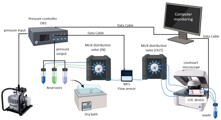 Figure 1
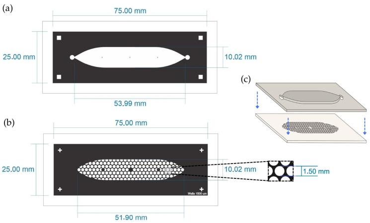 Figure 2
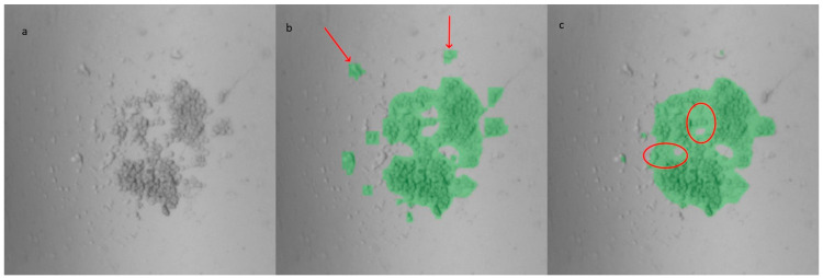 Figure 3
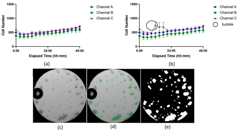 Figure 4
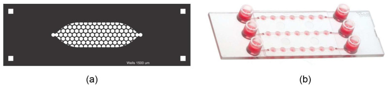 Figure 5
 Figure 6
 Figure 7
 Figure 8
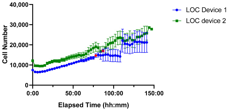 Figure 9
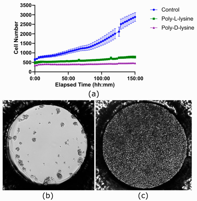 Figure 10
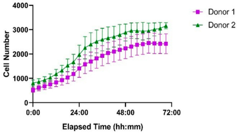 Figure 11
Peer Reviews
No public reviews on file for this paper yet. If you reviewed it on a platform where reviews are public (OpenReview, ICLR, NeurIPS, ICML), you can paste yours below so the community can read it here.
Videos
No videos yet. Explain this paper in a talk, walkthrough, or lecture? Add one.
Taxonomy
TopicsCell Image Analysis Techniques · Microfluidic and Bio-sensing Technologies · 3D Printing in Biomedical Research