Clinical, Radiological, and Pathological Features of Intraosseous Hibernoma: A Systematic Review of Case Reports and Case Series
Jawad Albashri, Ahmed Albashri, Muhannad Alhamrani, Abdulrahman Hassan, Hisham Shamah, Rayan Alhefzi, Najim Z. Alshahrani, Mohammed R. Algethami, Louis-Romée Le Nail, Ramy Samargandi

TL;DR
This review summarizes 62 cases of intraosseous hibernoma, a rare benign bone tumor that often mimics cancer, to improve diagnosis and avoid unnecessary treatments.
Contribution
The study provides a comprehensive synthesis of clinical, radiological, and pathological features of intraosseous hibernoma from case reports and series.
Findings
Intraosseous hibernoma commonly appears in the pelvis and spine and is often discovered incidentally during cancer staging.
Radiologically, it typically shows as a sclerotic lesion with variable PET/CT uptake and is confirmed by S100-positive histology.
Conservative management after biopsy is effective, with no reported cases of malignant transformation.
Abstract
Intraosseous hibernoma (IOH) is a rare benign tumor composed of brown fat within the bone. Its unusual imaging features often mimic metastatic disease or primary bone tumors, leading to unnecessary investigations or treatments. This systematic review synthesized all published case reports and case series to clarify its clinical, radiological, histopathological, and management characteristics. We identified 62 confirmed cases from 30 publications. Most lesions were found incidentally, commonly during cancer staging, with the pelvis and spine as the most frequent sites. Radiologically, IOH typically appears as a solitary sclerotic lesion with variable PET/CT uptake, while histology reveals brown adipose tissue with strong S100 positivity. Nearly all patients were managed conservatively following biopsy. Recognizing IOH as a benign mimicker is essential to avoid misdiagnosis and…
Genes, proteins, chemicals, diseases, species, mutations and cell lines named across the full text — each resolved to its canonical identifier and authoritative record.
Click any figure to enlarge with its caption.
 Figure 1
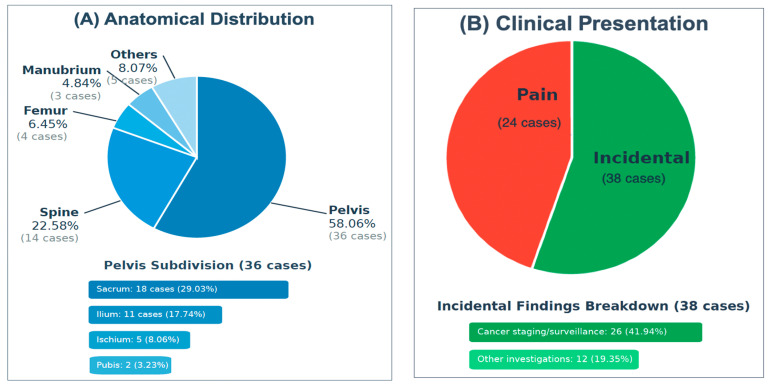 Figure 2
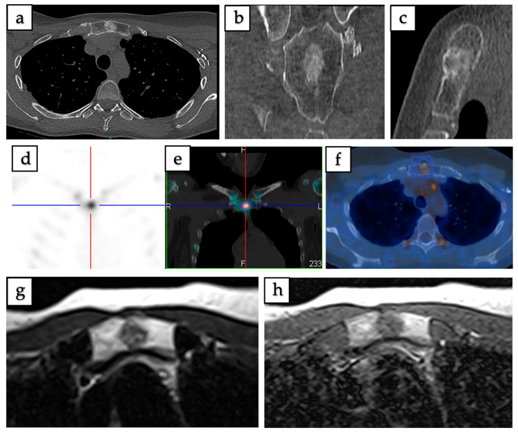 Figure 3
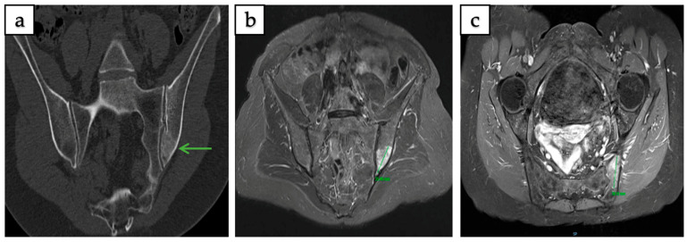 Figure 4
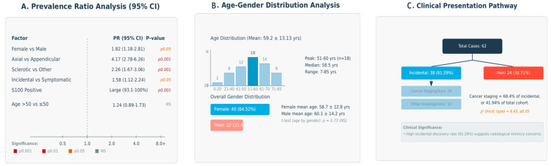 Figure 5
Peer Reviews
No public reviews on file for this paper yet. If you reviewed it on a platform where reviews are public (OpenReview, ICLR, NeurIPS, ICML), you can paste yours below so the community can read it here.
Videos
No videos yet. Explain this paper in a talk, walkthrough, or lecture? Add one.
Taxonomy
TopicsSalivary Gland Tumors Diagnosis and Treatment · Head and Neck Surgical Oncology · Pleural and Pulmonary Diseases