Radiomic Characterization and Automated Classification of Drusen Substructure Phenotype Associated with High-Risk Dry Age-Related Macular Degeneration
Scott W. Perkins, Neal Shah, Jon Whitney, Karen Matar, Hannah J. Yu, Charles C. Wykoff, Justis P. Ehlers

TL;DR
This study introduces automated radiomic metrics to classify drusen substructures in dry age-related macular degeneration, improving prediction of geographic atrophy risk.
Contribution
The novel use of radiomic features for automated drusen classification and GA risk prediction is introduced.
Findings
Radiomic features classified drusen phenotypes with AUC = 0.87–0.95.
H-type drusen show higher reflectivity and coarser texture compared to others.
Radiomic features predict GA conversion and growth rate with AUC = 0.59–0.74.
Abstract
Background/Objectives: Optical coherence tomography (OCT)-reflective drusen substructures (ODSs) are associated with the conversion of intermediate AMD to geographic atrophy (GA). However, ODSs must be manually identified, a laborious process introducing bias and variation. This study proposes objective radiomic metrics of drusen phenotypes and validates them for the prediction of GA development and GA growth rate. Methods: A total of 104 drusen with high-reflective cores (H-type), 105 with low-reflective cores (L-type), 129 conical drusen (C-type), and 101 normal drusen (N-type) were segmented from OCT images. Radiomic features were extracted from these drusen, and the most important features for drusen classification were extracted from the retinal pigment epithelium–Bruch’s membrane compartment of 743 OCT scans of eyes with dry AMD and used to predict GA conversion and fast growth.…
Genes, proteins, chemicals, diseases, species, mutations and cell lines named across the full text — each resolved to its canonical identifier and authoritative record.
Click any figure to enlarge with its caption.
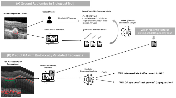 Figure 1
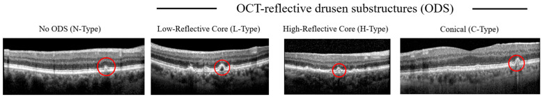 Figure 2
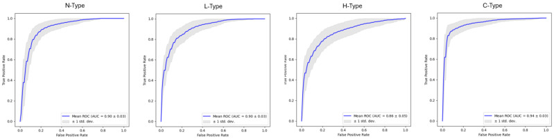 Figure 3
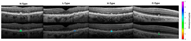 Figure 4
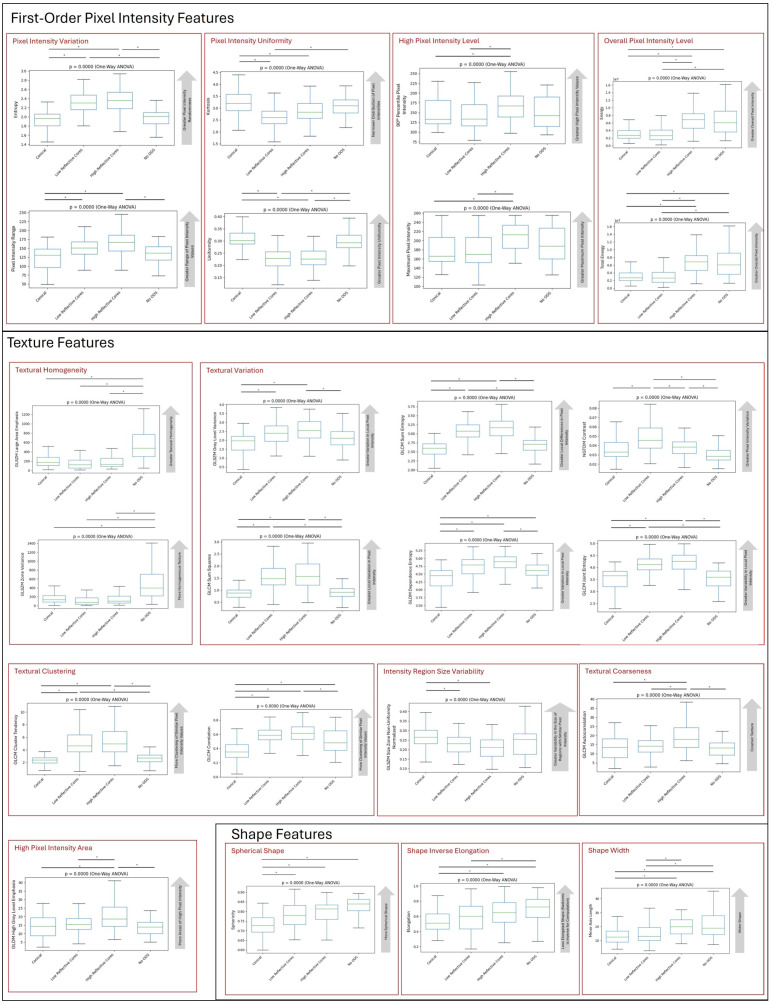 Figure 5
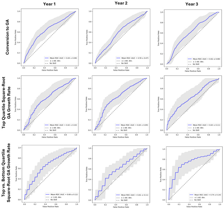 Figure 6
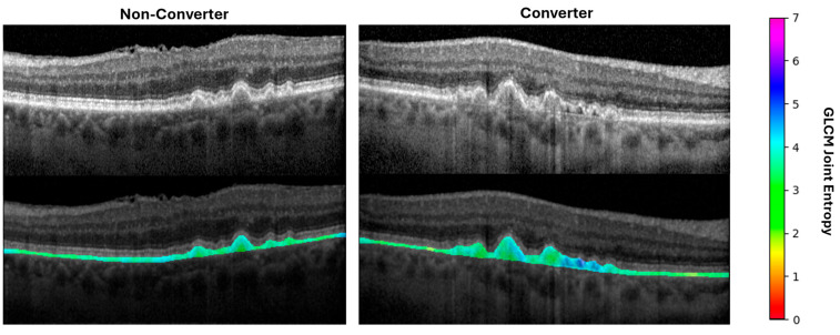 Figure 7
Peer Reviews
No public reviews on file for this paper yet. If you reviewed it on a platform where reviews are public (OpenReview, ICLR, NeurIPS, ICML), you can paste yours below so the community can read it here.
Videos
No videos yet. Explain this paper in a talk, walkthrough, or lecture? Add one.
Taxonomy
TopicsMRI in cancer diagnosis · Retinal Imaging and Analysis · Cerebral Venous Sinus Thrombosis