The Importance of the Interventional Pathologist in Fine Needle Aspiration and Core Needle Biopsy Procedures for Lymph Node Lesions: A Retrospective Analysis of Diagnostic Correlation and Proposal of a Classification System for Lymph Node Core Needle Biopsies
Montserrat de la Torre Serrano, Ana María Colino-Gallardo, Jesús Vega González, Maria Reyes Bergillos Giménez, Ramón Robledano Soldevilla, Teresa Iscar Galán, Julian Sanz Ortega, Maria del Mar Olmo Fernández, Santiago Nieto Llanos, Karen Villar Zarra

TL;DR
This study shows how pathologists using FNA and CNB can accurately diagnose lymph node lesions, suggesting a new classification system for better communication.
Contribution
The paper proposes a classification system for CNB in lymph node lesions and highlights the role of interventional pathologists in improving diagnostic accuracy.
Findings
FNA had a 98% diagnostic yield with 94% correlation to CNB results.
Ultrasound guidance improved lesion characterization in FNA procedures.
A new classification system for CNB is proposed to align with the Sydney system for FNA.
Abstract
Introduction: Pathology requires the integration of macroscopic and microscopic findings for an accurate diagnosis. Fine needle aspiration biopsy (FNA) traditionally became the domain of radiologists with the introduction of ultrasound; however, its increased accessibility has allowed other specialists, including pathologists, to incorporate it into their daily practice. Both ultrasound-guided FNA and core needle biopsy (CNB) performed by interventional pathologists have proven valuable tools in the diagnosis of lymph node lesions. Materials and Methods: An observational, descriptive, retrospective study was conducted at Hospital Universitario del Henares, analysing 134 FNABs and 31 CNBs of lymph node lesions between 2023 and 2024. The diagnostic yield of both techniques and their correlation were evaluated. Results: Ultrasound facilitated better lesion characterization. FNA…
Genes, proteins, chemicals, diseases, species, mutations and cell lines named across the full text — each resolved to its canonical identifier and authoritative record.
Click any figure to enlarge with its caption.
 Figure 1
 Figure 2
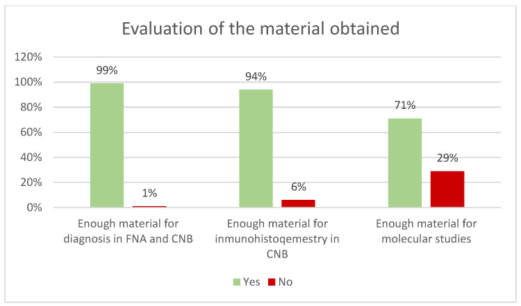 Figure 3
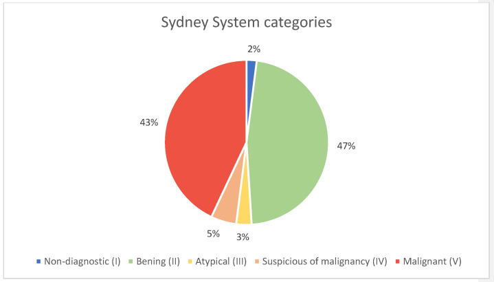 Figure 4
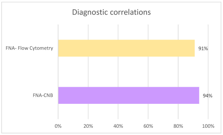 Figure 5
 Figure 6
 Figure 7
 Figure 8
Peer Reviews
No public reviews on file for this paper yet. If you reviewed it on a platform where reviews are public (OpenReview, ICLR, NeurIPS, ICML), you can paste yours below so the community can read it here.
Videos
No videos yet. Explain this paper in a talk, walkthrough, or lecture? Add one.
Taxonomy
TopicsLung Cancer Diagnosis and Treatment · Salivary Gland Tumors Diagnosis and Treatment · Head and Neck Anomalies