New Insights into Pathogenesis and Management of Keratoacanthoma: A Narrative Review
Mariafrancesca Hyeraci, Dario Didona, Damiano Abeni, Francesca Magri, Francesco Ricci, Chiara Bertagnin, Arianna Loregian, Giovanni Di Lella, Antonio Di Guardo, Annarita Panebianco, Camilla Chello, Claudio Conforti, Elena Dellambra, Luca Fania

TL;DR
This review explores the diagnosis and treatment of keratoacanthoma, emphasizing new imaging techniques and the role of HPV in its development.
Contribution
The paper highlights advances in non-invasive diagnostics and the emerging role of HPV in keratoacanthoma pathogenesis.
Findings
Non-invasive techniques like dermoscopy and confocal imaging improve KA diagnosis and differentiation from SCC.
β-genus HPV types are suggested as cofactors in KA carcinogenesis, especially under UV exposure.
Alternative therapies like radiotherapy and topical agents offer options when surgery is not feasible.
Abstract
Keratoacanthoma (KA) is a rapidly growing epithelial neoplasm characterized by clinical and histopathological features that often overlap with well-differentiated squamous cell carcinoma (SCC), posing diagnostic challenges. This review provides a comprehensive overview of KA, emphasizing advances in non-invasive diagnostic techniques such as dermoscopy, reflectance confocal microscopy (RCM), and line-field confocal optical coherence tomography (LC-OCT), which improve lesion characterization and differentiation from SCC. We discuss the histopathological phases of KA and highlight key features aiding in diagnosis. Furthermore, we explore the emerging role of human papillomavirus (HPV), particularly β-genus types, as a cofactor in KA carcinogenesis through modulation of apoptosis and DNA damage response pathways, especially under ultraviolet (UV) radiation exposure. Therapeutic strategies…
Genes, proteins, chemicals, diseases, species, mutations and cell lines named across the full text — each resolved to its canonical identifier and authoritative record.
Click any figure to enlarge with its caption.
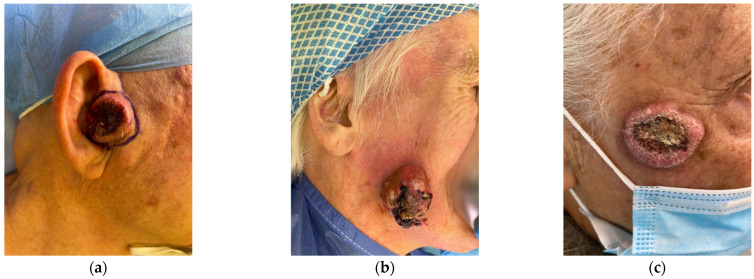 Figure 1
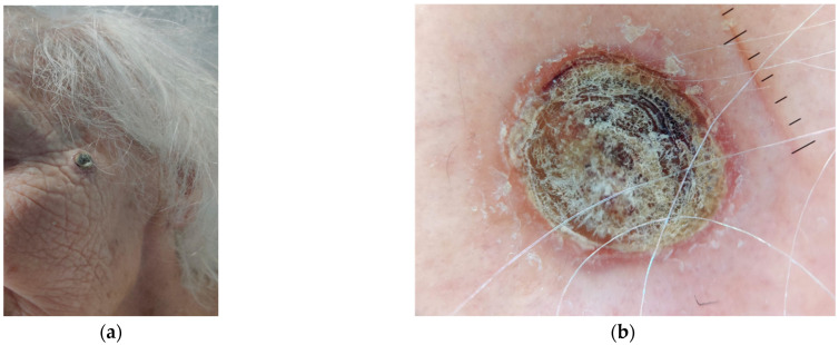 Figure 2
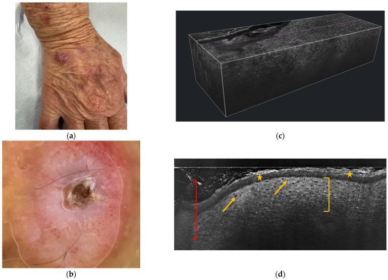 Figure 3
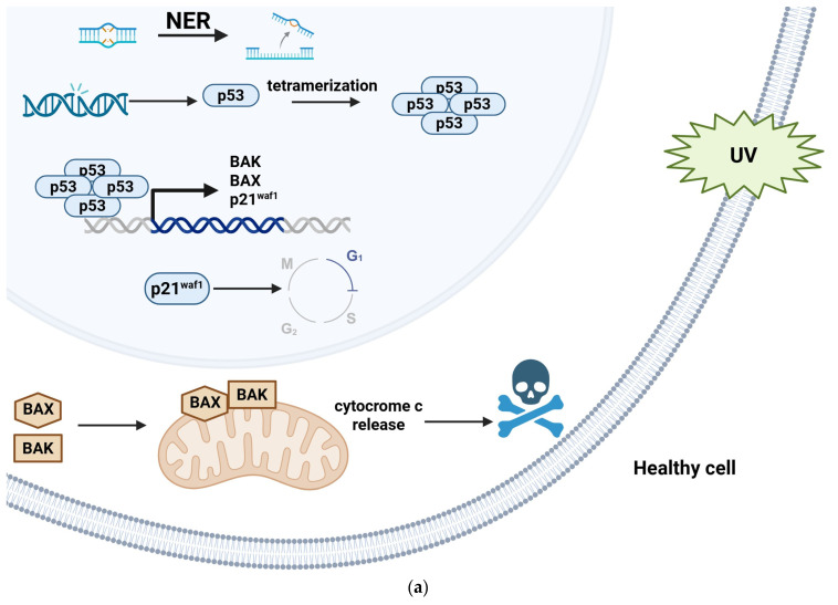 Figure 4
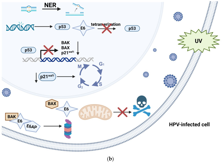 Figure 5
 Figure 6
 Figure 7
 Figure 8
 Figure 9
 Figure 10
 Figure 11
Peer Reviews
No public reviews on file for this paper yet. If you reviewed it on a platform where reviews are public (OpenReview, ICLR, NeurIPS, ICML), you can paste yours below so the community can read it here.
Videos
No videos yet. Explain this paper in a talk, walkthrough, or lecture? Add one.
Taxonomy
TopicsNonmelanoma Skin Cancer Studies · Cancer and Skin Lesions · Infectious Diseases and Mycology