Quantification of High-Resolution Contrast-Enhanced T1-Weighted Vessel Wall MRI for Predicting Disease Progression in Moyamoya Disease
Kateryna Goloshchapova, Patrick Haas, Daniel Vogl, Lucas Wiggenhauser, Helene Hurth, Florian Hennersdorf, Benjamin Bender, Till-Karsten Hauser, Marcos Tatagiba, Nadia Khan, Constantin Roder

TL;DR
This study shows that high-resolution MRI can reliably track moyamoya disease progression by measuring vessel wall contrast enhancement over time.
Contribution
The study establishes a reproducible method for quantifying disease activity in moyamoya using normalized vessel wall MRI signal intensity.
Findings
75% of patients showed vessel wall contrast enhancement, indicating active disease.
Signal variability was stable when normalized to the pituitary stalk (9.4% median variability).
Pituitary and temporal lobe signal changes were strongly correlated over time.
Abstract
Objective: In moyamoya disease (MMD), the internal carotid and proximal cerebral arteries narrow, potentially leading to stroke or hemorrhage from fragile collaterals. Disease activity and progression may be detected by contrast-enhanced (CE) high-resolution (HR) vessel wall imaging (CE-VWI) on T1-weighted MRI. However, this imaging approach needs standardization for the evaluation of signal intensity and longitudinal reproducibility. Methods: MMD patients with at least two separate CE-VWI examinations on the same and on different scanners were included. Signal intensity of the vessel wall, pituitary stalk, and temporal lobe white matter were measured and normalized using manually selected regions of interest. Intraindividual longitudinal reproducibility of MRI was analyzed and the clinical course was correlated with vessel wall enhancement data. Results: Eighty-seven patients were…
Genes, proteins, chemicals, diseases, species, mutations and cell lines named across the full text — each resolved to its canonical identifier and authoritative record.
Click any figure to enlarge with its caption.
 Figure 1
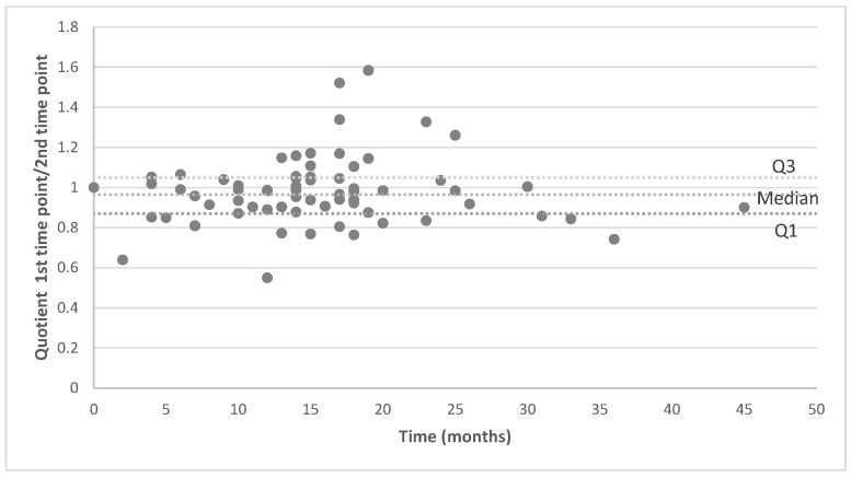 Figure 2
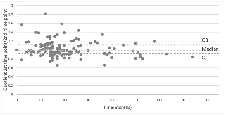 Figure 3
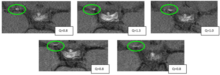 Figure 4
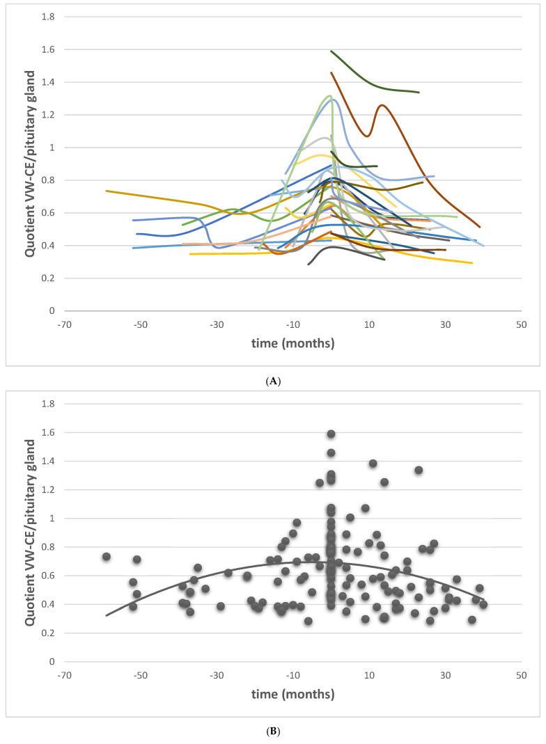 Figure 5
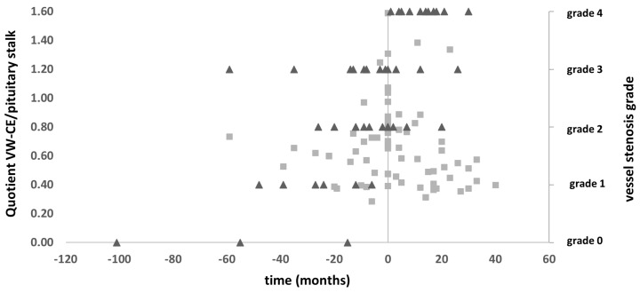 Figure 6
Peer Reviews
No public reviews on file for this paper yet. If you reviewed it on a platform where reviews are public (OpenReview, ICLR, NeurIPS, ICML), you can paste yours below so the community can read it here.
Videos
No videos yet. Explain this paper in a talk, walkthrough, or lecture? Add one.
Taxonomy
TopicsMoyamoya disease diagnosis and treatment · Cerebrovascular and Carotid Artery Diseases · Neurological Complications and Syndromes