Elucidating the Molecular Basis of Thermal Stress Response in Juvenile Turbot (Scophthalmus maximus) via an Integrative Transcriptome–Metabolome Approach
Xiatian Chen, Tao Gao, Ziwen Wang, Shuaiyu Chen, Nan Zhang, Xiaoming Zhang, Yudong Jia

TL;DR
This study explores how juvenile turbot respond to heat stress at the molecular level, using gene and metabolite analysis to identify key pathways involved in thermal adaptation.
Contribution
The study provides new insights into the molecular mechanisms of thermal stress response in turbot through an integrative transcriptome-metabolome approach.
Findings
Thermal stress significantly alters gene expression and metabolite profiles in turbot liver.
Key pathways like PI3K-Akt signaling and amino acid metabolism are crucial for thermal adaptation.
Genes such as CDH1, Col9a1, and DNAJB6 are linked to specific metabolic changes under heat stress.
Abstract
High temperatures can induce tissue damage and alter physiological processes in turbot (Scophthalmus maximus), yet the molecular mechanisms driving these changes remain largely unclear. In this study, we applied a combined transcriptomic and metabolomic approach to reveal the systemic responses of turbot under thermal stress. Our results revealed that heat stress leads to significant alterations in gene expression and metabolite profiles in the liver. Key genes were found to be closely linked to amino acid metabolism, particularly those involved in leucine, isoleucine, and valine pathways—as well as the metabolism of specific compounds such as galactonic acid. Pathway analysis further identified critical roles for the PI3K-Akt signaling pathway, protein transport, and protein processing in the thermal stress response. These pathways are essential for maintaining cellular homeostasis,…
Genes, proteins, chemicals, diseases, species, mutations and cell lines named across the full text — each resolved to its canonical identifier and authoritative record.
Click any figure to enlarge with its caption.
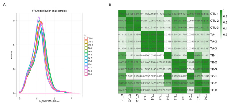 Figure 1
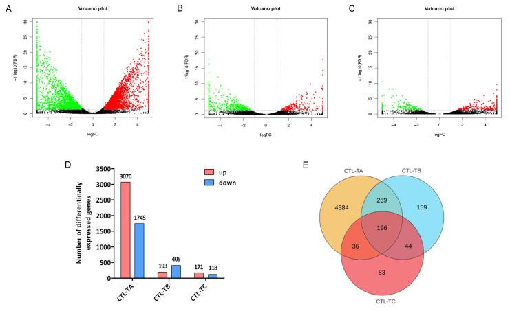 Figure 2
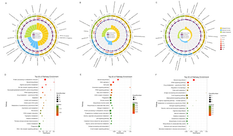 Figure 3
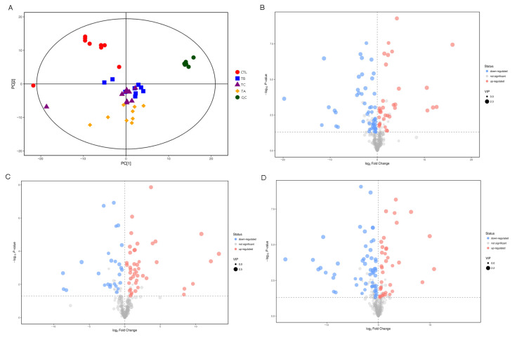 Figure 4
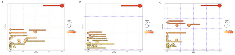 Figure 5
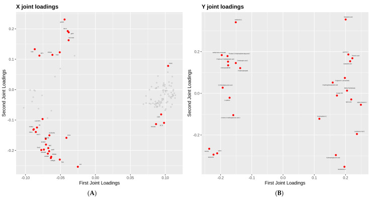 Figure 6
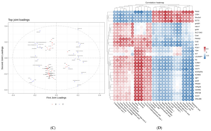 Figure 7
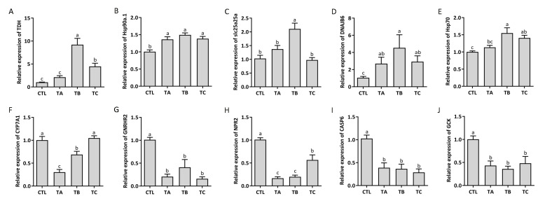 Figure 8
- —Central Public-interest Scientific Institution Basal Research Fund
- —Qingdao Postdoctoral Science Foundation under Grant
Peer Reviews
No public reviews on file for this paper yet. If you reviewed it on a platform where reviews are public (OpenReview, ICLR, NeurIPS, ICML), you can paste yours below so the community can read it here.
Videos
No videos yet. Explain this paper in a talk, walkthrough, or lecture? Add one.
Taxonomy
TopicsPhysiological and biochemical adaptations · Aquaculture disease management and microbiota · Aquaculture Nutrition and Growth
1. Introduction
The impact of increasing global seawater temperatures on marine life has become severe. Fish are ectotherms, meaning that their body temperature is primarily derived from the external environment. Consequently, the body temperature of most fish species conforms to that of the surrounding water. Their principal strategy for coping with temperature fluctuations is behavioral thermoregulation, such as moving between different water depths or habitats to find optimal temperatures. Many studies have reported that thermal stress can affect various aspects of a fish’s entire life process, including growth [1], oxidative response [2], immunity homeostasis [3] and epigenetic inheritance [4]. Under prolonged high-temperature conditions, Nile tilapia (Oreochromis niloticus) exhibited impaired growth performance and experienced stress-induced physiological damage [5]. Elevated temperatures affected both gill and skeletal muscle cellular morphology as well as immune regulation in black cuskeel (Genypterus maculatus), mediated through the upregulation of antibacterial peptides and pro-inflammatory cytokine expression [6]. Thermal stress can also alter the behavioral responses of fish, such as Atlantic salmon (Salmo Salar), including increasing inter-individual distances and modifying swimming depths [7]. Nevertheless, how elevated temperatures influence fish growth and development requires further in-depth research.
With the deepening understanding of nucleic acid molecules and the advancement of sequencing technologies, a pivotal methodology of RNA sequencing (RNA-seq) has been developed for transcriptome analysis and gene expression profiling [8]. Nowadays, RNA-seq methodologies enable the comprehensive investigation of diverse RNA biological features, including transcriptional dynamics, translational regulation, and structural characteristics [9,10,11]. Metabolomics is the analysis method of metabolites and is commonly used to discover the systemic effects of metabolites. In addition, owing to the high sensitivity of metabolomic approaches, even minor perturbations in biological pathways can be precisely identified, thereby providing mechanistic insights into diverse physiological states and pathological processes [12,13,14]. Integrated transcriptomic and metabolomic analyses enable the simultaneous investigation of biological systems from both gene regulation and metabolic response perspectives, which could facilitate the in-depth exploration of macroscopic developmental processes while elucidating the complexity and systemic integrity of biological phenomena [15,16,17]. Critically, this integrated approach is powerful for bridging the gap between genetic potential and phenotypic manifestation, allowing for the identification of key regulatory nodes that would remain hidden in single-omics studies.
The turbot (Scophthalmus maximus) is also called “Duobao fish” in China and is the major marine culture species in Northern China because of its high nutritional and economic value. At present, the breeding areas of turbot are mainly distributed in the northern coastal areas along the Bohai and Yellow Sea coasts. Our previous study has demonstrated that thermal stress could impair liver function in turbot by inducing apoptosis and injuring the hepatic capacity [18]. Yang et al. have found that high temperature stress could disturb the metabolic process in turbot, with a concomitant significant upregulation in creatinine and cortisol levels [19]. Zhao et al. reported significant alterations in hepatic lipid metabolism in turbot under thermal stress conditions [20]. However, the specific signaling pathways that are centrally involved, the coordinated interplay between gene expression and metabolic flux, and the key regulatory genes and metabolites that drive the thermal stress response in turbot remain largely unexplored and poorly integrated [21,22].
This study employed an integrated transcriptomic and metabolomic approach to elucidate the molecular mechanisms underlying the thermal stress response in turbot. By analyzing differentially expressed genes (DEGs) and metabolites, we aimed to precisely identify the core regulatory pathways and construct a gene–metabolite interaction network to uncover the systemic adaptive strategy of turbot under thermal stress. We expect that our findings will identify critical candidate genes and metabolic pathways that could serve as potential therapeutic targets for enhancing thermotolerance in turbot.
2. Materials and Methods
2.1. Study Design and Sample Collection
Healthy turbots were collected from Tianyuan Aquaculture Co., Ltd., Yantai, Shandong Province, China. In total, 120 fish (five months old) of similar weight (106.96 ± 15.71 g) and standard length (18.8 ± 0.9 cm) were randomly divided into four groups with three replicates per group and 10 fish per replicate (180 L/tank). The fish were acclimated in the tank for one week before the experiment and were fed a commercial pelleted diet twice a day until satiation at 9:00 and 15:00. Water quality parameters were measured and maintained at a salinity of 27‰–30‰, dissolved oxygen content was higher than 6.0 mg/L, pH was 7.5–8.0, and ammonia nitrogen content was less than 0.1 mg/L. Based on our previous results, we found that rapid warming can cause large-scale mortality of turbot [18]. The thermal acclimation protocol was implemented in a stepwise manner: first increasing from ambient 18 °C to 24 °C at 1 °C increments per 24 h period, then maintaining a slower escalation rate of 0.5 °C per 24 h period until the final test temperature of 27 °C was attained. The temperature was reduced from 27 °C to 18 °C at 1 °C per 1 h under flow-through conditions, with sampling conducted after 48 h of recovery at 18 °C. The controls were marked CTL, TA represented the group in 27 °C at 0 h, TB represented the group in 27 °C at 6 h, and TC represented the recovery group.
Following euthanasia with MS-222 (tricaine methane sulfonate, 100 mg/L; Sigma, St. Louis, MO, USA), hepatic tissues were aseptically excised using sterile surgical instruments and flash-frozen in liquid nitrogen [23].
2.2. RNA Isolation and Sequencing
Trizol reagent was used for RNA extraction from liver tissues. In brief, after tissue fragmentation with 1 mL of Trizol, 200 μL of chloroform was added, and centrifuged at 4 °C, 12,000 rpm, and 15 min. The supernatant was placed in a new tube with isopropanol added and then centrifuged at 4 °C, 12,000 rpm, and 10 min. RNA was dissolved in DEPC water. RNA quality was assessed using a NanoDrop (2000, Thermo Fisher Scientific, Waltham, MA, USA). mRNAs were enriched by magnetic beads and fragmented to 200–500 bp through the M220 Focused-ultrasonicator (Covaris M220, Covaris Inc., Waltham, MA, USA), using enzymes and random primers as templates to synthesize double-stranded cDNA, followed by repaired ends and adding the poly (A) and adapters. The sequencing was analyzed on the Illumina HiSeq platform of Gene Denovo Biotechnology (Guangzhou, China). The Trinity (https://github.com/trinityrnaseq (accessed on 10 May 2024)), BUSCO (http://busco.ezlab.org (accessed on 10 May 2024)), TransRate (http://hibberdlab.com/transrate/ (accessed on 10 May 2024)), and CD-HIT (http://weizhongli-lab.org/cd-hit/ (accessed on 10 May 2024)) platforms were used to reconstruct and evaluate the transcripts. Transcript abundance was quantified and normalized via the FPKM metric. The genes with fold change (FC) ≥ 2 and false discovery rate (FDR) < 0.05 were considered significant differences.
2.3. Gene Ontology and KEGG Analysis
The Gene Ontology (GO) database was used to classify and annotate DEGs according to their participation in molecular function, biological process and cellular component. KEGG function analysis was performed by KOBAS (http://bioinfo.org/kobas/ (accessed on 10 May 2024)).
2.4. Metabolites Extraction and Analysis
The liver samples used for this metabolomic analysis were collected from the same individuals as those used for transcriptomic profiling. Liver tissue samples of approximately 50 mg were homogenized in 1.5 mL centrifuge tubes with a precise volume of L-2-chlorophenylalanine (as an internal standard) and methanol. The mixture was incubated at −20 °C for 10 min and mechanically homogenized. Subsequently, it was subjected to ultra-sonication in an ice-water bath for 10 min, followed by incubation at −20 °C for 30 min. The samples were then centrifuged at 13,000× g for 10 min at 4 °C. The resulting supernatant was transferred to a new vial and lyophilized in a vacuum concentrator at ambient temperature. The dried extract was reconstituted in an appropriate volume of methanol and centrifuged again under the same conditions (13,000× g, 10 min, 4 °C), and the final supernatant was injected into a gas chromatography-mass spectrometry system (GeneDenovo Biotechnology, Guangzhou, China). Data acquisition was performed using Chroma TOF 4.3X software (LECO), and metabolites were filtered with a quality control criterion of relative standard deviation < 30% in the quality control samples.
2.5. Pathway Analysis
The differentially expressed metabolites were annotated on the established biochemical databases (KEGG and PubChem). Subsequently, the metabolic pathways were mapped in the database of corresponding species (Cynoglossus semilaevis) on the MetaboAnalyst website.
2.6. Association Analysis of Metabolome and Transcriptome
Two-way orthogonal partial least squares (O2PLS) is a generalized OPLS that can be used for bidirectional modeling and prediction in two data matrices. With this analysis, the internal connections between the two omics can be explored, the degree of association between the two omics data can be determined, and the main genes, metabolites, or proteins that cause this association can be identified. The DEGs-metabolites network was performed on the OmicShare platform (https://www.omicshare.com/tools/ (accessed on 17 May 2024), Gene Denovo) [24].
2.7. Real-Time Quantitative PCR (qPCR)
One μg RNA was reversed into cDNA using the cDNA synthetis kit (Cat#R323, Vazyme, Nanjing, China). The expression of genes was detected by the Real-time Quantitative Thermal Cycler (MA-6000, Molarray, Suzhou Molarray Biological Technology Co., Ltd., Suzhou, China), and SYBR Green reagent (Cat#R711, Vazyme, Nanjing, China) was used for progress tracking. The cycler reaction was composed of 1 × SYBR Green mix, 200 nM primer (forward/reverse), and 10 ng cDNA template, with nuclease-free water added to 20 μL volume. The thermal protocol was as follows: 95 °C for 30 s, followed by 40 cycles (including 95 °C for 10 s and 60 °C for 30 s) and then 95 °C for 15 s, 60 °C for 1 min, and 95 °C for 15 s. The data were analyzed using the 2^−ΔΔCt^ method [18,25]. GAPDH was set as an internal reference gene. All primer pairs were designed with Primer 3 software (the sequences are provided in Table 1).
2.8. Statistical Analysis
All data were shown as mean ± standard deviation. After verifying assumptions of normality and the homogeneity of variances, inter-group differences were assessed by one-way ANOVA. Post hoc comparisons were made using the least significant difference test, with findings further validated by Tukey’s test. Statistical analyses were performed with IBM SPSS Statistics (Version 27.0.1.0). p < 0.05 was considered a significant difference between the groups.
3. Results
3.1. Transcriptome Sequencing and Data Analysis
After comparing and analyzing the sequencing results, a total of approximately 85.83 Gb bases and 581.23 Mb reads were obtained from 12 libraries. The average data of sequencing quality to Q20 and Q30 were 98.37% and 94.92%, respectively. The average of GC content was 54.52% (Table 2). The distribution of gene expression abundance showed a normal distribution of gene expression in each group of samples (Figure 1A). Correlation analysis revealed significant divergence between control and experimental groups, with the TA group exhibiting particularly distinct clustering patterns (Figure 1B). These results indicate that the quality of sequencing was verified, and the data were reliable.
3.2. Differentially Expressed Genes Under High Temperatures in Turbot
Overall, 5702 genes were differently expressed in the three groups. Among them, there were 3070, 193, and 171 upregulated mRNA and 1745, 405, and 118 downregulated mRNA in the CTL vs. TA, CTL vs. TB, and CTL vs. TC groups, respectively (Figure 2A–D, Supplementary Tables S1–S3). The top 10 DEGs of the three groups are shown in Table 3, Table 4 and Table 5. Venn diagram analysis showed that there are 395 common genes between the CTL vs. TA group and the CTL vs. TB group, 170 common genes between the CTL vs. TB group and the CTL vs. TC group, 162 common genes between the CTL vs. TC group and the CTL vs. TA group, and 126 common genes among the three groups (Figure 2E).
3.3. Enrichment Analysis of DEGs
In the CTL vs. TA group, the DEGs were mainly enriched in the metabolic process, localization, immune system process, biological adhesion, single-organism process, cellular component organization or biogenesis, and developmental process in the BP category. In the CC category, the DEGs were associated with the membrane part, organelle part, cell part, and macromolecular complex. In the MF category, the DEGs were involved in molecular signal transduction, catalytic activity, and binding transcription factor activity (Figure 3A, Supplementary Table S4).
The DEGs in the CTL vs. TB group were mainly involved in the cellular process, biological regulation, metabolic process, and response to stimulus in the BP category. Moreover, those genes were involved in the cell, cell part, organelle, and membrane processes in the CC part. In the MF category, the DEGs were significantly involved in catalytic activity and complex binding (Figure 3B, Supplementary Table S5). In the CTL vs. TC group, the process of those dysregulated genes was similar to the above group, such as molecular signal transduction, catalytic activity, stimulus response and binding (Figure 3C, Supplementary Table S6).
KEGG analysis results revealed that the DEGs in the CTL vs. TA group were mainly correlated with protein processing, arginine and proline metabolism, the Toll signaling pathway, the TCA cycle, the PPAR signaling pathway, metabolism processes and apoptosis (Figure 3D). In the CTL vs. TB group, the dysregulated genes were associated with cell cycle, DNA replication, steroid biosynthesis, chemokine signaling pathway, amino acids biosynthesis, the PPAR signaling pathway, ECM-receptor interaction, and fatty acid metabolism (Figure 3E). In the CTL vs. TC. group, the dysregulated genes were associated with steroid biosynthesis, autophagy regulation, the PPAR signaling pathway, adipocytokine signaling, fatty acid metabolism and FoxO signaling pathway (Figure 3F).
3.4. Differential Metabolites Under High Temperatures in Turbot
The PCA analysis exhibited that there was significant distribution among the samples (Figure 4A). The volcano plot showed that 64 metabolites were detected in the CTL vs. TA group, of which 27 were upregulated, and 37 were downregulated. There were 56 differential metabolites in the CTL vs. TB group, of which 37 were upregulated and 19 were downregulated. Moreover, there were 69 differential metabolites in the CTL vs. TC group, of which 30 were upregulated and 39 were downregulated (Figure 4B–D). The metabolome results showed that the most dysregulated metabolites associated with high temperature stress included 4-hydroxy-3-methoxybenzoic acid, 2-amino-3-methoxybenzoic acid, 1, 2, 3-dimethylsuccinic acid, lipoic acid, and L-glutamic acid (Supplementary Tables S7 and S8). However, the most dysregulated metabolites were trans-4-hydroxy-L-proline 1, 3-hydroxy-3-methylglutaric acid, lactobionic acid 1, α-santonin 1, and methyl palmitoleate in the CTL vs. TC group (Supplementary Table S9). Of particular interest, we detected a signal in the liver metabolome of turbot that corresponds to α-santonin 1. Since this compound is unlikely to be of endogenous origin, the observed signal may represent an as-yet-unidentified endogenous metabolite sharing structural or chemical similarities with α-santonin. Alternatively, it could have been derived from plant-based components in the formulated diet and subsequently accumulated in the liver through dietary intake.
3.5. Pathway Analysis of Metabolites
The results of pathway analysis showed that although there were different metabolites between the high temperature and recovery groups, these metabolites were involved in the same pathways, such as amino acid biosynthesis and metabolism (e.g., valine, leucine, alanine, aspartate), glyoxylate and dicarboxylate metabolism, glycerolipid metabolism, and the TCA cycle (Figure 5, Supplementary Table S10).
3.6. Integrated Transcriptomic and Metabolomic Profiling
Thirty dysregulated genes and 27 metabolites were loaded in the analysis, including DNAJB6, CDH1, TDH, slc25a25a, CDHR2, PCK1, leucine, valine, galactonic acid and isoleucine (Figure 6A,B). Integrated analysis identified significant correlations between DEGs and metabolites. As shown in Figure 6C, valine, isoleucine, leucine, galactonic acid, guanine 2, and methionine 1 showed higher correlation with DEGs.
Moreover, we analyzed the correlations between these differential metabolites and differential genes and the correlation coefficient absolute values of >0.5 were plotted on the heat map (Figure 6D, Supplementary Table S11). The four downregulated genes (Plch2, CISH, Slco2a1, ECT2) were negatively correlated with the 13 upregulated metabolites, including 4-aminolhenol 1, valine, isoleucine, leucine, L-cysteine, 2,3-dimethylsuccinic acid, and methionine 1. Correlation analysis revealed significant positive associations between CDH1, Col9a1, and ECT2 genes and leucine/isoleucine metabolism. The expression levels of Plch2 and Col9a1 genes exhibited significant regulatory effects on valine metabolic pathways. Moreover, CISH and Plch2 genes showed strong correlations (r > 0.8) with leucine and isoleucine metabolism, while the gene cluster comprising DNAJB6, Gcnt1, and trim71 was significantly involved in the metabolic regulation of galactonic acid.
3.7. Identification of the Expression of DEGs by qPCR
The expression of 10 dysregulated key genes correlated with the metabolism was validated in the liver of turbot (Figure 7). The expression of TDH, Hsp90a.1, slc25a25a, DNAJB6, and Hsp70 in the liver was higher than in the control under the heat stress, and CYP7A1, GNRHR2, NPR2, CASP6 and GCK were downregulated. The results were consistent with the transcriptomic sequencing data. However, in the recovery group, the expression of most genes, except for slc25a25a, DNAJB6, and CYP7A1, still showed significant differences compared to the control group.
4. Discussion
As a predominant abiotic stressor, thermal elevation beyond optimal ranges disrupts homeostasis in cold-water fish, triggering cascading effects from molecular to organismal levels. Elucidating the molecular mechanisms underlying thermal stress responses is fundamental for developing conservation strategies to mitigate climate change impacts on fisheries. In this study, we identified many DEGs by using transcriptome sequencing. A total of 5413 genes were dysregulated in fish under heat stress, including 126 common genes, which can provide targets for subsequent research on resistance to high temperature stress. We found that fibulin-7 (FBLN7) was downregulated in thermal stress and exhibited the highest differential expression at 27 °C for 0 h. FBLN7 is an extracellular matrix (ECM) adhesion protein that mediates interactions with various ECM components, including structural proteins, cell surface receptors, and growth factors [26]. Sarangi et al. found that it can regulate the migration and infiltration of monocytes and macrophages and reduce the expression of inflammatory factors [27]. We considered that FBLN7 may participate in regulating the occurrence of inflammation in fish under high temperature stress. During the stress process, animals break down their own tissues to generate energy, which is directed toward specific tissues while also reducing the energy supplied to other tissues to resist damage caused by stress stimulation [28,29,30].
GCK plays a vital role in intracellular glucose uptake and utilization, not only initiating all major pathways of glucose utilization but also maintaining the gradient concentration required to promote glucose entry into cells [31,32]. The expression of GCK was downregulated in the liver of turbot under heat stress, according to the results from qPCR and transcriptome analysis. SET-1 is a class of epigenetic modification enzymes containing SETD, mainly affecting gene expression by modifying the methylation of histones H3K4, H3K9, H3K36, and H4K20 [33]. SETD can also catalyze the methylation of non-histones, thereby affecting signal transduction and participating in DNA repair and other processes [34,35,36]. In the present study, SET-1 expression was among the top 10 upregulated genes in turbot under thermal stress, suggesting a potentially high level of methylation activity. Epigenetic regulation—including histone modification and DNA methylation—has been increasingly recognized as a key mechanism in fish stress responses, with heat-induced changes reported in species such as zebrafish and salmon [37,38]. In fish, temperature stress can influence membrane fluidity, gene expression, and metabolic processes, and epigenetic mechanisms like those mediated by SET-1 are thought to help orchestrate these adaptations. Potential approaches, such as ChIP-seq to assess genome-wide histone methylation patterns or DNA methylation assay, are needed to clarify whether SET-1-mediated methylation directly influences stress-responsive gene expression in turbot. Moreover, research in other models indicates that SET1 protein levels can be regulated by factors such as the APC/C^Cdh1^ complex and Cla4 kinase [39], suggesting that the post-translational regulation of SET-1 may also be important in fish’s thermal response.
Amino acids are the basic building blocks of biological functional macromolecular proteins [40,41]. They are raw materials used in the body to manufacture antibody proteins, hemoglobin, enzyme proteins, hormone proteins, and neurotransmitter substances and can even be used to provide energy sources for living organisms. In this study, we found that the metabolic processes were accelerated under high temperatures, including the biosynthesis of amino acids, the TCA cycle process, CoA synthesis, and lipid metabolism. Steroids are considered to be the analog of glucocorticoids that can effectively activate the body’s immune defense system [42,43]. Numerous studies have shown that steroids can inhibit intracellular mediated immune responses, and the signaling of steroids is activated to maintain the body’s homeostasis [44,45,46]. The pathway of steroid biosynthesis was significantly enriched in turbot under heat stress, which was similar to the previous studies. Cheng et al. reported that supplementing steroids with largemouth bass can effectively alleviate heat stress-induced immune damage and metabolic disorders [47]. Moreover, Zhang et al. found that the synthesis of steroids was impaired under salinity stress, accompanied by a decrease in reproductive and immune function and an increased risk of infection [48].
The body will increase its metabolic response to resist the influence caused by external pressure stimulation, and then, the respiratory metabolism of mitochondria will be accelerated, resulting in significant ROS generation and insufficient clearance, resulting in the generation of oxidative stress [49,50,51]. Moreover, oxidative stress was associated with the apoptosis process. In the previous study, we found that thermal stress could induce the apoptosis of hepatic cells in turbot [18]. Liu et al. reported that thermal stress significantly alters the activity profile of hepatic antioxidant enzymes (e.g., SOD, CAT) while initiating programmed cell death pathways in Clarias fuscus liver tissue [52]. In this study, we found that the dysregulated genes and metabolites were related to the process of oxidative stress, autophagy, and apoptosis, including the PPAR signaling pathway, amino metabolic, biosynthesis, and the FoxO signaling pathway. PPAR is the nuclear receptor involved in the regulation of energy metabolism, cell development and differentiation [53,54,55]. In addition, Li et al. revealed that PPAR participated in lipid metabolism and the response to stress stimulation during the embryonic development of fish [56]. Specifically, the PPAR pathway could help maintain energy homeostasis by regulating fatty acid oxidation and lipid metabolism, a conserved mechanism observed in species like large yellow croaker (Larimichthys crocea) and Nile tilapia (Oreochromis niloticus) under temperature fluctuations [57,58]. Forkhead box proteins represent a conserved family of transcription factors that regulate apoptotic pathways across species by upregulating key pro-apoptotic factors, including Dally-like protein, Bcl-2 antagonist/killer, and tumor necrosis factor-related apoptosis-inducing ligand [59]. The FoxO signaling pathway detected here is a central regulator of cellular stress response, promoting the expression of antioxidant enzymes (e.g., SOD, CAT) to mitigate oxidative damage and inducing autophagy to maintain cellular homeostasis, as similar studies in zebrafish (Danio rerio) and common carp (Cyprinus carpio) reveal [60,61,62]. These results showed that when fish underwent the thermal stress, apoptosis and inflammatory pathways were mainly activated and enriched.
Through associated analysis of transcriptome and metabolome, it was found that some genes and metabolites exhibit correlations under high temperature stress, such as TDH, slc25a25a, Plch1, CISH, and PCK1. Moreover, we selected and validated the expression of those key genes in turbot liver, and the results were consistent with the sequencing. Correlation analysis revealed that genes (e.g., COL9A1, PCK1, SGK1, and DNAJB6) were significantly associated with the metabolism of leucine, galactonic acid, isoleucine, and valine, suggesting their potential regulatory roles in amino acid metabolism under high temperature stress. Furthermore, these DEGs (e.g., COL9A1, PCK1, SGK1, DNAJB6) were primarily enriched in intracellular protein folding, nuclear transport, and the PI3K-Akt signaling pathway. The PI3K-Akt pathway serves as a critical hub coordinating cell survival, growth, and metabolism. Its activation under heat stress likely represents a protective mechanism to inhibit apoptosis and support energy-demanding repair processes. These findings imply that the above signaling pathway may mediate the physiological response of turbot to thermal stress. Collectively, the enrichment of the PPAR, FoxO, and PI3K-Akt pathways highlights a coordinated transcriptional and metabolic reprogramming in turbot liver aimed at balancing energy allocation, mitigating oxidative injury, and determining cell fate under thermal stress. While these core pathways are evolutionarily conserved, the specific gene–metabolite interactions identified here, such as the strong association between COL9A1 and branched-chain amino acid metabolism, may represent species-specific regulatory features in turbot’s adaptation to high temperature.
5. Conclusions
In summary, the synergistic application of multi-omics approaches has elucidated the systemic responses of turbot to thermal stress, identifying coordinated dysregulation of transcriptional networks and metabolic pathways critical for thermal tolerance. Moreover, joint analysis revealed significant positive associations between CDH1, Col9a1, and ECT2 genes and leucine/isoleucine metabolism. The expression levels of Plch2 and Col9a1 genes exhibited significant regulatory effects on valine metabolism. CISH and Plch2 genes showed strong correlations with leucine and isoleucine, while the gene cluster comprising DNAJB6, Gcnt1, and trim71 was significantly involved in the metabolic regulation of galactonic acid. These dysregulated genes and metabolites were mainly involved in ABC transporters, the PI3K-Akt signaling pathway, and protein transport and processing. The identified biomarkers could guide the selective breeding of thermotolerant fish, while the metabolic insights inform the development of targeted management practices to mitigate heat stress, thereby enhancing the industry’s resilience to rising temperatures. These findings offer valuable insights for addressing environmental stressors in turbot aquaculture. However, it should be noted that as the fish in this study were sourced from a commercial processing company, the potential genetic diversity within the population may introduce variability in thermal response, which represents a limitation of the present study. Thus, further validation and multi-generational studies are required to validate the findings of this study.
The reference list from the paper itself. Each links out to its DOI / PubMed record.
- 1Hu Q.X. Lu J.M. Yang Y. Li D.P. Liu J.Y. Acute Thermal Stress Reduces Skeletal Muscle Growth and Quality in Gibel Carp (Carassius gibelio)Water 202315270610.3390/w 15152706 · doi ↗
- 2da Silva D.O. Ratko J. Côrrea A.P.N. da Silva N.G. Pereira D.M.C. Schleger I.C. Neundorf A.K.A. de Souza M. Herrerias T. Donatti L. Assessing physiological responses and oxidative stress effects in Rhamdia voulezi exposed to high temperatures Fish Physiol. Biochem.20245061763310.1007/s 10695-023-01294-238175338 · doi ↗ · pubmed ↗
- 3Lee D. Kim K.H. Park J.W. Lee J.H. Kim J.H. High water temperature-mediated immune gene expression of olive flounder, Paralichthys olivaceus according to pre-stimulation at high temperatures Environ. Toxicol. Pharmacol.202310110415910.1016/j.etap.2023.10415937245611 · doi ↗ · pubmed ↗
- 4Mateus A.P. Costa R.A. Sadoul B. Bégout M.L. Cousin X. Canario A.V. Power D.M. Thermal imprinting during embryogenesis modifies skin repair in juvenile European sea bass (Dicentrarchus labrax)Fish Shellfish Immunol.202313410864710.1016/j.fsi.2023.10864736842641 · doi ↗ · pubmed ↗
- 5Abu-Zahra N.I.S. Atia A.A. Elseify M.M. Abass M.E. Soliman S. Dietary Pelargonium Sidoides extract mitigates thermal stress in Oreochromis niloticus: Physiological and immunological insights Vet. Res. Commun.20254915210.1007/s 11259-025-10705-z 40126735 PMC 11933177 · doi ↗ · pubmed ↗
- 6Becerra S. Arriagada-Solimano M. Escobar-Aguirre S. Palomino J. Aedo J. Estrada J.M. Barra-Valdebenito V. Zuloaga R. Valdes J.A. Dettleff P. High temperature induces oxidative damage, immune modulation, and atrophy in the gills and skeletal muscle of the teleost fish black cusk-eel (Genypterus maculatus)Dev. Comp. Immunol.202516410533210.1016/j.dci.2025.10533239892682 · doi ↗ · pubmed ↗
- 7Korus J. Filgueira R. Grant J. Influence of temperature on the behaviour and physiology of Atlantic salmon (Salmo Salar) on a commercial farm Aquaculture 202458974097810.1016/j.aquaculture.2024.740978 · doi ↗
- 8Schreibing F. Anslinger T.M. Kramann R. Fibrosis in Pathology of Heart and Kidney: From Deep RNA-Sequencing to Novel Molecular Targets Circ. Res.20231321013103310.1161/CIRCRESAHA.122.32176137053278 · doi ↗ · pubmed ↗