Are Reusable Dry Electrodes an Alternative to Gelled Electrodes for Canine Surface Electromyography?
Ana M. Ribeiro, I. Brás, L. Caldeira, J. Caldeira, C. Peham, H. Plácido da Silva, João F. Requicha

TL;DR
This study shows that reusable dry electrodes can work as well as traditional gelled ones for measuring muscle activity in dogs, without needing skin prep.
Contribution
The paper pioneers the use of reusable dry electrodes for canine surface EMG without hair clipping.
Findings
Dry electrodes provided reliable frequency-domain data and higher amplitude than gelled electrodes.
Reusable dry electrodes reduced preparation time and avoided hair clipping.
Signal quality from dry electrodes was broadly comparable to gelled ones despite minor differences.
Abstract
Non-invasive assessment of muscle activity in veterinary patients can be highly advantageous, especially when dealing with neuromuscular disorders. A primary test for such a purpose is electromyography (EMG), particularly surface EMG (sEMG); however, its use in small animals is limited by the need for skin preparation and disposable electrodes. To our best knowledge, with this study, we evaluated for the first time whether reusable dry electrodes constitute a feasible alternative to conventional gel-based electrodes, through signals collected from 12 dogs during dynamic treadmill walking. Our findings show that, even without hair clipping, dry electrodes yield reliable frequency-domain data and higher amplitude readings comparatively to gel-based electrodes, offering a practical, faster, and more sustainable alternative for clinical and research use. Our results pave the way for a more…
Genes, proteins, chemicals, diseases, species, mutations and cell lines named across the full text — each resolved to its canonical identifier and authoritative record.
Click any figure to enlarge with its caption.
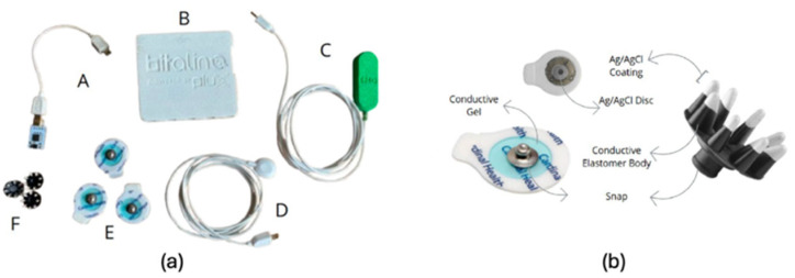 Figure 1
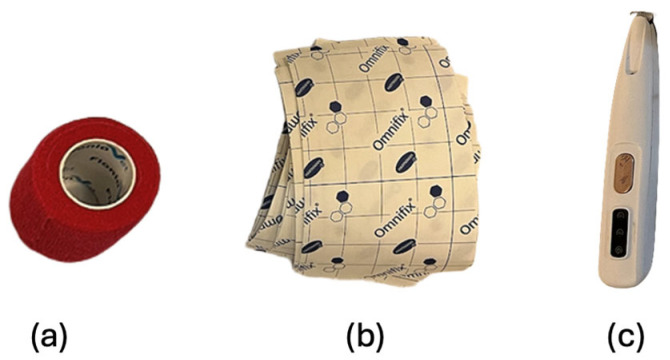 Figure 2
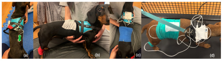 Figure 3
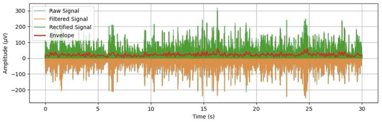 Figure 4
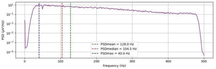 Figure 5
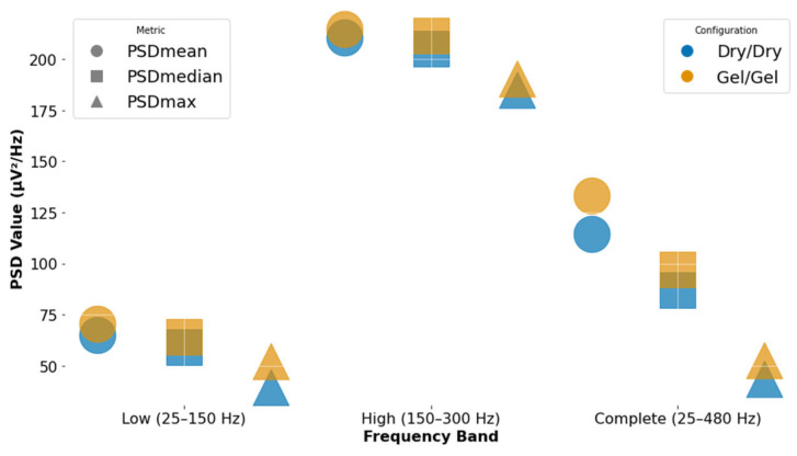 Figure 6
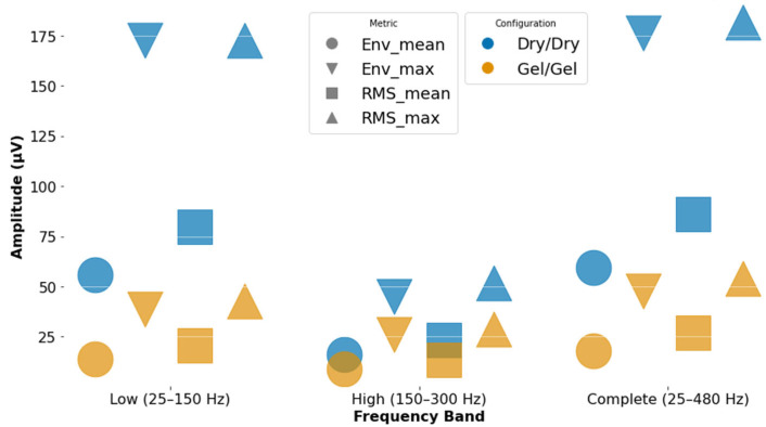 Figure 7
- —Portuguese Foundation for Science and Technology (FCT)
Peer Reviews
No public reviews on file for this paper yet. If you reviewed it on a platform where reviews are public (OpenReview, ICLR, NeurIPS, ICML), you can paste yours below so the community can read it here.
Videos
No videos yet. Explain this paper in a talk, walkthrough, or lecture? Add one.
Taxonomy
TopicsMuscle activation and electromyography studies · Motor Control and Adaptation · Advanced Sensor and Energy Harvesting Materials
1. Introduction
Surface electromyography (sEMG) is a non-invasive diagnostic tool that records the electrical activity of muscles [1,2,3,4,5]. Measuring electrical signals generated by motor units’ action and muscle fiber recruitment offers meaningful insights into the mechanisms underlying muscle contraction and neuromuscular control [6]. sEMG has proven to be a valuable technique for assessing neuromuscular disorders, monitoring treatment progress, and evaluating therapeutic interventions, allowing clinicians to evaluate muscle function in dynamic activities; it is used in various disciplines, including basic research, clinical diagnostics, sports medicine, and rehabilitation [6,7,8,9,10,11]. Studies by Balbinot et al. 2021 and Mitchell et al. 2015 suggest that sEMG can be a useful tool in conjunction with current standard protocols (International Standards for Neurological Classification of Spinal Cord Injury—ISNCSCI, American Spinal Cord Association Impairment Scale—AIS and Graded Redefined Assessment of strength, sensibility and Prehension—GRASSP) for clinical assessment of individuals with spinal cord injury (SCI) [12,13]. sEMG amplitude has been associated with a measure of muscle vitality, is a reliable indicator of muscle recovery, and can identify residual muscle activity in patients with no observable movement below a spinal lesion [13]. Although, to the best of our knowledge, it is still unexplored in applications, beyond clinical assessment, EMG opens new avenues for the control of robotic devices, prosthetics, and exoskeletons in human healthcare [5,6,14,15].
In the context of veterinary rehabilitation, sEMG has emerged as a promising tool for the non-invasive measurement of muscular function in conscious patients, used increasingly in research of both equine and companion animal medicine [16,17,18].
Our study focuses on using sEMG in veterinary medicine, motivated by the lack of non-invasive and objective tools to assess neuromuscular function in animals with spinal cord disorders such as intervertebral disk disease (IVDD) undergoing rehabilitation medicine treatments. To our best knowledge, this study provides the first direct comparison of soft polymeric dry electrodes and gel-based electrodes for canine dynamic sEMG, addressing a key barrier to wider adoption of objective neuromuscular assessment tools in veterinary rehabilitation. With the use of sEMG, clinicians can develop targeted rehabilitation strategies [6] tailored to the individual needs of each patient, based on their neurological status and tolerance [19].
According to the Consensus for Experimental Design in Electromyography (CEDE)—an international initiative aimed at guiding best practices for the recording, analysis, and interpretation of EMG data [9,10,20,21], bipolar surface electromyography (sEMG) involves the differential amplification of signals detected by pairs of surface electrodes; the bipolar configuration is particularly advantageous, as it can better mitigate the effects of common mode noise. This technique is widely used for assessing superficial and easily accessible muscles, with the estimated activation level representing the activity of a relatively large number of muscle fibers. Furthermore, CEDE highlights that, as the interface between the tissue and the recording system, the selection of the appropriate EMG electrodes is of utmost importance.
In human sEMG, conventional silver/silver chloride (Ag/AgCl) electrodes are typically placed on the skin overlaying the target muscle [9]. However, in veterinary applications, sEMG presents additional challenges, including the need for skin preparation (i.e., trichotomy), to ensure adequate electrode adhesion and better manage behavioral constraints during data collection [16,17,18]. Most of the previous literature on animal sEMG primarily focuses on kinematics and locomotion function on muscles of horses and dogs, specifically limb muscles [22,23,24,25,26,27]; there is a gap in what concerns the evaluation of neuromuscular diseases. As such, our main objective was to evaluate the practical efficiency of sEMG in the paraspinal muscle region, using a completely different set of electrodes.
Particularly, intervertebral disk disease (IVDD) is a common condition in dogs that, among others, can lead to several serious myelopathies such as intervertebral disk herniation (IVDH), degenerative lumbosacral stenosis, or cervical spondylomyelopathy [28], which in turn can significantly impair quality of life and life expectancy. In fact, IVDD underlies the most common forms of intervertebral disk herniation (IVDH) [29], and it is estimated to account for approximately 21% of all neurological cases reported in domestic dogs. The overall incidence of this pathology across all domestic canine breeds is estimated at 2% to 3.5% [30]. A previous pilot study developed by Ribeiro et al. 2024 [22] involving IVDD patients and healthy controls, suggested a decrease in sEMG amplitude and an increase in sEMG frequency in exercises featuring more dynamic muscle activation in the group of animals affected with IVDD. This further reinforces that sEMG can be an added value tool to assess neuromuscular function, as it has the potential to categorize EMG signals between healthy animals and the ones affected with IVDD. Also, a study by Schwartz et al. 2024 [31], using sEMG in dogs that had undergone hemilaminectomy, concluded that post-hemilaminectomy dogs had greater hind limb activation when compared with normal dogs. Their results support the idea that sEMG can be valuable to evaluate muscle activity in dogs recovering from myelopathies/spinal decompression surgery.
Still, the broader use of sEMG in small animals has been limited by trichotomy, signal instability, and the need for specialized training in signal analysis [16,17,18]. Our study seeks to analyze the feasibility of soft polymeric dry electrodes for sEMG applications, comparatively to conventional gel-based electrodes in canine subjects. If successful, this procedure would greatly simplify the clinical use of sEMG in this context.
In general, soft polymeric dry electrodes produced higher amplitude and a larger root mean square error (RMSE). Regarding power spectral density (PSD) values, both dry and pre-gelled electrodes were similar, although the pre-gelled electrodes tended to have slightly higher spectral power. Our results indicate that dry electrodes offer practical advantages in terms of easier use and reduced preparation time, without compromising signal integrity, but pre-gelled electrodes remain advisable whenever possible.
2. Materials and Methods
2.1. Pre-Study Evaluation
The animal study protocol was approved by the Ethics Committee of Trás-os-Montes e Alto Douro University (Doc106-CE-UTAD-2024) for studies involving animals.
Before participating in any of the experimental procedures, all studied dogs underwent a clinical pre-assessment conducted by a Certified Canine Rehabilitation Veterinarian (Canine Certified Canine Practitioner—CCRP). This evaluation served not only to ensure that each animal was clinically fit to perform the planned tasks without discomfort or risk of injury, but also to confirm the absence of musculoskeletal or neurological conditions that could compromise the validity of the results. This pre-assessment consisted of a general health evaluation and a thorough orthopedic and neurological assessment [32,33,34].
2.2. Animal Selection
To isolate breed as a variable, only Dachshunds were selected. The inclusion of this breed in this study was based on two principal criteria: (i) its increasing popularity as a companion animal [35,36,37]; and (ii) its well-documented genetic predisposition to a broad spectrum of orthopedic and neurological disorders, with particular emphasis on IVDD which is estimated to affect 19–31% of these dogs during their lifetime, with the highest incidence occurring between 4 and 6 years of age [38,39,40,41]. This breed-specific anatomical and physiological predisposition frequently results in significant neuromuscular impairment, necessitating extended therapeutic interventions and an intensive requirement for physiotherapeutic and functional rehabilitation [42].
The main characteristics of the dogs recruited are summarized in Table 1.
From the 13 dogs initially recruited, one dog was excluded after clinical evaluation due to the presence of pain at vertebral column palpation. From the 12 dogs included in the study, four were females, three of which had ovariohysterectomy, and one was intact; the remaining eight were males, of which two were neutered and six were intact. The mean age of the animals was 3 ± 1.7 years (median of 3 years). The average body condition score was 5 ± 1 based on a 9-point scale, with individual scores ranging from 4/9 to 7/9.
Inclusion criteria required that animals had no history of lameness or orthopedic issues and exhibited normal findings on both orthopedic and neurological examinations. Furthermore, no animals under the age of one year or older than 7 years were included, as this could compromise the sEMG readings [43,44].
2.3. Clinical Evaluation
For the participants included in the study, a more comprehensive assessment was carried out in eight stages [32,33,34]:
- Collection of medical history;
- General health inspection;
- Standing inspection to assess muscle asymmetry, musculoskeletal deformities, and even weight distribution across limbs;
- Gait inspection with special attention to signs of lameness;
- Palpation of limb joints to confirm absence of crepitus, edoema, instability, or pain;
- Palpation of the spine and surrounding musculature, including careful manipulation of the cervical spine through its full range of motion (ROM) to ensure absence of pain;
- Proprioceptive testing and spinal reflexes on all four limbs;
- Specific orthopedic tests, such as the drawer test and tibial thrust.
2.4. Data Acquisition Setup
The experimental setup was composed of a BITalino (r)evolution Core BT board (PLUX^®^, Lisbon, Portugal), one BITalino assembled sEMG sensor (PLUX^®^, Lisbon, Portugal), and a 1-lead electrode cable (for the reference electrode). Both pre-gelled Ag/AgCl (Kendall™H124SG, Leeds, UK) surface electrodes and soft polymeric dry electrodes (SoftPulse^TM^ Flex, Datwyler^®^, Altdorf, Switzerland) were used for signal acquisition (Figure 1). Specific characteristics of both electrode types and applications are described in Table 2. A BITalino accelerometer (ACC) sensor (PLUX^®^, Lisbon, Portugal) was also integrated into the setup and physically attached to the BITalino unit, which was secured to a harness worn by the dog. The accelerometer was used to monitor the animal’s movement and to assist in identifying stable signal segments in the subsequent sEMG processing stages. Data collection and visualization were performed using a laptop running the OpenSignals v2.2.5 (r)evolution software (PLUX^®^, Lisbon, Portugal).
For sEMG device and electrode fixation purposes co-adhesive flexible bandage 10 × 4.5 cm (Vet-flex, Kruuse^®^, Langeskov, Denmark) was used, as well as Omnifix 10 × 10 cm elastic adhesive (Hartmann^®^, Heindenheim, Germany) (Figure 2). Lastly, when trichotomy was necessary, a professional trimmer (IPX5 waterproof, Yiwu, China) for small areas was used, to minimize the amount of hair clipped, the trimmer chosen had a blade of proximally 20 mm width and a rounded R-type design that is believed to prevent skin scratching (Figure 2).
2.5. Skin Preparation and Exercise
Measurements from all 12 participants were performed twice, first with the soft polymeric dry electrodes and untrimmed hair conditions, and afterwards with the pre-gelled Ag/Ag electrodes and trimmed hair conditions. Electrodes were placed on the same anatomical location, overlaying the longissimus dorsi muscle between thoracic vertebra 12 (T12) and lumbar vertebra 2 (L2), 0.5 cm lateral to the spinous processes on the right side of the body, and on the same day. In both evaluations, the reference electrode (grounding)—Ag/Ag electrodes were used—was positioned on a bony prominence—medial aspect of the tarsus (medial tibial malleolus)—to minimize interference and provide a stable baseline. These electrodes were secured using a co-adhesive flexible bandage. Before positioning, trichotomy was performed, and skin was cleaned using 70% alcohol wipes.
In the first stage (untrimmed hair condition), two dry electrodes were applied to the longissimus dorsi muscle (Figure 3), ensuring firm contact with the skin and proper parallel alignment with the muscle fibers. To guarantee adequate adhesion and reduce skin impedance, before electrode placement, the skin was cleaned using 70% alcohol wipes. After placement, electrodes were secured in place with Omnifix elastic adhesive and, to guarantee minimal displacement during walking, 10 × 4.5 cm co-adhesive flexible bandage was also applied. To ensure consistency, whenever the electrode setup shifted or fell out of place during signal acquisition, the preparation process was restarted entirely from the beginning. This was performed to maintain uniformity and avoid introducing artifacts or inconsistencies in the data. Our goal was to minimize the effects of movement as much as possible.
Each participant was placed on a professional veterinary treadmill (FitFurLife^®^ Professional Treadmill, Surrey, UK) and encouraged to walk at a constant, comfortable speed of 1.93 km/h to promote regular and consistent muscle activation of the spinal muscles. To ensure stable gait cycles and minimize motion artifacts, each dog was allowed to walk on the treadmill for a few seconds until a steady and repeatable walking pattern was achieved. Once a regular gait was confirmed, signal acquisition was performed in two stages. First, data was collected while the dog walked steadily on the treadmill for approximately 60 s. After this recording, the experiment proceeded to the second stage (trimmed condition), in which the hair in the electrode placement area was carefully trimmed (trichotomy), maintaining the same position and alignment of the electrodes relative to the muscle fibers.
The location for trichotomy was guided by the faint imprint left by the dry electrodes on the fur, which enabled precise trimming in the same spot used during the untrimmed condition; this ensured consistency in electrode positioning across both stages of the experiment. The skin was again cleaned with alcohol wipes, and pre-gelled Ag/AgCl electrodes were applied using the exact same technique previously used; the ground treadmill exercise was then performed under the same conditions and for the same duration to allow a direct comparison between electrode types.
An accelerometer was used simultaneously and synchronously recorded with the sEMG sensor to monitor the dog’s movement, enabling the identification of stable gait cycles and signal segments suitable for amplitude and spectral analysis. Signals were also acquired at 1 kHz using the BITalino system and OpenSignals software (Section 2.3), with sessions labeled by electrode type and hair condition for analysis and date (e.g., SH3/flex/notricho/07-05 vs. SH3/Ag/AgCl/tricho/07-05).
2.6. Signal Processing
All raw sEMG signals acquired in both settings were processed using the same pipeline to ensure consistency and comparability across electrode types and hair conditions. Each raw signal was first filtered using a 6th-order digital band-pass filter with a passing band between 25 and 480 Hz to isolate the relevant muscle activity and remove unwanted noise; this specific frequency range is chosen because it encompasses the typical frequencies produced by muscle contractions, while simultaneously attenuating low-frequency motion artifacts and high-frequency noise [45,46].
Additionally, a notch filter centered at 50 Hz with a quality factor of 30 (which indicates the filter’s sharpness in attenuating this specific frequency) was applied to suppress powerline interference, i.e., unwanted electrical noise at the frequency of the power grid in Portugal [47]. When cardiac interference (i.e., electrocardiography contamination) was visually detected in the sEMG signal, typically characterized by low-frequency periodic components, an additional high-pass filter with a cutoff frequency of 60 Hz was applied to minimize this effect [1,48,49,50].
After filtering, the signals were full-wave rectified and smoothed using a 4th-order low-pass filter with a cutoff frequency of 5 Hz to generate the sEMG envelope [1,6]. This envelope reflects the amplitude modulation of muscle activation over time, providing an overall measure of the signal’s intensity/trend. From the envelope, descriptive statistics were extracted, including mean, standard deviation, and peak amplitude for each acquisition condition (Figure 4).
To further characterize the signals in the frequency domain, the Fast Fourier Transform (FFT) was applied to the filtered (non-rectified) data, as it can be seen in Figure 5 [6]. From the resulting power spectral density, which provides a statistical representation of a signal’s power distribution across different frequencies, the maximum (PSDmax), mean (PSDmean), and median (PSDmedian) frequencies were calculated, providing insight into the frequency decomposition of the sEMG signals and allowing comparison of noise profiles across electrode configurations.
In addition to the full frequency band (25–480 Hz), spectral analysis was also performed separately for two sub-bands: low-frequency (25–150 Hz) and high-frequency (150–300 Hz) [22,46]. The rationale for these frequency bands was derived from existing literature and the spectral regions most relevant for distinguishing signal and noise [22,46].
3. Results
Key metrics from both frequency and time domains were extracted for comparison between the dry and gel configurations during treadmill walking. This dynamic task allowed us to assess signal behavior under more realistic, movement-involved conditions. All computed metrics were compiled and organized for subsequent comparison and visualization of signal quality under the different hair conditions (with and without trichotomy); metrics were also stratified by electrode configuration and frequency band. A summary of the results is provided in Table 3.
As expected in a dynamic task, greater variability was observed in amplitude-related features, particularly for the dry configuration. Full-band Power spectral density (PSD) values serve as a reference for overall spectral content during treadmill walking; Figure 6 shows the PSD metrics for each configuration across the three frequency bands. Both dry and gel electrodes yielded broadly comparable spectral features. However, the gel configuration showed slightly higher PSDmean, PSDmedian, and PSDmax values in all bands, particularly in the full-band (25–480 Hz), which may suggest more effective capture of neuromuscular activity with reduced spectral distortion. The dry setup exhibited slightly lower PSD values, which could reflect reduced sensitivity or increased noise filtering due to less optimal skin contact. Nonetheless, the consistency of the values across bands indicates that dry electrodes can still provide meaningful spectral information during dynamic conditions.
Figure 7 summarizes the time-domain results. Here, the dry configuration yielded higher amplitude values across most metrics, notably in Max Env and Max RMS, which may indicate either stronger signal capture or increased susceptibility to motion artifacts caused by treadmill movement. In contrast, the gel electrodes produced lower but more stable amplitude values, especially in Mean Env and Mean RMS, suggesting improved signal stability under dynamic conditions. This trade-off between amplitude and stability should be considered when selecting electrodes for dynamic tasks.
4. Discussion
This work showed that soft polymeric dry electrodes can provide comparable signal quality to the one obtained with gelled electrodes in both static and dynamic conditions. The obtained results are consistent with previous research in veterinary sEMG, reporting the feasibility of dynamic muscle activity recording during treadmill locomotion [22,27]. However, by introducing dry electrodes into this context, the authors address an important gap identified in reviews such as Fuchs et al. (2022) [17], which highlighted the need for more practical, standardized, and less invasive approaches in clinical sEMG.
Hence, our results reinforce the feasibility of using dry electrodes for dynamic canine sEMG acquisition, although gelled electrodes may still offer slight advantages in terms of contact stability and signal clarity during movement. While gelled electrodes produced slightly higher spectral metrics, dry electrodes offer practical advantages, including reduced preparation time, greater animal tolerance, and environmental sustainability through reusability. In a study conducted by Steenbergen et al. (2023) [14], dry conductive elastomer (CE) electrodes also demonstrated performance comparable to traditional wet Ag/AgCl electrodes in human subjects, consistent with the results obtained in the present work. Unlike Ag/AgCl electrodes, which require gel and are limited by skin preparation, dermatological reactions, and short-term usability, CE electrodes offer flexibility, stretchability, and reduced mechanical mismatch without an electrolyte [14,51].
Electrochemical and functional sEMG testing showed that CE electrodes had lower impedance at relevant frequencies, which in this study translated into cleaner sEMG signals with reduced noise and motion artifacts. Classification using a backpropagation neural network yielded high accuracy (99.57% for CE-trained data and 94.30% for Ag/AgCl-trained data), demonstrating that CE electrodes can be effectively integrated into movement classification frameworks [14]. These findings support the use of solid, reusable CE electrodes as a viable long-term alternative to wet Ag/AgCl electrodes.
Our amplitude findings align with observations in human rehabilitation sEMG studies, where dry electrodes often yield higher peak values due to altered skin-electrode impedance and motion artifacts [7]. The slightly reduced PSD values did not compromise the extraction of key frequency metrics, in agreement with Suo et al. (2024) [6], who showed that spectral features can remain stable across electrode types if placement and fixation are consistent.
From a clinical perspective, the ability to obtain reliable sEMG data without trichotomy is particularly relevant for veterinary rehabilitation. Many canine patients, especially those undergoing long-term physiotherapy for IVDD, may require repeated sEMG assessment sessions; avoiding hair clipping can improve owner acceptance and reduce stress for the animal.
5. Conclusions
To our best knowledge, this study is the first to show that reusable soft polymeric dry electrodes can capture reliable paraspinal sEMG signals in dogs without hair clipping, offering comparable performance to gel-based electrodes. Their practicality, reusability, and animal-friendly application could expand the use of objective neuromuscular assessment in veterinary rehabilitation, especially for intervertebral disk disease. However, there are limitations to consider in future studies. Our study involved only one breed; despite reducing the number of potential confounding variables, it may limit generalizability to dogs with different coat types or body conformations. Future work should address this matter to permit the comparison between different breeds and myopathies, as it will permit a broader use of sEMG. In addition, the sample size was relatively small, and all subjects were healthy. While the assessment of animals with intervertebral disk disease was beyond the scope of the present study, future investigations will focus on this clinical population to further evaluate the applicability and generalizability of the findings. Long-term durability and repeated use performance of dry electrodes were not assessed. Future work should evaluate dry electrodes in multi-session rehabilitation monitoring, investigate optimal fixation techniques to minimize motion artifacts, and explore integration with wearable sEMG systems for remote physiotherapy monitoring. If validated in patient populations, this approach could expand the use of sEMG as an objective measure in veterinary rehabilitation, enabling more precise and individualized therapy adjustments. Considering the dynamic context of the text subjects (i.e., monitored during treadmill walking, and naturally excited and mobile), studying in greater detail the susceptibility of dry electrodes to noise and motion artifacts is of utmost importance.
The reference list from the paper itself. Each links out to its DOI / PubMed record.
- 1Merletti R. Muceli S. Tutorial. Surface EMG Detection in Space and Time: Best Practices J. Electromyogr. Kinesiol.20194910236310.1016/j.jelekin.2019.10236331665683 · doi ↗ · pubmed ↗
- 2Meekins G.D. So Y. Quan D. American Association of Neuromuscular & Electrodiagnostic Medicine Evidenced-Based Review: Use of Surface Electromyography in the Diagnosis and Study of Neuromuscular Disorders Muscle Nerve 2008381219122410.1002/mus.2105518816611 · doi ↗ · pubmed ↗
- 3Cram J.R. The History of Surface Electromyography Appl. Psychophysiol. Biofeedback 200328819110.1023/A:102380240713212827987 · doi ↗ · pubmed ↗
- 4Hermens H.J. Freriks B. Disselhorst-Klug C. Rau G. Development of Recommendations for SEMG Sensors and Sensor Placement Procedures J. Electromyogr. Kinesiol.20001036137410.1016/S 1050-6411(00)00027-411018445 · doi ↗ · pubmed ↗
- 5Cheng L. Li J. Guo A. Zhang J. Recent Advances in Flexible Noninvasive Electrodes for Surface Electromyography Acquisition NPJ Flex. Electron.202373910.1038/s 41528-023-00273-0 · doi ↗
- 6Suo M. Zhou L. Wang J. Huang H. Zhang J. Sun T. Liu X. Chen X. Song C. Li Z. The Application of Surface Electromyography Technology in Evaluating Paraspinal Muscle Function Diagnostics 202414108610.3390/diagnostics 1411108638893614 PMC 11172025 · doi ↗ · pubmed ↗
- 7Al-Ayyad M. Owida H.A. De Fazio R. Al-Naami B. Visconti P. Electromyography Monitoring Systems in Rehabilitation: A Review of Clinical Applications, Wearable Devices and Signal Acquisition Methodologies Electronics 202312152010.3390/electronics 12071520 · doi ↗
- 8Alcan V. Zinnuroğlu M. Current Developments in Surface Electromyography Turk. J. Med. Sci.2023531019103110.55730/1300-0144.566738813041 PMC 10763750 · doi ↗ · pubmed ↗