Antihelix ulcer in anti-MDA5-positive dermatomyositis
Wenhan Huang, Lin Tang

Abstract
Genes, proteins, chemicals, diseases, species, mutations and cell lines named across the full text — each resolved to its canonical identifier and authoritative record.
Click any figure to enlarge with its caption.
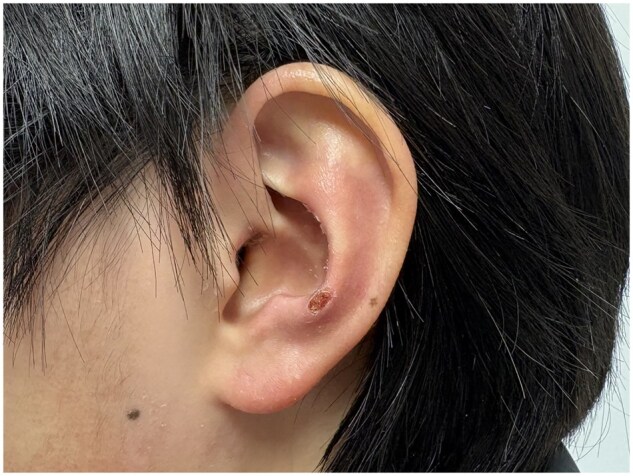 Figure 1
Peer Reviews
No public reviews on file for this paper yet. If you reviewed it on a platform where reviews are public (OpenReview, ICLR, NeurIPS, ICML), you can paste yours below so the community can read it here.
Videos
No videos yet. Explain this paper in a talk, walkthrough, or lecture? Add one.
Taxonomy
TopicsInflammatory Myopathies and Dermatomyositis · Eosinophilic Disorders and Syndromes · Skin Diseases and Diabetes
A 44-year-old woman presented to the rheumatology clinic with a 2-month history of cutaneous eruption, arthralgia and fatigue. Physical examination revealed Gottron’s papules, mechanic’s hands, V-neck sign and antihelix erythema with ulcer formation (Fig. 1). Proximal muscle strength was 4 according to the Medical Research Council scale. Laboratory findings revealed elevated ESR (52 mm/h) and serum ferritin (991.4 ng/ml). The level of creatine kinase was 487 U/l (reference range: 38–174). Test for anti-melanoma differentiation-associated protein 5 (MDA5) antibodies and anti-Ro52 antibodies were positive. Chest CT showed interstitial lung disease. MRI showed a high signal of thigh muscles on the T2-weighted image. The diagnosis of anti-MDA5-positive dermatomyositis was made. Treatment with methylprednisolone (i.v. therapy, 80 mg/d), cyclophosphamide (i.v. therapy, 0.4 g once/week) and tacrolimus (oral, 1 mg twice a day; the blood trough concentration was maintained at 5–10 ng/ml) was initiated.
Interstitial lung disease and skin involvement were two significant features of anti-MDA5 dermatomyositis. Apart from common manifestations, such as Gottron’s papules, V-neck and shawl sign, skin lesions in other areas were gradually being recognized as well. Antihelix and helix erythema were relatively rare in anti-MDA5 dermatomyositis. On the one hand, it is regarded as microvascular injury induced by pressure, just like decubitus [1]. On the other hand, it has been reported that persistent endoplasmic reticulum stress and unfolded protein response induced by the combination of MDA5 protein and protein kinase RNA-like endoplasmic reticulum kinase might be an important mechanism of vascular damage in anti-MDA5 dermatomyositis patients [2]. This patient has developed antihelix ulcers, indicating more severe vasculitis. Early combined immunotherapy is helpful to control the development of anti-MDA5 dermatomyositis.
The reference list from the paper itself. Each links out to its DOI / PubMed record.
- 1Okiyama N , Inoue S, Saito A et al Antihelix/helix violaceous macules in Japanese patients with anti-melanoma differentiation-associated protein 5 (MDA 5) antibody-associated dermatomyositis. Br J Dermatol 2019;180:1226–7.30431155 10.1111/bjd.17431 · doi ↗ · pubmed ↗
- 2Zhao LQ , Yang XQ, Niu Q et al MDA 5 protein mediating persistent ER stress/unfolded protein response contributes to endothelial-mesenchymal-transition of lung microvascular endothelial cell in dermatomyositis. Cell Commun Signal 2025;23:149.40122798 10.1186/s 12964-025-02159-2PMC 11930013 · doi ↗ · pubmed ↗