Gallbladder Rupture in an Adult Weimaraner Dog
Armands Vekšins, Ilze Dūzena, Olga Rabočaja

TL;DR
A Weimaraner dog was diagnosed with a ruptured gallbladder after initial suspicion of a stomach issue, leading to successful surgical treatment.
Contribution
The case highlights the importance of advanced imaging in diagnosing gallbladder rupture in dogs.
Findings
Radiography failed to confirm GDV but showed peritoneal effusion.
CT scan revealed gallstones, thickened gallbladder, and suspected rupture.
Surgical confirmation and treatment resolved the peritonitis.
Abstract
A 6-year-old spayed female Weimaraner weighing 47 kg was referred to the University Veterinary Hospital with suspected acute gastric dilatation and volvulus (GDV). Radiographic examination did not confirm GDV, but a mild peritoneal effusion was diagnosed. Abdominal ultrasound confirmed the diagnosis of peritoneal effusion, but the cause of these changes was not clarified. An increase in leukocytosis and a worsening of the clinical condition were quickly noted. It was decided to perform a CT scan, and localized ileus, gallstones, and the gallbladder showed marked irregular contours and thickening, with suspected rupture as the cause of the peritonitis. A laparotomy confirmed the diagnosis, and surgical treatment was performed.
Genes, proteins, chemicals, diseases, species, mutations and cell lines named across the full text — each resolved to its canonical identifier and authoritative record.
Click any figure to enlarge with its caption.
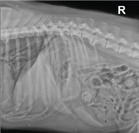 Figure 1
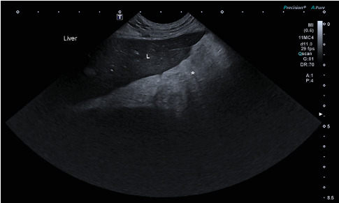 Figure 2
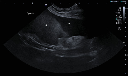 Figure 3
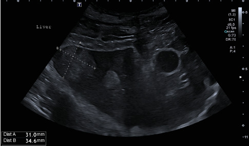 Figure 4
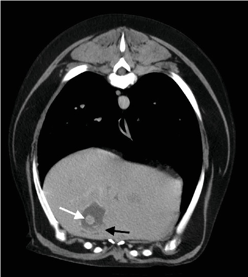 Figure 5
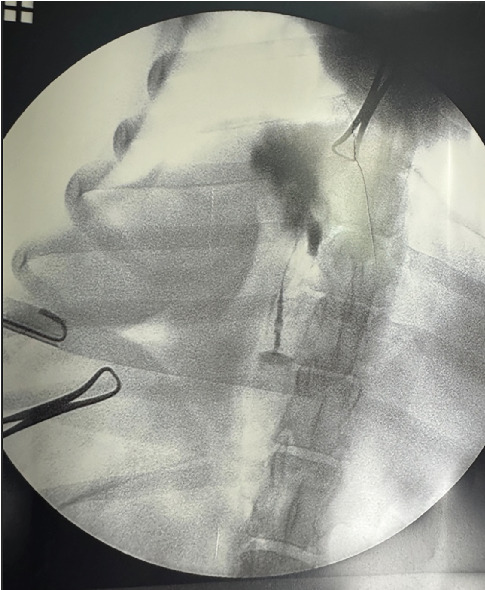 Figure 6
 Figure 7
Peer Reviews
No public reviews on file for this paper yet. If you reviewed it on a platform where reviews are public (OpenReview, ICLR, NeurIPS, ICML), you can paste yours below so the community can read it here.
Videos
No videos yet. Explain this paper in a talk, walkthrough, or lecture? Add one.
Taxonomy
TopicsCongenital Anomalies and Fetal Surgery · Pediatric Hepatobiliary Diseases and Treatments · Animal Virus Infections Studies