Rapid 3D phenotyping of chick embryo liver development at HH22-HH41 embryonic stages using X-ray microcomputed tomography with PTA staining
Igor Rzhepakovsky, Sergei Piskov, Svetlana Avanesyan, Marina Sizonenko, Lyudmila Timchenko, Magomed Shakhbanov, Gloria Nassali, Idrisa Kiryowa, Andrey Nagdalian, Ayman Swelum, Ayman Swelum, Ayman Swelum

TL;DR
This study uses X-ray microcomputed tomography to visualize and analyze chick embryo liver development, providing detailed 3D images and growth patterns.
Contribution
A refined PTA staining protocol enables high-resolution 3D imaging of chick embryo liver development, offering normative data for developmental research.
Findings
Liver growth peaks between incubation days 6 and 9 (HH29–HH35), followed by vascular stabilization by day 10 (HH36).
µCT with PTA staining achieves image quality comparable to histology for visualizing liver microstructures and vascular networks.
3D µCT renderings simplify structural orientation and highlight the technique's utility for developmental and toxicological studies.
Abstract
Animal models, particularly the chicken embryo (CE), remain crucial for advancing developmental biology and medicine. Liver embryogenesis, a complex and tightly regulated process, is particularly susceptible to developmental abnormalities. This study presents a comprehensive 2D and 3D µCT analysis of CE liver development (HH22–HH41) using a refined 1% phosphotungstic acid (PTA) staining protocol. Our methodology yielded high-resolution visualization of liver microstructures, including intricate vascular networks, with image quality and contrast comparable to histological analysis. Quantitative assessment revealed a critical period of rapid liver growth between incubation days 6 and 9 (HH29–HH35), followed by the stabilization of hepatic vascular volume by day 10 (HH36). The ease of structural orientation in µCT datasets, enhanced by 3D renderings, further underscores the technique’s…
Genes, proteins, chemicals, diseases, species, mutations and cell lines named across the full text — each resolved to its canonical identifier and authoritative record.
Click any figure to enlarge with its caption.
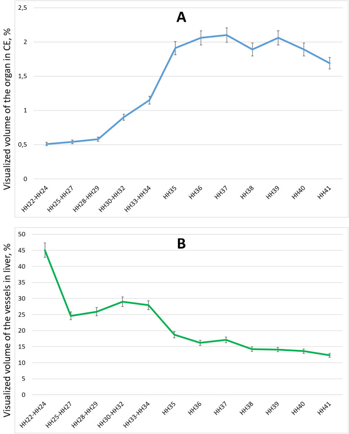 Figure 1
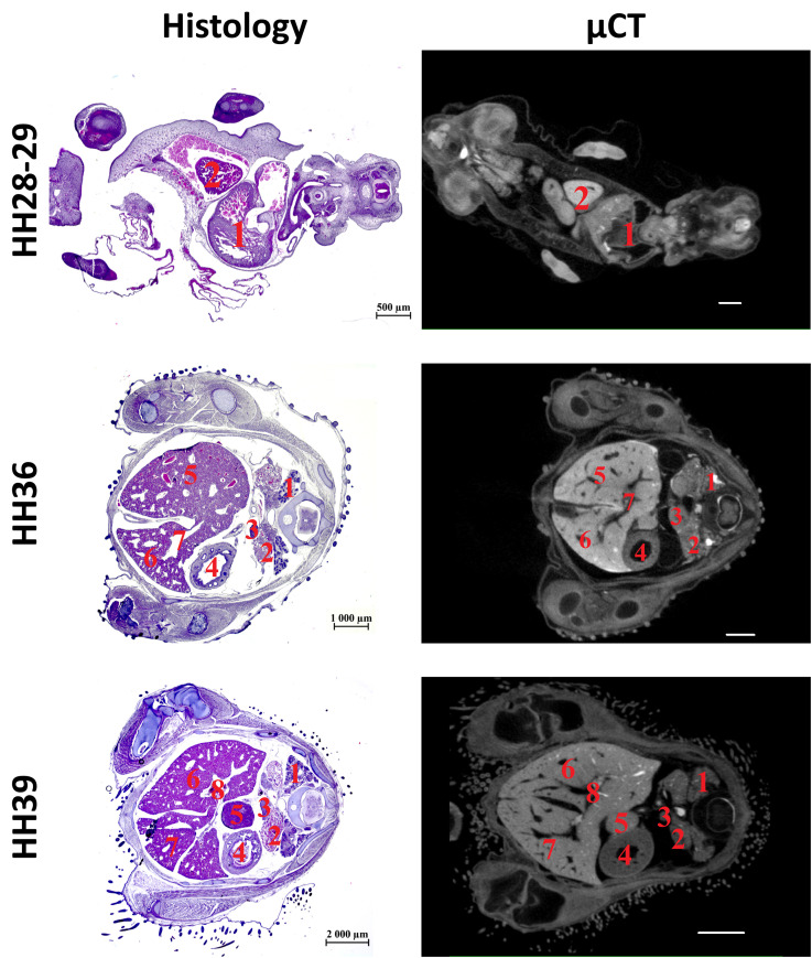 Figure 2
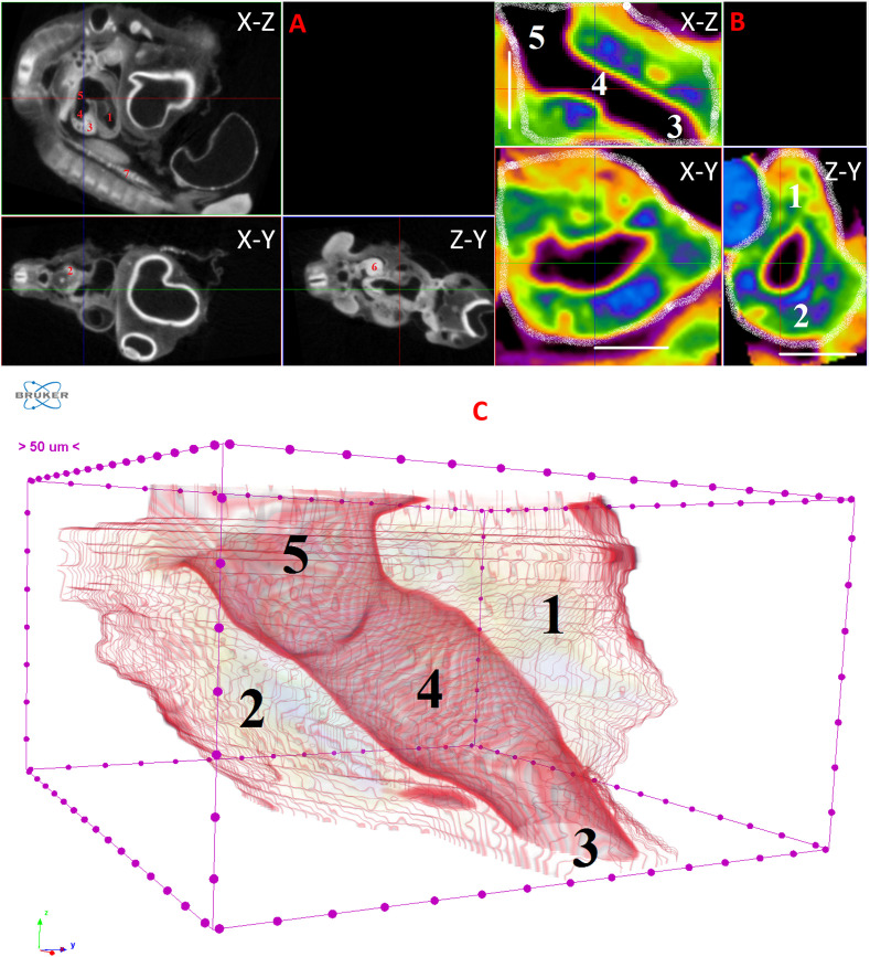 Figure 3
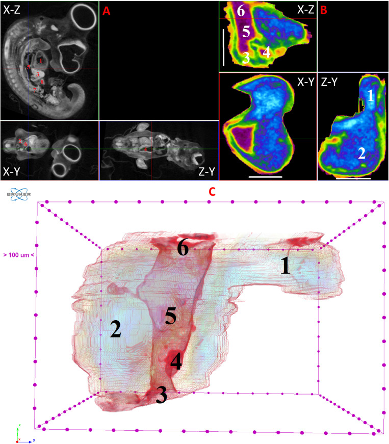 Figure 4
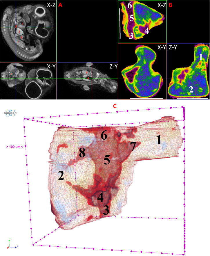 Figure 5
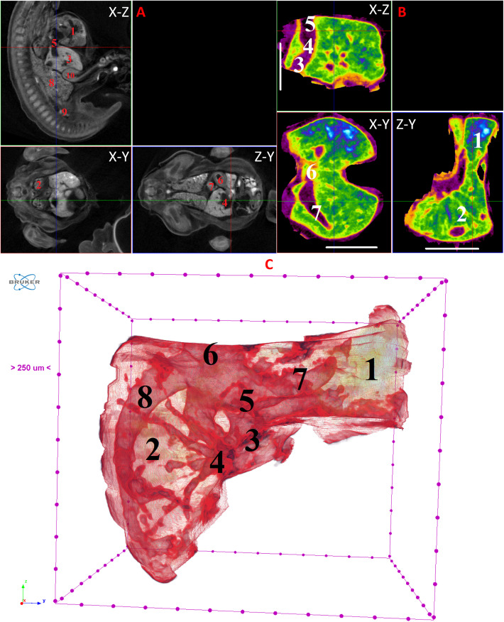 Figure 6
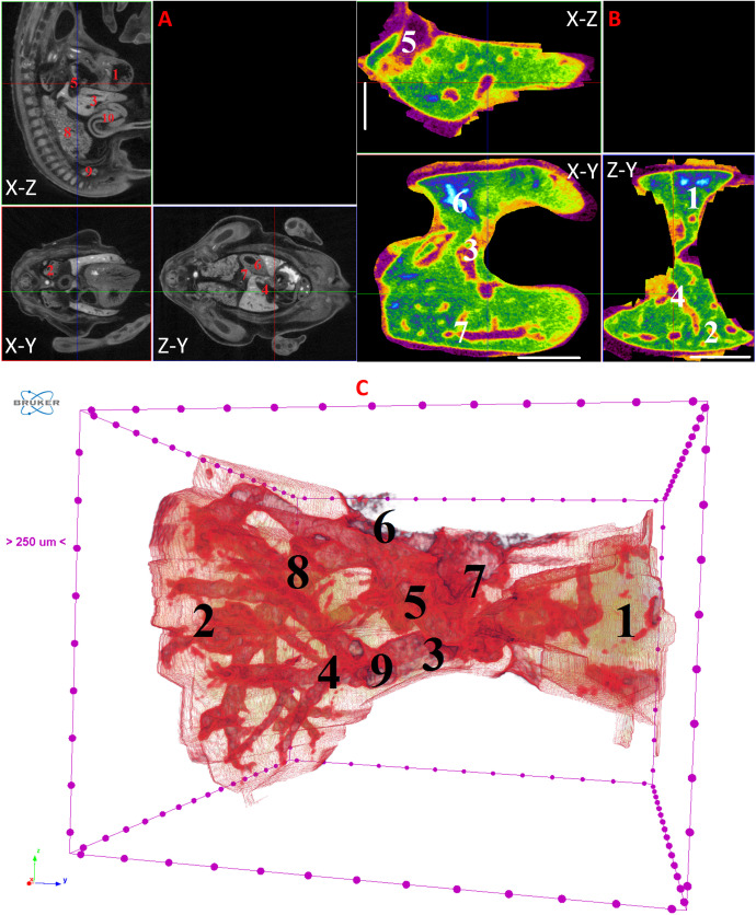 Figure 7
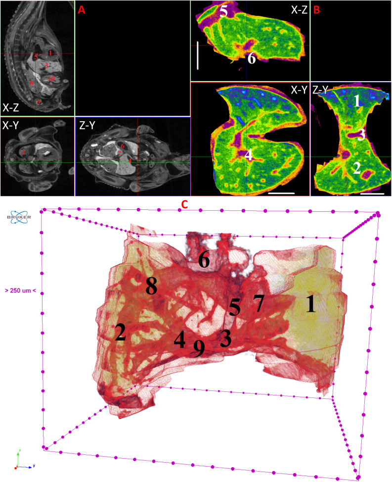 Figure 8
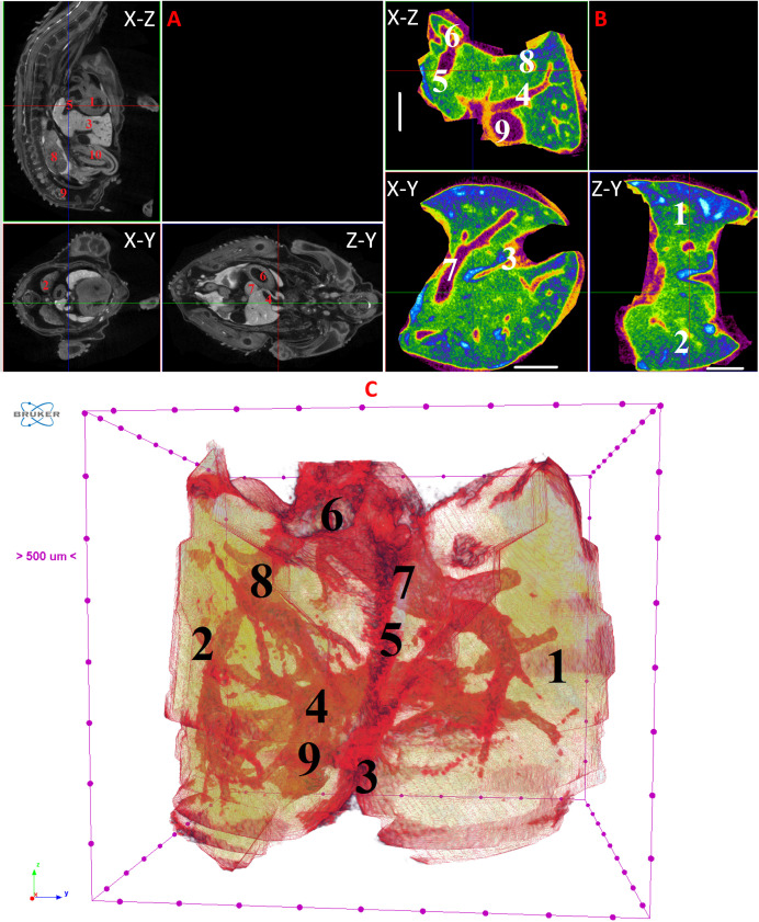 Figure 9
 Figure 10
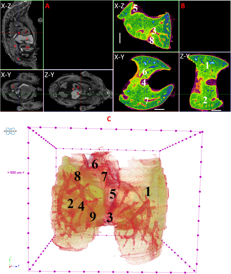 Figure 11
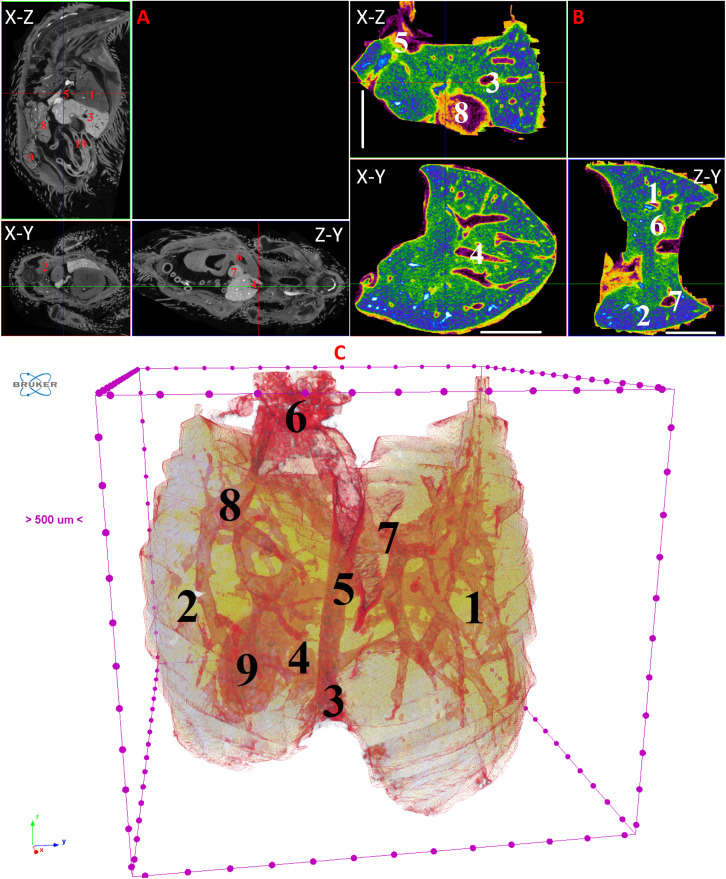 Figure 12
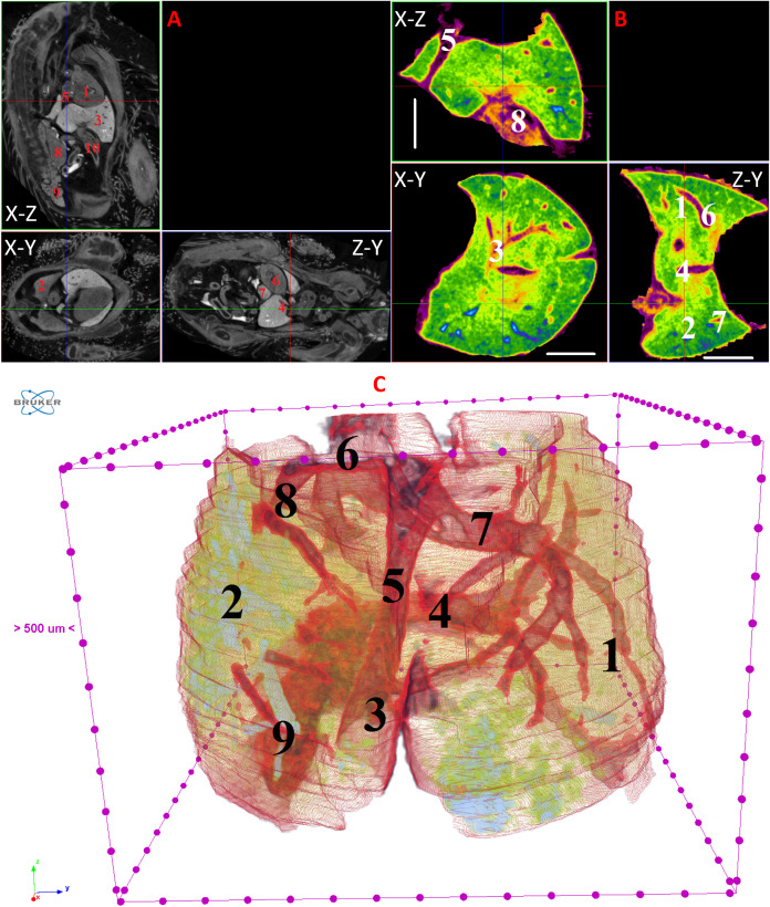 Figure 13
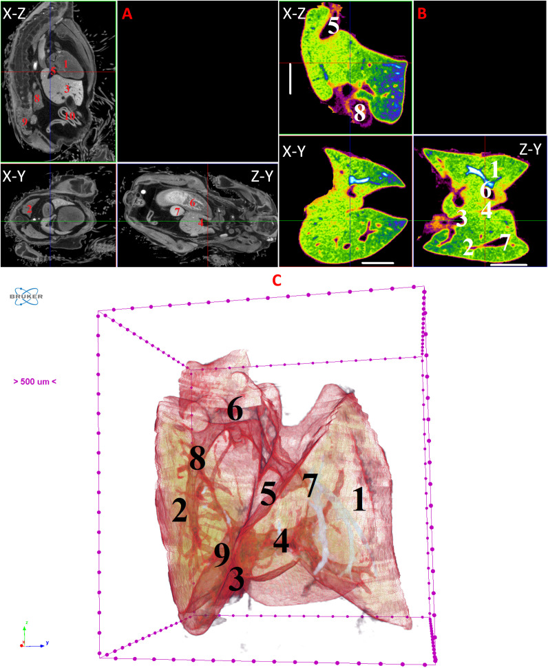 Figure 14
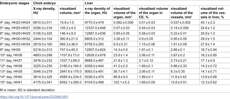 Figure 15
Peer Reviews
No public reviews on file for this paper yet. If you reviewed it on a platform where reviews are public (OpenReview, ICLR, NeurIPS, ICML), you can paste yours below so the community can read it here.
Videos
No videos yet. Explain this paper in a talk, walkthrough, or lecture? Add one.
Taxonomy
TopicsCongenital heart defects research · Birth, Development, and Health