Imaging insights in veno-venous and veno-arterial extracorporeal membrane oxygenation (ECMO): CT protocols, underlying pathophysiology, and main complications
Francesco Lauriero, Giuseppe Cicchetti, Alessio Perazzolo, Silvia De Vizio, Daniele Perla, Agostino Meduri, Riccardo Marano, Anna Rita Larici, Luigi Natale

TL;DR
This paper reviews how imaging, especially CT scans, should be adapted for patients on ECMO to ensure accurate diagnosis and management of complications.
Contribution
The paper introduces tailored CT imaging protocols specific to ECMO configurations and hemodynamic factors to improve diagnostic accuracy.
Findings
Tailored CT protocols are essential to account for ECMO-induced hemodynamic changes and ensure accurate diagnosis.
Approximately 50% of ECMO patients develop complications such as hemorrhage, thromboembolic disease, and vascular injury.
Non-invasive imaging is crucial for evaluating cannula placement and identifying complications in ECMO patients.
Abstract
Extracorporeal membrane oxygenation (ECMO) is a vital life support technique employed in patients experiencing pulmonary or cardiopulmonary failure. This procedure entails the use of a pump to replace heart function and an oxygenator to ensure adequate blood oxygenation. ECMO systems are categorized into two main configurations: veno-venous (VV) and veno-arterial (VA) circuits. VV-ECMO is employed for isolated respiratory failure, while VA-ECMO provides temporary mechanical circulatory support for patients with cardiogenic shock or cardiac arrest. A less common alternative, veno-arterial-venous (VAV) ECMO, may be used in complex cases, reducing left ventricular afterload, leading to an improvement of pulmonary edema. Imaging plays a pivotal role in ECMO management, particularly in confirming proper cannula placement, detecting malposition or migration, and identifying complications such…
Genes, proteins, chemicals, diseases, species, mutations and cell lines named across the full text — each resolved to its canonical identifier and authoritative record.
Click any figure to enlarge with its caption.
 Figure 10
 Figure 11
 Figure 12
 Figure 1
 Figure 2
 Figure 3
 Figure 4
 Figure 5
 Figure 6
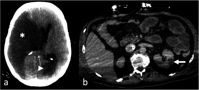 Figure 7
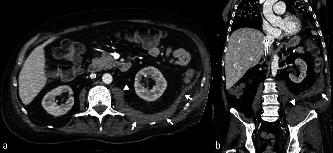 Figure 8
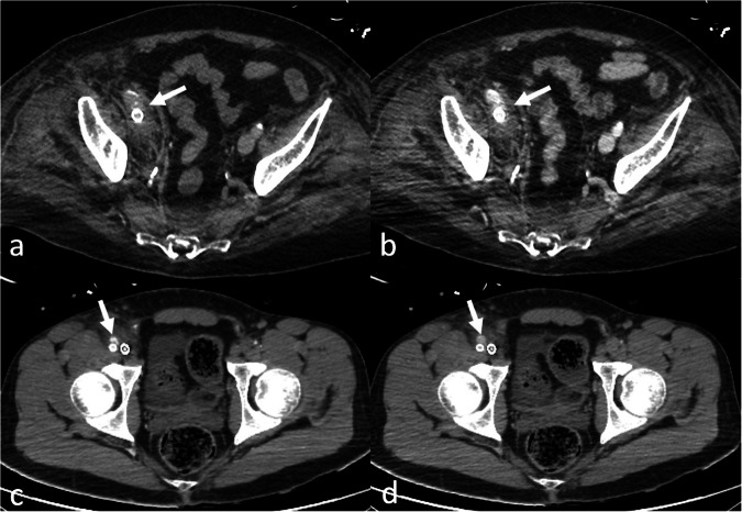 Figure 9
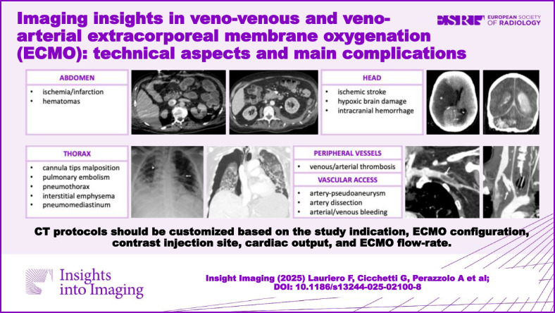 Figure 13
Peer Reviews
No public reviews on file for this paper yet. If you reviewed it on a platform where reviews are public (OpenReview, ICLR, NeurIPS, ICML), you can paste yours below so the community can read it here.
Videos
No videos yet. Explain this paper in a talk, walkthrough, or lecture? Add one.
Taxonomy
TopicsMechanical Circulatory Support Devices · Central Venous Catheters and Hemodialysis · Cardiac Structural Anomalies and Repair