Joint modeling and marker set selection significantly influence functional biomechanics in end-stage knee osteoarthritis: evidence from the sit-to-stand task
Giovanni Spallone, Letizia Mancini, Arianna Carnevale, Stefano Campi, Emiliano Schena, Pieter D’Hooghe, Michael T. Hirschmann, Rocco Papalia, Umile Giuseppe Longo

TL;DR
This study shows that different motion capture protocols significantly affect biomechanical measurements in knee osteoarthritis patients during a sit-to-stand task.
Contribution
The study reveals that marker set and modeling choices critically influence knee biomechanics measurements in end-stage osteoarthritis.
Findings
Peak variability in knee flexion angle reached 23.99° across different protocols.
Cluster-based marker sets showed reduced variability compared to anatomical-based ones.
Differences were most pronounced during movement initiation and stabilization phases.
Abstract
The sit-to-stand (STS) movement represents a mechanically demanding task, particularly informative in patients with knee osteoarthritis. While three-dimensional optoelectronic motion capture is the gold standard for analyzing joint biomechanics, the influence of protocol selection remains poorly characterized in the context of STS. This study investigated protocol-induced variability in knee kinematics and kinetics by evaluating two widely used marker sets: the anatomical-based IOR and the cluster-based CAST, each combined with either inverse kinematics or a six degrees-of-freedom joint model. Twenty-four patients (mean age of 67 ± 5 years and BMI of 28.9 ± 3.8 kg/m2) with end-stage KOA (Kellgren-Lawrence grade 3 or 4) performed three STS trials, and biomechanical outputs were compared across the four resulting protocols using Mean Absolute Variability (MAV), Mean Absolute Differences…
Genes, proteins, chemicals, diseases, species, mutations and cell lines named across the full text — each resolved to its canonical identifier and authoritative record.
Click any figure to enlarge with its caption.
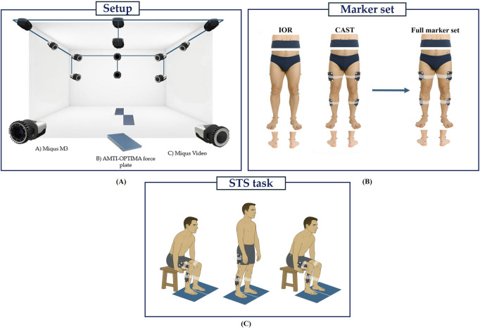 Figure 1
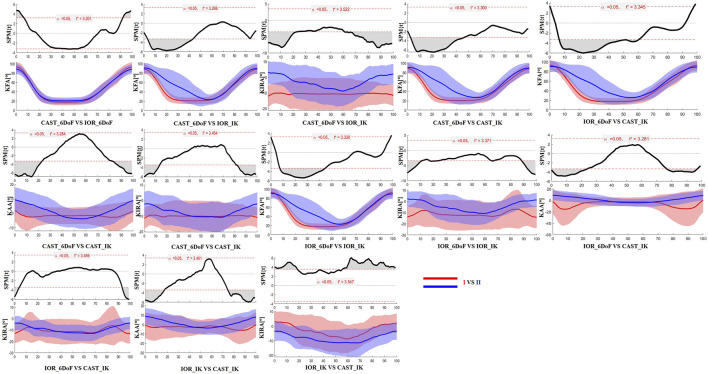 Figure 2
 Figure 3
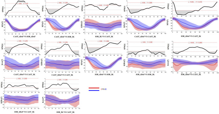 Figure 4
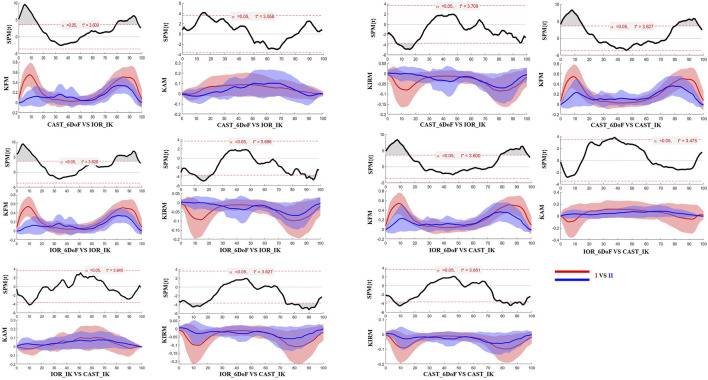 Figure 5
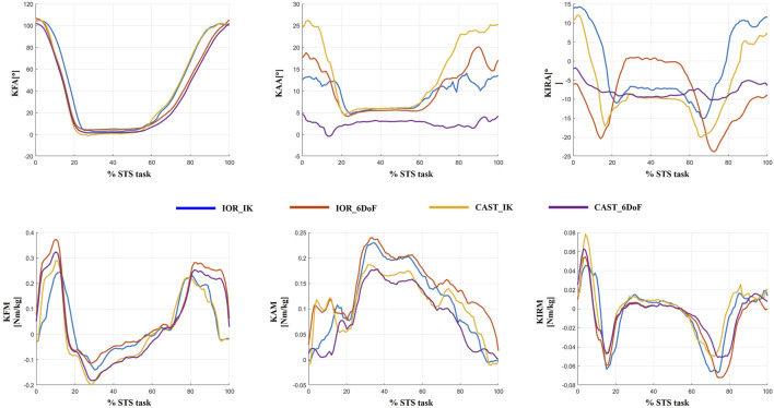 Figure 6
Peer Reviews
No public reviews on file for this paper yet. If you reviewed it on a platform where reviews are public (OpenReview, ICLR, NeurIPS, ICML), you can paste yours below so the community can read it here.
Videos
No videos yet. Explain this paper in a talk, walkthrough, or lecture? Add one.
Taxonomy
TopicsLower Extremity Biomechanics and Pathologies · Osteoarthritis Treatment and Mechanisms · Knee injuries and reconstruction techniques