Oral Lesions With Identical Clinical Presentation and Different Histopathological Diagnoses: A Case Series of Mucocele, Schwannoma, and Hamartoma
Cristina Suaza, Lenin Torres-Osorio, Jaime E Plazas Román, Adel Martinez Martinez, Antonio Diaz, Carlos M Ardila

TL;DR
This case series shows how similar-looking oral lesions can have very different microscopic diagnoses, emphasizing the need for histopathological analysis to avoid misdiagnosis.
Contribution
The study highlights the diagnostic importance of histopathology in distinguishing clinically similar oral lesions through three distinct cases.
Findings
A lower lip lesion in a 20-year-old was diagnosed as a mucocele with mucin extravasation and chronic inflammation.
A dorsal tongue mass in a 68-year-old was identified as a schwannoma with encapsulated biphasic patterns.
A lower lip lesion in a 65-year-old was diagnosed as a fibrovascular lipomatous hamartoma with adipose tissue and vascular elements.
Abstract
Oral lesions with similar clinical presentation often conceal distinct histopathological profiles, creating diagnostic challenges and potential risks of misdiagnosis. This report presents the cases of three patients whose oral lesions appeared macroscopically alike but revealed markedly different microscopic characteristics. The first case involved a 20-year-old man with a persistent lower lip lesion that had recurred after two prior interventions; histopathological analysis confirmed a mucocele characterized by mucin extravasation and chronic inflammatory infiltrate. The second case was of a 68-year-old woman with a dorsal tongue mass that produced discomfort in speech and mastication; biopsy revealed a benign schwannoma with the classic biphasic Antoni A and B pattern, encapsulated and without malignant features. The third case was of a 65-year-old woman with a slow-growing,…
Genes, proteins, chemicals, diseases, species, mutations and cell lines named across the full text — each resolved to its canonical identifier and authoritative record.
Click any figure to enlarge with its caption.
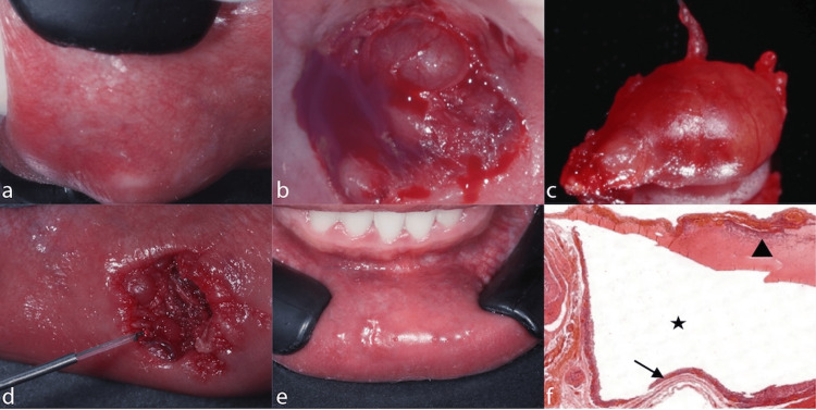 Figure 1
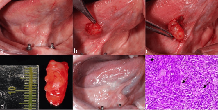 Figure 2
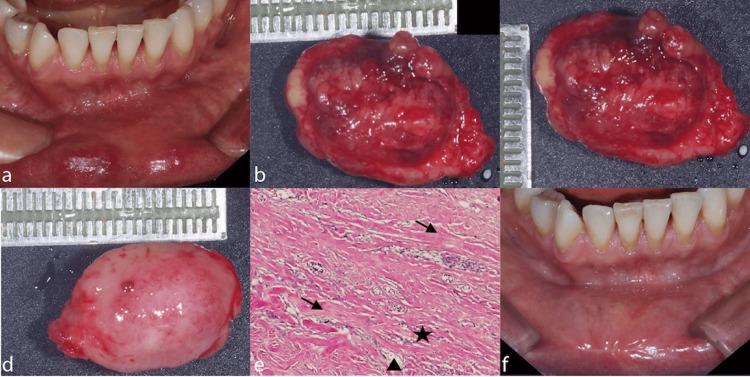 Figure 3
Peer Reviews
No public reviews on file for this paper yet. If you reviewed it on a platform where reviews are public (OpenReview, ICLR, NeurIPS, ICML), you can paste yours below so the community can read it here.
Videos
No videos yet. Explain this paper in a talk, walkthrough, or lecture? Add one.
Taxonomy
TopicsSalivary Gland Tumors Diagnosis and Treatment · Oral and Maxillofacial Pathology · Neurofibromatosis and Schwannoma Cases