Optimized Protocol for Isolation and Culture of Primary Human Corneal Epithelial Cells
Rongshan Yan, Feeling Y. Chen, Ethan S. Lindgren, Qi Gao, Yien-Ming Kuo, Danielle M. Robertson, Onur Cil, Matilda F. Chan, Neel D. Pasricha

TL;DR
This paper provides a detailed protocol for isolating and culturing high-purity human corneal epithelial cells, which can be used for drug testing and disease research.
Contribution
The study introduces an optimized and reliable protocol for culturing primary human corneal epithelial cells with preserved functionality.
Findings
High-purity primary HCECs with strong proliferative capacity and preserved morphology were successfully isolated and cultured.
Immunofluorescence staining confirmed the presence of limbal stem cells and differentiated corneal epithelial cells in vitro.
Cultured HCECs retained the ability to respond to external Ca²⁺ stimuli, demonstrating functional integrity.
Abstract
To establish a reliable method for isolating and culturing high-purity primary human corneal epithelial cells (HCECs) for ophthalmic drug testing. We present a detailed, step-by-step protocol for the efficient isolation and culture of primary HCECs. This protocol includes the characterization of HCEC morphological responses to varying Ca2+ concentrations in the culture medium. Additionally, immunofluorescence staining with well-established markers is used to identify and confirm the cell types in vitro. A Ca2+ assay is performed to validate the functionality of the cultured primary HCECs. By following the procedures detailed in this protocol, high-purity primary HCECs with strong proliferative capacity and preserved morphological integrity are obtained. Immunofluorescence staining confirms the presence of both limbal stem cells and differentiated corneal epithelial cells in vitro.…
Genes, proteins, chemicals, diseases, species, mutations and cell lines named across the full text — each resolved to its canonical identifier and authoritative record.
Click any figure to enlarge with its caption.
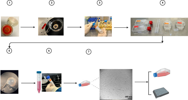 Figure 1
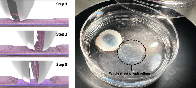 Figure 2
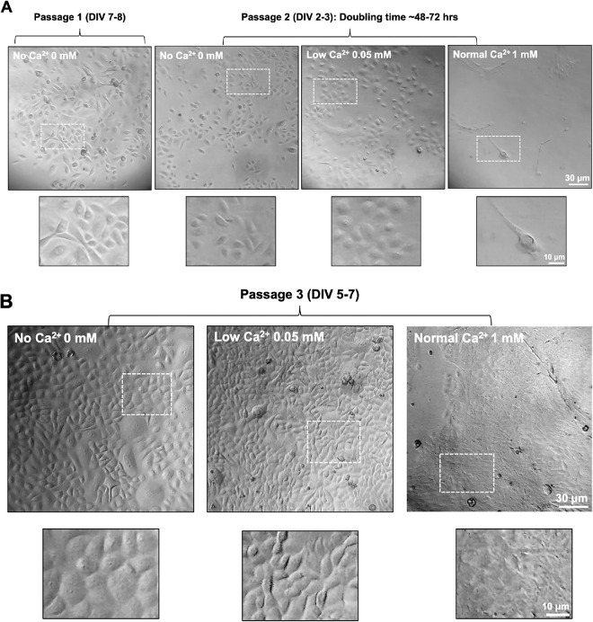 Figure 3
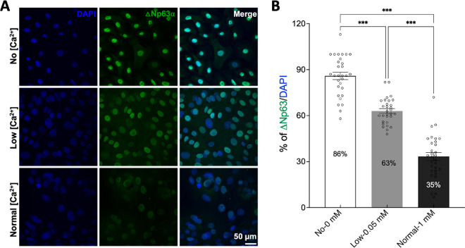 Figure 4
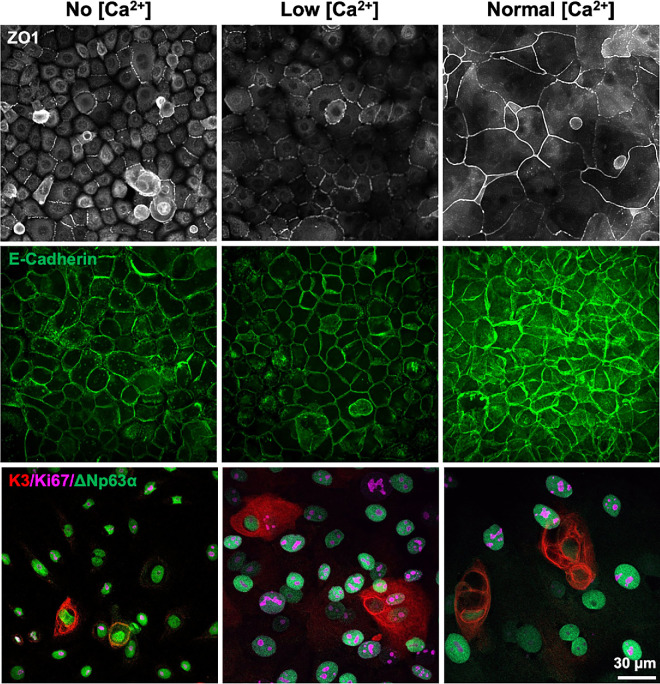 Figure 5
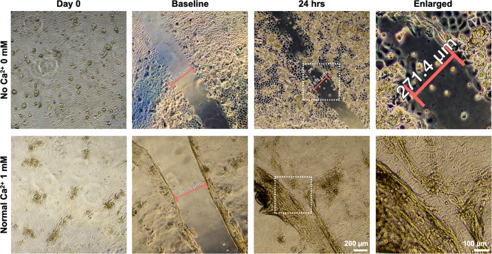 Figure 6
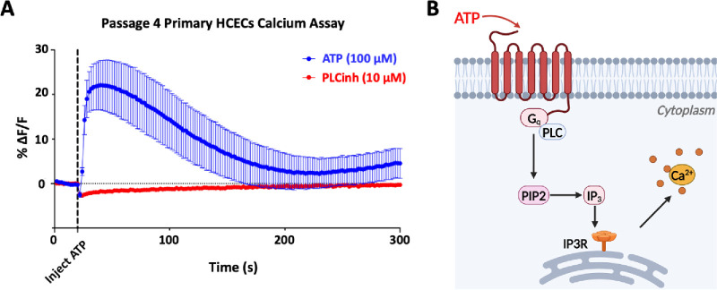 Figure 7
Peer Reviews
No public reviews on file for this paper yet. If you reviewed it on a platform where reviews are public (OpenReview, ICLR, NeurIPS, ICML), you can paste yours below so the community can read it here.
Videos
No videos yet. Explain this paper in a talk, walkthrough, or lecture? Add one.
Taxonomy
TopicsCorneal Surgery and Treatments · Ocular Surface and Contact Lens · Proteoglycans and glycosaminoglycans research