Volumetric MRI of dorsal root ganglia as a biomarker for disease progression and response to AAV treatment in a mouse model of Fabry disease
Fuqiang Zhao, Shipeng Yuan, Charalambos Kaittanis, Mugdha Deshpande, Abirami Kugadas, Katayoun Derakhchan, Wanida Ruangsiriluk, Rizwana Islam, Natalia Boukharov, Paul McQuade, Johannes Tauscher, Christopher T. Winkelmann, Talakad G. Lohith

TL;DR
This study shows that MRI can track disease progression and treatment response in a mouse model of Fabry disease by measuring changes in dorsal root ganglion volume.
Contribution
The study introduces a noninvasive MRI method to monitor Gb3 accumulation and treatment effects in a mouse model of Fabry disease.
Findings
DRG enlargement in Fabry mice was detectable as early as 8 weeks of age.
AAV gene therapy normalized DRG size within 4 weeks and sustained the effect up to 24 weeks.
MRI findings were supported by behavioral and histological analyses.
Abstract
Noninvasive and objective biomarkers for disease-associated pathology are critical for clinical trials. For Fabry disease, one important pathological change due to the deficiency of the lysosomal enzyme α-galactosidase A (α-GAL) caused is accumulation of globotriaosylceramide (Gb3) in dorsal root ganglion (DRG) neurons, which manifests as the overall DRG hypertrophy. Magnetic resonance imaging (MRI) has been successfully used to noninvasively measure DRG enlargement in Fabry patients, and DRG volumetric MRI can be a potential noninvasive biomarker for Gb3 accumulations in DRG neurons in clinical trials. To evaluate disease progression and treatment response in preclinical proof-of-concept studies, we developed an in vivo MRI method to measure DRG size in the G3Stg/GLA knockout mouse model of Fabry disease. Compared to the wild type mice, the DRG enlargement in the Fabry mice was…
Genes, proteins, chemicals, diseases, species, mutations and cell lines named across the full text — each resolved to its canonical identifier and authoritative record.
Click any figure to enlarge with its caption.
 Figure 1
 Figure 2
 Figure 3
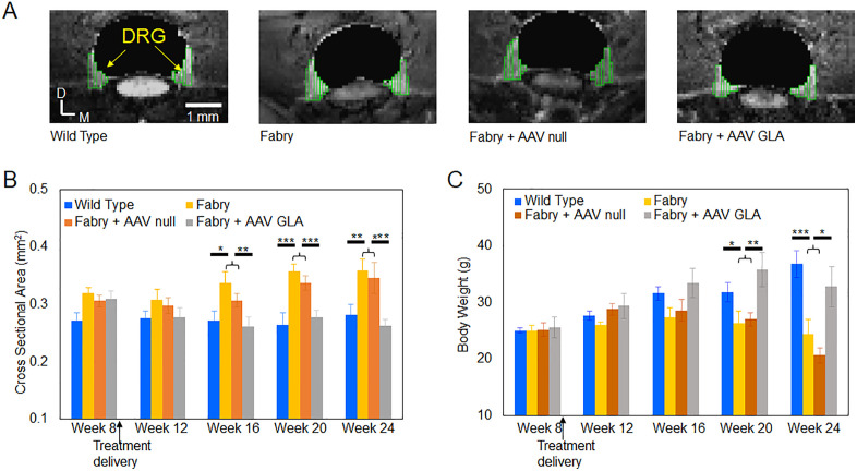 Figure 4
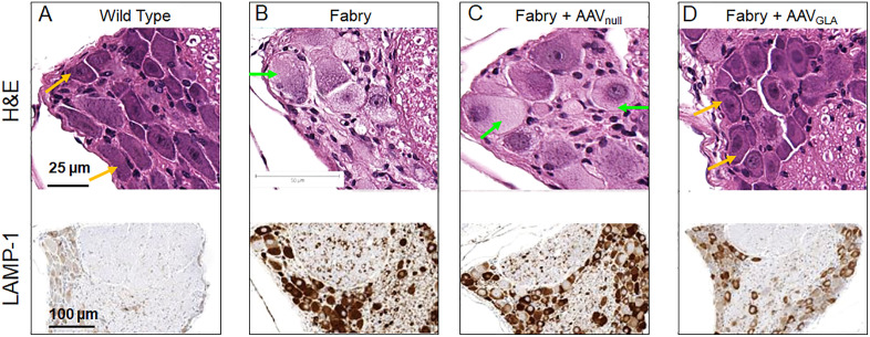 Figure 5
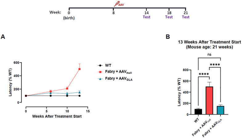 Figure 6
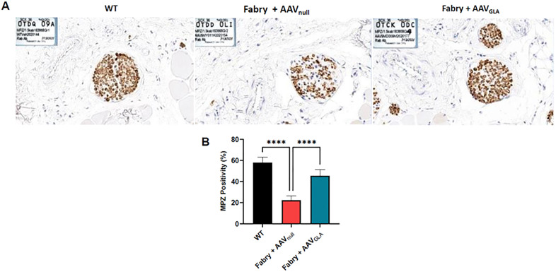 Figure 7
 Figure 8
 Figure 9
Peer Reviews
No public reviews on file for this paper yet. If you reviewed it on a platform where reviews are public (OpenReview, ICLR, NeurIPS, ICML), you can paste yours below so the community can read it here.
Videos
No videos yet. Explain this paper in a talk, walkthrough, or lecture? Add one.
Taxonomy
TopicsLysosomal Storage Disorders Research · Cellular transport and secretion · Parkinson's Disease Mechanisms and Treatments