Correlation of Genotype-Phenotype of Congenital Hypothyroidism Cohort Diagnosed by Newborn Screening: A Long-Term Observational Study
Yajie Su, Xifeng Lei, Ayijiamali Muhetaer, Jinfeng He, Long Li

TL;DR
This study explores how genetic variations in congenital hypothyroidism affect treatment needs and outcomes in children.
Contribution
The study identifies significant genotype-phenotype correlations in congenital hypothyroidism, particularly highlighting the role of DUOX2 mutations.
Findings
Patients with DUOX2 variants required lower L-T4 doses at 12 months compared to those with TPO or TSHR variants.
TPO and TSHR variants were associated with more severe clinical phenotypes and thyroid enlargement.
No significant differences were found in disease severity based on the number of DUOX2 alleles affected.
Abstract
This long-term observational study aimed to define the spectrum of genetic variation in a congenital hypothyroidism (CH) cohort and investigate the correlations between specific genotypes and clinical phenotypes, including treatment requirements and outcomes. We analyzed the maintenance dose of L-thyroxine (L-T4) at 6, 12, 18, and 24 months, alongside clinical outcomes after 3 years. Data were collected from the Neonatal Disease Screening Center at our hospital between January 2011 and March 2024. Of 247 patients with confirmed CH, 119 had available genetic testing and complete clinical information. The genetic positivity rate was 56.3% (67/119). DUOX2 was the most frequently mutated gene (28.57%), followed by TPO, TG, and TSHR. Phenotypic correlation analysis revealed that patients with DUOX2 variants had significantly lower initial screening TSH levels and required lower L-T4…
Genes, proteins, chemicals, diseases, species, mutations and cell lines named across the full text — each resolved to its canonical identifier and authoritative record.
Click any figure to enlarge with its caption.
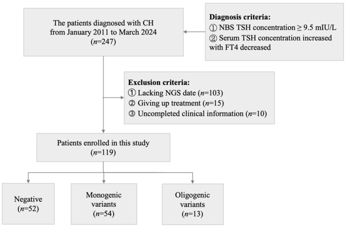 Figure 1
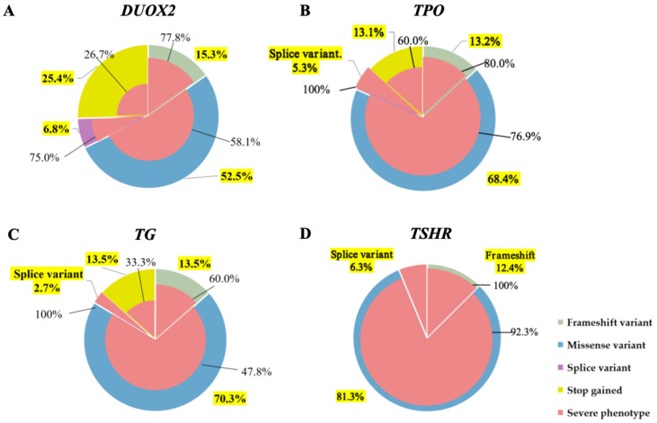 Figure 2
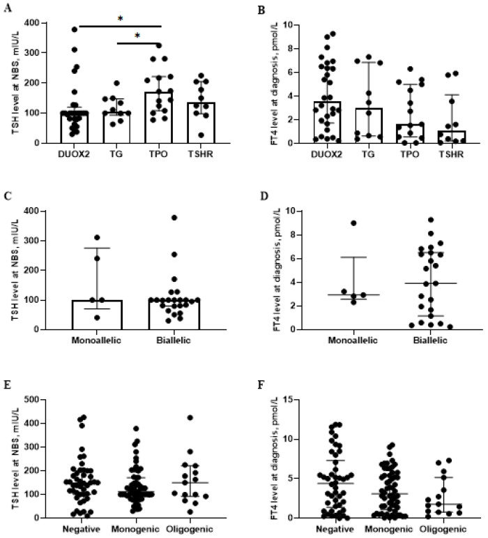 Figure 3
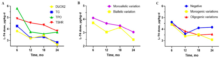 Figure 4
- —Natural Science Foundation of Xinjiang Uygur Autonomous Region
- —National Natural Science Foundation of China
Peer Reviews
No public reviews on file for this paper yet. If you reviewed it on a platform where reviews are public (OpenReview, ICLR, NeurIPS, ICML), you can paste yours below so the community can read it here.
Videos
No videos yet. Explain this paper in a talk, walkthrough, or lecture? Add one.
Taxonomy
TopicsThyroid Disorders and Treatments · Neonatal Health and Biochemistry · Metabolism and Genetic Disorders
1. Introduction
Congenital hypothyroidism (CH) is a prevalent and preventable endocrine disorder worldwide, with an increasing annual incidence of primary CH now estimated at 1/3000–1/2000 [1]. CH arises from defects in thyroid gland development or hormone biosynthesis, traditionally classified as thyroid dysgenesis (TD) or dyshormonogenesis (DH) [2]. Patients with CH lack specific clinical symptoms in the early stage, such as enlarged fontanel, delayed regression of jaundice, umbilical hernia, abdominal distension, and constipation [3]. If untreated in early infancy, CH can result in irreversible mental and physical growth retardation [4,5,6].
Targeted genetic testing identified variants is up to 66.5% in patients with CH [7,8,9,10]. Over 20 genes are associated with CH, such as TSHR, TTF-1/NKX2-1, PAX-8, TTF-2/NKX2-5, FOXE1, DUOX2, DUOXA2, TG, TPO, SLC5A5/NIS, SLC26A4/PDS, and IYD [11]. The prevalence of specific gene variants varies among populations; DUOX2 variants are common in Chinese cohorts, while TPO variants predominate in Caucasian populations [12]. However, available genetic and clinical data show a lack of genotype–phenotype correlation and significant phenotypic heterogeneity among patients with the same genotype. While CH is often considered as monogenic disorder, more studies and several pedigrees confirmed the potential oligogenic origin of CH [9,13]. The pathogenic contribution of these genes and their relationship with the clinical phenotype require further clarification [13].
This study conducted a comprehensive analysis of clinical phenotypes, maintenance L-T4 doses, clinical outcomes, and genotypes of infants diagnosed with CH via newborn screening over 13 years. The objectives were to establish the genetic variation spectrum of CH and elucidate genotype–phenotype relationships to facilitate precise patient management.
2. Materials and Methods
2.1. Study Design
A retrospective study was conducted to analyze the correlation of genotype–phenotype in children diagnosed with CH according to the Consensus Guidelines [14] at the Newborn Screening Center in our hospital between January 2011 and March 2024. A total of 119 patients with CH were enrolled in this study (Figure 1) and written informed consent was obtained from their parents for the collection of samples and publication of medical data. This study was approved by the Ethics Board of Children’s Hospital of Xinjiang Uygur Autonomous Region (KY2022031207).
2.2. Participants and Data Collection
The diagnosis of CH was made according to the Chinese national consensus statement and European guidelines. [1,14] A pretreatment blood thyrotropin stimulating hormone (TSH) concentration at newborn screening (NBS) of ≥ 9.5 mIU/L on initial screening was required. The diagnosis of CH was confirmed by elevated serum TSH levels and FT4 levels below the reference range.
Patients were further filtered based on the following exclusion criteria: (1) individuals lacking next-generation sequencing (NGS) data of CH-related genes, and (2) treatment with L-T4 was abandoned after diagnosis, and (3) clinical information was including pretreatment clinical presentation, thyroid ultrasonography, therapeutic dose of L-T4 at 6, 12, 18 and 24 months.
2.3. Laboratory Assessments
The screening TSH levels from dried blood spots were measured using a fluorometric assay with the Neonatal TSH kit (Fenghua, China) and an Auto Fluoroimmunoassay Analyzer (Auto TRFIA-2, Fenghua, China). Serum TSH and FT4 concentration were detected using a Roche electrochemiluminometric analyzer (Cobas-e601 analyzer, Roche, Mannheim, Germany).
2.4. Thyroid Ultrasonography Examination
Thyroid ultrasonography was performed on all patients using a 12 MHz small-parts linear transducer connected to an EnVisor scanner (Philips, Amsterdam, The Netherlands). The infants were placed in a supine position with their necks hyperextended without sedation. The length, breadth, and depth of the thyroid gland were measured, and the thyroid volume of each lobe was calculated as volume = length × breadth × depth × 0.479. The sum of the left and right thyroid lobe volumes was considered the total volume, excluding the isthmus. Gland volume was defined as enlarged, normal, or hypoplastic compared with published data for Chinese infants aged 0 to 12 months [15].
2.5. Targeted Next-Generation Sequencing and Variant Annotations
Peripheral blood samples were collected, and genomic DNA was extracted using the QIAamp DNA Blood Mini Kit (Qiagen, Hilden, Germany) according to the manufacturer’s instructions. DNA fragments were enriched for clinical exome sequencing using the ClearSeq Inherited Disease panel kit (Agilent Technologies, Santa Clara, CA, USA), which covered 2742 genes and included 21 genes related to CH. Sequencing was performed on a HiSeq 2500, HiSeq X10, or NovaSeq 6000 platform (Illumina, San Diego, CA, USA).
The pathogenicity of each variant was reassessed according to the ClinVar annotations https://www.ncbi.nlm.nih.gov/clinvar/ (accessed on 16 April 2025). Novel variants and variants of uncertain significance in the ClinVar were reassigned based on the guidelines of the ACMG [16].
2.6. Classification of Variants in Patients with CH
The genetic classification was defined into three groups based on the pattern of detected variants. Negative group: no pathogenic (P)/likely pathogenic (LP) variant was detected in CH-related genes; patients with variants of uncertain significance (VUS), benign or likely benign based on ACMG guideline were included here. Monogenic variation group: only one CH-related gene was detected and the P/LP variants were consistent with the mode of inheritance. The information on CH-related genes that we detected is shown in Supplementary Table S1. Oligogenic variation group: two or more CH-related genes were detected and the single P/LP heterozygous variant of one gene with autosomal recessive inherited mode was also included.
2.7. Definition of Phenotypic Indicators
Clinical presentation: the clinical manifestations at diagnosis (pretreatment), including jaundice, rough skin, constipation, etc. Severe CH was defined by a very low pretreatment serum FT4 < 5 pmol/L. Therapeutic dose of L-T4 (μg/kg·d) was recorded at the age of 6 ± 1 months, 12 ± 1 months, 18 ± 1 months and 24 ± 1 months. To determine clinical outcome, L-T4 therapy was temporarily withdrew after 3 years. Thyroid function was re-examined at 1 month, 2 months and 10 months, post-withdrawal. If TSH and FT4 levels remained normal, the patients were diagnosed as transient CH (TCH). If the results were abnormal, therapy was resumed, and the patient was diagnosed as permanent CH (PCH). Patients under 3 years of age were defined as “undiagnosed classification” [17].
2.8. Statistical Analyses
Statistical analysis was performed using Stata 16.0 software. Continuous variables were presented as mean ± standard deviation or median (Q1, Q3), while categorical variables were expressed as frequency and percentage. The normality of continuous variables was determined using the Shapiro–Wilk test. For comparisons among multiple groups, the Kruskal–Wallis test was used for continuous variables, and the chi-square or Fisher’s exact tests were used for categorical variables. If the Kruskal–Wallis test indicated a statistically significant difference (p < 0.05), post hoc pairwise comparisons were conducted using Dunn’s test with a Bonferroni correction for multiple comparisons. The Wilcoxon rank sum test was used to compare the intergroup differences for continuous variables. Group differences were evaluated using the chi-square or Fisher’s exact tests. Statistical significance was set at a p value less than 0.05. Graphs were created using GraphPad Prism 8 software.
3. Results
3.1. Genetic Spectrum of 119 Patients with CH by Next-Generation Sequencing
Genetic testing and complete clinical information were obtained for 119 out of 247 patients diagnosed with CH. In this cohort, 52 patients were classified into the negative group, as no P/LP variants in CH-related genes were identified. Monogenic variants consistent with Mendelian inheritance patterns were identified in 56 cases (47.06%, 56/119), which constituted the monogenic variants group. Meanwhile, 13 patients were found to carry P/LP variants in at least two CH-associated genes, which constituted the Oligogenic variants group. The genetic positivity rate was established at 56.3% (67/119) (Figure 1).
A total of 102 diverse variants were detected in the 14 genes examined in 119 patients, 82 were P/LP variants detected in the following genes: DUOX2 in 34 patients (28.57%, 34/119), TPO in 20 patients (16.81%, 20/119), TG in 13 patients (10.92%, 13/119), TSHR in 11 patients (9.24%, 11/119), DUOXA2. SLC26A4 and SLC5A5 in 2 patients each (1.68%, 2/119), GNAS, NKX2-1, PAX8, JAG1, NKX2-5 in one patient each (0.84%,1/119) (Supplementary Table S4).
3.2. Relationship Between Different Genes and Clinical Phenotypes
The four genes with the highest variation frequencies were selected to correlate their variation types with patients’ clinical phenotypes. For the DUOX2 gene, missense variants were the most common variant (52.5%), among which 58.1% of patients presented with severe phenotypes (pretreatment serum FT4 < 5 pmol/L). Notably, only 26.7% of those with nonsense variants exhibited severe clinical manifestations (Figure 2A). For TPO gene, missense variants accounted for 68.4%, and 76.9% of these cases were severe phenotypes. Additionally, all patients with splice variants (5.3%) showed severe phenotypes (Figure 2B). For TG gene, missense variants predominated (70.3%), with 56.5% leading to severe phenotypes. Nonsense and frameshift variants each accounted for 13.5%, while splice variants constituted 2.7%; all patients with splice variants exhibited severe phenotypes (Figure 2C). For TSHR gene, missense variants were most common (81.3%), followed by frameshift (12.5%) and splice variations (6.3%). Except for 7.7% of patients with missense variants who had mild CH, all other patients presented with severe phenotypes (Figure 2D).
Each pie chart consists of two layers: the first is the distribution of variation types, with green indicating frameshift variants, blue for missense variants, purple for splice variants, and yellow for nonsense variants. The yellow-labeled numbers indicate the proportion of each variation type. The second shows the proportion of patients with severe clinical phenotypes, depicted in dark pink, with black numerals specifying the exact percentage. Panels A–D present DUOX2, TPO, TG, and TSHR, respectively.
Further analysis was performed on the baseline characteristics and clinical phenotypes associated with variations in the four genes. The Kruskal–Wallis test revealed a statistically significant difference in initial screening TSH levels across the four genetic variant groups (p = 0.022). Numerically, the median TSH level in the DUOX2 group was lower than that in the TPO group, although post hoc testing with Bonferroni correction did not identify significant pairwise comparisons (100.0 (79.8, 120.0) vs. 172.1 (108.2, 221.6), p = 0.018). (Figure 3A). In addition, a statistically significant difference in the L-T4 dose was observed between the DUOX2 and TSHR groups at 12 months 3.2 (1.2) vs. 4.5 (0.7), (p = 0.042) (Figure 4A). No significant correlation was observed between various genotypes and clinical outcomes, and PCH was the predominant outcome in our cohort. Patients with TPO and TSHR variation exhibited severe phenotypes compared to those with DUOX2 variations (DUOX2 vs. TPO: p = 0.011. f DUOX2 vs. TSHR: p = 0.006). Similarly, a significantly higher prevalence of enlarged thyroid ultrasound was observed in patients with TG and TPO variants compared with TSHR variants. (TG vs. TSHR: p = 0.025. and TPO vs. TSHR: p = 0.011) (Table 1).
3.3. The Differences Between “DUOX2 Monoallelic Variations” and “DUOX2 Biallelic Variations”
The analysis focused on the most common DUOX2 variations, involving 5 patients with monoallelic variations and 23 with biallelic variations. No significant differences were observed in the biochemical data, maintenance L-T4 doses at different ages, and clinical prognosis between the DUOX2 monoallelic and DUOX2 biallelic variation groups (Figure 3C,D and Figure 4B) (Supplementary Table S2).
A–B show the differences in the biochemical data between DUOX2, TG, TPO, and TSHR gene variations groups. C–D show the differences in the biochemical data between “DUOX2 monoallelic variations” and “DUOX2 biallelic variations” groups. E–F show the differences in the biochemical data between “Negative”, “Monogenic variations” and “Oligogenic variations” groups. The data are presented as the median with interquartile range, with each scatter point representing an individual patient; only statistically significant differences are indicated, using asterisks (p < 0.05).
3.4. The Differences Between “Negative”, “Monogenic Variations” and “Oligogenic Variations” Groups
For the between-group analysis, 52 patients were classified as negative 54 as monogenic variations, and 13 as oligogenic. The information on oligogenic variations patients is showed in Table 2. No significant differences in biochemical characteristics or L-T4 doses were found among the negative, monogenic, and oligogenic variation groups. However, the negative group was noted to have fewer cases of enlarged thyroid tissue at diagnosis and fewer cases of PCH after 3 years of age (Figure 3E,F and Figure 4C) (Supplementary Table S3).
4. Discussion
In this study, we screened for CH-related genes and genotype–phenotype relationships in 119 patients with CH. Biochemical data, thyroid morphology, drug maintenance dosages, and clinical prognosis were compared across different genetic groups. Previous studies indicated that while 85% of CH cases have historically been attributed to TD [18], an increased proportion of CH cases caused by DH has been reported recently [19,20].
In our cohort, the genetic positivity rate was 56.3%, with DUOX2 being the most frequently mutated gene, a finding consistent with previous reports in East Asian populations [21,22]. However, TPO has been reported as the most frequently mutated genes in Western Caucasian populations [12].The high-frequency variants p.K530X and p.R1110Q were identified in the DUOX2 gene, with the p.R1110Q variant having been previously demonstrated to significantly reduce H_2_O_2_ production in vitro [23] with p.K530X being the most common variation in southern and central Chinese populations also [7,24,25]. A study in Shandong Province, China, did not detect the p.K530X variant but identified the p.R1110Q variant as high-frequency [26]. In Korean populations, the common DUOX2 variant is p.G488R [21], further highlighting genetic variations across diverse populations.
In the genotype–phenotype analysis, missense variants were identified as the most common type of variation among the four frequently occurring mutant genes. In the present study, TSHR variations mainly present with severe hypothyroidism, while DUOX2 variations were less common. Zhang et al. [27] indicated that patients with TSHR variations resulted in milder hypothyroidism compared to DUOX2 variations. The discrepancy in findings may be related to sample selection. Moreover, TSHR variations can lead to irreversible damage by causing receptor dysfunction, which disrupts thyroid gland development and interrupts the stimulating signal for hormone synthesis, thereby predisposing affected individuals to PCH.
Based on biochemical data and maintenance doses, patients with DUOX2 variations generally exhibited milder clinical phenotypes than those with variations in other genes. The critical role of TPO in thyroid hormone synthesis, for which no functional substitute is known, may explain why TPO variations often lead to more severe symptoms and necessitate higher L-T4 doses [18,28]. Consistently, in our cohort, patients with TPO and TSHR variants were observed to exhibit more severe clinical phenotypes compared to the DUOX2 group. Accordingly, the DUOX2 group was found to require a lower L-T4 dosage at 12 months of age. We hypothesize that milder presentation in DUOX2 variation patients could be attributed to partial functional compensation by DUOX1.
We observed phenotypic variations not only across different genes but also among different allelic variants of the same gene, such as DUOX2, an essential component of the thyroid H_2_O_2_ generation system, which plays a pivotal role in thyroid hormone production [29]. DUOX2 variations, a primary cause of thyroid hormone production disorders, predominantly exhibit an autosomal recessive inheritance pattern. However, pathogenic variants can arise even from single allele variations, contributing to considerable genotype–phenotype variability. Moreno et al. [30] report that PCH is associated with biallelic inactivating DUOX2 variations, while TCH is linked to monoallelic variations. Some studies suggest that monoallelic DUOX2 variations typically correlate with mild to moderate phenotypes [24,31]. However, subsequent research suggests that the permanence or transience of CH is not directly linked to the number of DUOX2 alleles, and the relationship between DUOX2 genotype and CH phenotype remains unclear [32,33,34,35]. In our cohort, significant differences were not observed in biochemical data, L-T4 doses, and clinical outcomes between DUOX2 monoallelic variations and biallelic variations. Nonetheless, we found that the L-T4 maintenance doses were generally higher in patients with DUOX2 monoallelic variations than those with DUOX2 biallelic variations. This further highlights the genetic and phenotypic heterogeneity of DUOX2 variations, emphasizing the need for additional in vitro functional experiments to elucidate their relationship.
Considerable phenotypic variability was also noted among different allelic variations in the same gene [29]. While biallelic inactivating DUOX2 variations have been associated with PCH and monoallelic variations with TCH in some studies, this relationship remains unclear [36] and further functional studies are necessary to validate these underlying mechanisms [3]. In our cohort, no significant differences in biochemical data, L-T4 doses, or clinical outcomes were found between patients with DUOX2 monoallelic and biallelic variations, highlighting the genetic and phenotypic heterogeneity of DUOX2 variations and underscoring the need for functional studies to elucidate these relationships.
Several limitations of this study are acknowledged. First, thyroid scintigraphy was performed on only a few patients, which may have affected the accurate classification of CH into TD or DH. Second, in the analysis of clinical outcomes, only definitive PCH and TCH diagnoses after re-evaluation were considered. It is possible that some patients classified as PCH in this study might successfully discontinue treatment at an older age.
5. Conclusions
We detected 102 different variants across 14 CH-related genes in neonatal CH cohort and elucidated genotype–phenotype relationships. We found a high genetic diagnosis rate of 56.3% among neonates with CH who underwent genetic testing, and DUOX2 as the most frequently mutated gene. Genotype–phenotype analysis provides clinicians with valuable insights for the precise follow-up management of CH.
The reference list from the paper itself. Each links out to its DOI / PubMed record.
- 1Van Trotsenburg P. Stoupa A. Léger J. Rohrer T. Peters C. Fugazzola L. Cassio A. Heinrichs C. Beauloye V. Pohlenz J. Congenital Hypothyroidism: A 2020-2021 Consensus Guidelines Update-An ENDO-European Reference Network Initiative Endorsed by the European Society for Pediatric Endocrinology and the European Society for Endocrinology Thyroid 20213138741910.1089/thy.2020.033333272083 PMC 8001676 · doi ↗ · pubmed ↗
- 2Peters C. van Trotsenburg A.S.P. Schoenmakers N. DIAGNOSIS OF ENDOCRINE DISEASE: Congenital hypothyroidism: Update and perspectives Eur. J. Endocrinol.2018179 R 297R 31710.1530/EJE-18-038330324792 · doi ↗ · pubmed ↗
- 3Zhang T. Shen Y. Xu Y. Wu D. Chen C. Yang R. Clinical, biochemical characteristics and genotype-phenotype analysis of congenital hypothyroidism diagnosed by newborn screening in China Clin. Chim. Acta 202354711745910.1016/j.cca.2023.11745937390946 · doi ↗ · pubmed ↗
- 4Nilsson M. Fagman H. Development of the thyroid gland Development 20171442123214010.1242/dev.14561528634271 · doi ↗ · pubmed ↗
- 5Dimitropoulos A. Molinari L. Etter K. Torresani T. Lang-Muritano M. Jenni O.G. Largo R.H. Latal B. Children with congenital hypothyroidism: Long-term intellectual outcome after early high-dose treatment Pediatr. Res.20096524224810.1203/PDR.0b 013e 31818 d 203018787501 · doi ↗ · pubmed ↗
- 6Grosse S.D. Van Vliet G. Prevention of intellectual disability through screening for congenital hypothyroidism: How much and at what level?Arch. Dis. Child.20119637437910.1136/adc.2010.19028021242230 · doi ↗ · pubmed ↗
- 7Tan M. Huang Y. Jiang X. Li P. Tang C. Jia X. Chen Q. Chen W. Sheng H. Feng Y. The prevalence, clinical, and molecular characteristics of congenital hypothyroidism caused by DUOX 2 mutations: A population-based cohort study in Guangzhou Horm. Metab. Res.20164858158810.1055/s-0042-11222427557340 · doi ↗ · pubmed ↗
- 8Long W. Lu G. Zhou W. Yang Y. Zhang B. Zhou H. Jiang L. Yu B. Targeted next-generation sequencing of thirteen causative genes in Chinese patients with congenital hypothyroidism Endocr. J.2018651019102810.1507/endocrj.EJ 18-015630022773 · doi ↗ · pubmed ↗