Real-time probe-based confocal laser endomicroscopy visualization of dual differentiation features in mixed-type gastric adenocarcinoma
Zhixia Dong, Bo Tian, Shan Wu, Yueqin Qian, Qian Zhuang, Xinjian Wan

Abstract
Genes, proteins, chemicals, diseases, species, mutations and cell lines named across the full text — each resolved to its canonical identifier and authoritative record.
Click any figure to enlarge with its caption.
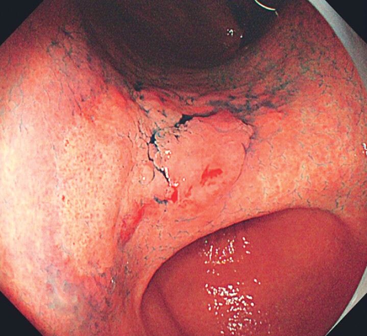 Fig. 1
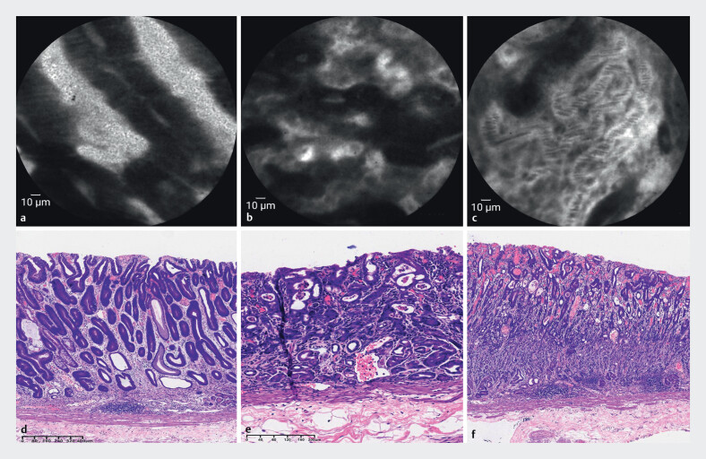 Fig. 2
- —National Key Research and Development Program of China10.13039/501100012166
Peer Reviews
No public reviews on file for this paper yet. If you reviewed it on a platform where reviews are public (OpenReview, ICLR, NeurIPS, ICML), you can paste yours below so the community can read it here.
Videos
No videos yet. Explain this paper in a talk, walkthrough, or lecture? Add one.
Taxonomy
TopicsGastric Cancer Management and Outcomes · Esophageal Cancer Research and Treatment · Helicobacter pylori-related gastroenterology studies
Probe-based confocal laser endomicroscopy (pCLE) has emerged as a revolutionary endoscopic tool, offering real-time, in vivo histological imaging that transforms our understating of complex gastric malignancies 1 2 . We present a case of mixed-type gastric adenocarcinoma in which pCLE played a pivotal role in precise visualization and diagnosis during endoscopic assessment ( Video 1 ).
Probe-based confocal laser endomicroscopy (pCLE) scan of the lesion.Video 1
A 40-year-old woman with Helicobacter pylori infection underwent esophagogastroduodenoscopy. White-light endoscopy (WLE) with indigo carmine chromoendoscopy revealed a 3.5×4 cm erythematous lesion at the gastric angle, featuring a superficial flat-elevated morphology with well-demarcated margins ( Fig. 1 ). It was considered a cancerous lesion according to diagnosis criteria under WLE: 1) well-demarcated border; and 2) irregularity in color/ surface pattern 3 . To further evaluate the lesion, pCLE (1000×magnification, BIOPSEE, Viestar Medical Technology, Wuhan, China) was employed, which revealed distinct microarchitectural patterns: elevated areas showed distorted glands with thickened epithelia ( Fig. 2 a ), whereas flat regions exhibited chaotic branching glands and stenosed lumens ( Fig. 2 b ). Dedifferentiation was characterized by complete glandular disorganization with dense neovascular networks ( Fig. 2 c ). Based on the Miami classification 4 and discriminable glandular structures 5 , mixed adenocarcinoma with dual differentiation was confirmed. Following informed consent, endoscopic submucosal dissection was performed. Real-time pCLE findings correlated perfectly with histopathology: microscopy confirmed both differentiated and undifferentiated components ( Fig. 2 d , Fig. 2 e ).
WLE images showed a 3.5×4 cm superficial flat and elevated lesion in the gastric antrum.
Microstructural imaging characteristics of distinct differentiation components under pCLE. a Chaotic glandular arrangement with increased branching and stenosed lumens. b Disordered cell arrangement in which normal glandular structure was almost unrecognizable. c Development of a dense network of neovascularization. d Histopathology showed well-differentiated adenocarcinoma, e moderately differentiated adenocarcinoma, and f poorly differentiated adenocarcinoma.
This case highlights pCLE's capacity to eliminate diagnostic ambiguity through immediate microscopic pattern recognition, reducing reliance on multiple biopsies. The technology's real-time delineation of heterogeneous tumor components represents a significant advancement toward optical biopsy-driven precision diagnostics for complex malignancies. This case report underscores pCLE's potential to transform endoscopic practice by enabling instant, high-resolution characterization of neoplastic lesions, thereby optimizing therapeutic decision-making.
The reference list from the paper itself. Each links out to its DOI / PubMed record.
- 1Wallace M Lauwers GY Chen Y Miami classification for probe-based confocal laser endomicroscopy Endoscopy 20114388289110.1055/s-0030-125663221818734 · doi ↗ · pubmed ↗
- 2Zuo XL Li Z Li CQ Probe-based endomicroscopy for in vivo detection of gastric intestinal metaplasia and neoplasia: a multicenter randomized controlled trial Endoscopy 2017491033104210.1055/s-0043-11538228753702 · doi ↗ · pubmed ↗
- 3Yao K The endoscopic diagnosis of early gastric cancer Ann Gastroenterol 201326112224714327 PMC 3959505 · pubmed ↗
- 4Wallace M Lauwers GY Chen Y Miami classification for probe-based confocal laser endomicroscopy Endoscopy 20114388289110.1055/s-0030-125663221818734 · doi ↗ · pubmed ↗
- 5Jeon SR Cho WY Jin SY Optical biopsies by confocal endomicroscopy prevent additive endoscopic biopsies before endoscopic submucosal dissection in gastric epithelial neoplasias: a prospective, comparative study Gastrointest Endosc 20117477278021802680 10.1016/j.gie.2011.05.005 · doi ↗ · pubmed ↗