A heart shaped coronary aneurysm
Charlotte Snik, Mustafa Koksu-Ilhan, Saman Rasoul

Abstract
Genes, proteins, chemicals, diseases, species, mutations and cell lines named across the full text — each resolved to its canonical identifier and authoritative record.
Click any figure to enlarge with its caption.
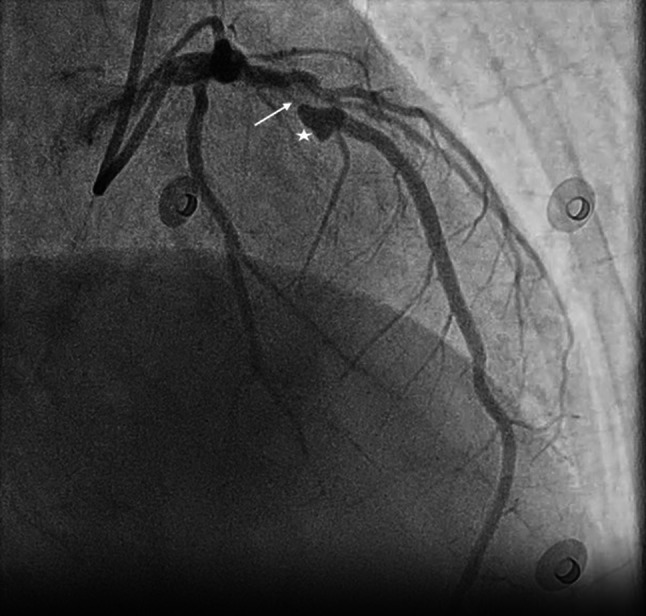 Figure 1
Peer Reviews
No public reviews on file for this paper yet. If you reviewed it on a platform where reviews are public (OpenReview, ICLR, NeurIPS, ICML), you can paste yours below so the community can read it here.
Videos
No videos yet. Explain this paper in a talk, walkthrough, or lecture? Add one.
Taxonomy
TopicsKawasaki Disease and Coronary Complications · Coronary Artery Anomalies · Cardiac Structural Anomalies and Repair
A 72-year-old woman with a history of hypertension and recently diagnosed heart failure underwent invasive coronary angiography, revealing a significant lesion of the left anterior descending with a heart-shaped aneurysm (Fig. 1). Following multidisciplinary Heart Team discussion, she underwent a successful percutaneous coronary intervention.Fig. 1. Heart-shaped coronary artery aneurysm indicated by a star, left anterior descending indicated by an arrow
Coronary artery aneurysm (CAA), a focal dilation of coronary segments of at least 1.5 times the adjacent normal segment, is found in up to 5% of patients undergoing coronary angiography [1] and is associated with poor long-term outcome [2]. CAA is usually found incidentally on cardiac imaging; however, it may present with stable angina or acute coronary syndrome [3]. Two forms exist: saccular and fusiform aneurysms. The pathogenesis is not well known. Genetic factors, atherosclerotic risk factors, certain vasculitides, and iatrogenic factors may be the cause [3]. Management is challenging and includes medical therapy, surgical excision, coiling, percutaneous coronary intervention, and coronary bypass grafting [4].
Supplementary Information
Figure series from the case studies.