Perioperative changes in inflammatory biomarkers and underlying molecular mechanisms in patients with trigeminal neuralgia undergoing surgical interventions
Cheng-Jie Qiu, Zhi-Ye Cui, Qi Zhang, Si-Jian Pan, Ben-Gen Pei

TL;DR
This paper reviews inflammatory biomarkers in trigeminal neuralgia patients before and after surgery, exploring their potential as indicators of treatment outcomes and disease mechanisms.
Contribution
The study identifies specific inflammatory biomarkers modulated by surgical interventions and links them to molecular pathways in trigeminal neuralgia.
Findings
Pro-inflammatory cytokines like IL-1β, IL-6, and TNF-ɑ are elevated in TN patients and can be reduced by percutaneous balloon compression.
Microvascular decompression reverses TRAIL levels but not TNF-β, indicating differential biomarker responses to surgical techniques.
Inflammatory biomarker changes are influenced by signaling pathways such as MAPK and P2X7, offering insights into TN pathogenesis.
Abstract
Trigeminal neuralgia (TN) is a very painful neurological condition with unilateral and electric shock-like pain attacks. The accurate diagnosis of the disease is of extreme importance for the determination of subsequent therapeutic strategies and clinical management. Surgical interventions including peripheral neurectomy, microvascular decompression (MVD), percutaneous balloon compression (PBC) and stereotaxic radiosurgery (SRS) are options for refractory patients. The utilization of proper perioperative biomarkers in serum, CSF and saliva may help in tracking the safety, efficacy and prognosis after surgical treatments. This narrative review aimed to identify potential inflammatory biomarkers that reflected perioperative changes in clinical practice and explored contributions of inflammation to pathogenesis of the disease. A total of 142 records and 95 clinical trials were identified…
Genes, proteins, chemicals, diseases, species, mutations and cell lines named across the full text — each resolved to its canonical identifier and authoritative record.
Click any figure to enlarge with its caption.
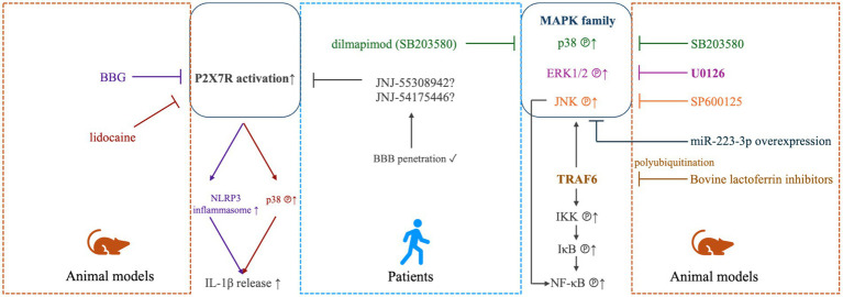 Figure 1
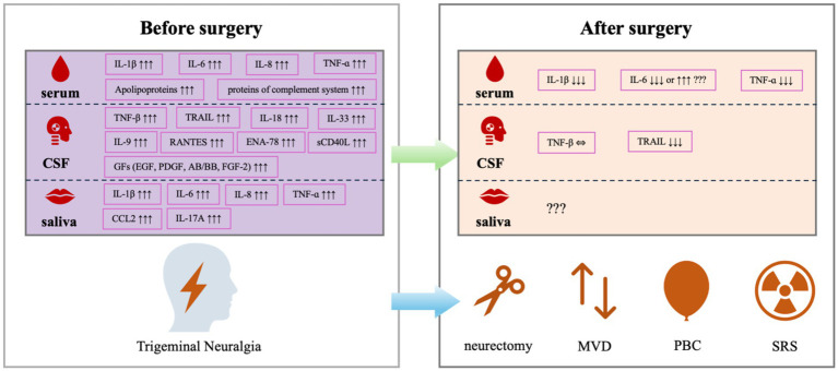 Figure 2
| Groups | Sample source | Biomarker | Preoperative change | Postoperative change | Conclusion | Ref. |
|---|---|---|---|---|---|---|
| Microvascular decompression (MVD) | ||||||
| ① Primary TN patients submitted to MVD ( | Serum | IL-1β | ↑↑↑ | / | Vascular compression in TN induces an increase in a variety of cytokines. | ( |
| IL-6 | ↑↑↑ | / | ||||
| IL-8 (CXCL8) | ↑↑↑ | / | ||||
| TNF-ɑ | ↑↑↑ | / | ||||
| ① TN patients submitted to MVD ( | CSF | TNF-β | ↑↑↑ | ⇔ | TNF-β did not seem to change after surgery. | ( |
| TRAIL | ↑↑↑ | ↓↓↓ | TRAIL level in TN may be reversible by surgery. | |||
| ① TN patients submitted to MVD ( | CSF collected one day before MVD | Apolipoproteins | ↑↑↑ | / | Inflammation components participate in the in the pathophysiology of TN. | ( |
| CO8B, CO8G, C5 (proteins involved in the complement system) | ↑↑↑ | / | ||||
| ① TN patients submitted to MVD ( | Serum | IL-1β | ⇔ | ↓↓↓ | MVD is effective to significantly reduce the pro-inflammatory cytokines at postoperative 3 days and 5 days compared with preoperation. | ( |
| IL-6 | ⇔ | ↓↓↓ | ||||
| TNF-ɑ | ⇔ | ↓↓↓ | ||||
| ① TN patients ( | Saliva | IL-1β | ↑↑↑ | / | Increased levels of chemokines play a vital role in TN pathogenesis. | ( |
| TNF-ɑ | ↑↑↑ | / | ||||
| CCL2 | ↑↑↑ | / | ||||
| IL-17A | ↑↑↑ | / | ||||
| IL-6 | ↑↑↑ | / | ||||
| IL-8 (CXCL8) | ↑↑↑ | / | ||||
| ① TN patients submitted to MVD ( | Serum | IL-6 | ⇔ | ↑↑↑ (compared to preoperative value in MVD group) | Greater surgical trauma from MVD. | ( |
| ① TN patients submitted to MVD ( | CSF collected during MVD | IL-18, IL-33 and IL-9 (pro-inflammatory cytokines) | ↑↑↑ | / | Neuro-inflammation is present in TN. | ( |
| RANTES and ENA-78 (chemokines) | ↑↑↑ | / | ||||
| TRAIL and sCD40L (TNF superfamily) | ↑↑↑ | / | ||||
| EGF, PDGF-AB/BB, FGF-2 (growth factors) | ↑↑↑ | / | ||||
| Percutaneous balloon compression (PBC) | ||||||
| ① TN patients submitted to MVD ( | Serum | IL-1β | ⇔ | ↓↓↓ | PBC is effective to significantly reduce the pro-inflammatory cytokines at postoperative 3 days and 5 days compared with preoperation. | ( |
| IL-6 | ⇔ | ↓↓↓ | ||||
| TNF-ɑ | ⇔ | ↓↓↓ | ||||
| ① TN patients submitted to MVD ( | Serum | IL-6 | ⇔ | ↑ | Postoperative levels of IL-6 was significantly increased in MVD group but not in PBC group. | ( |
| *ID | Title | Sponsor | Participants | Groups | Outcome | Status |
|---|---|---|---|---|---|---|
| Comparing Clinical Benefits of Gamma Knife and Microvascular Decompression for Trigeminal Neuralgia | University of Baghdad | Primary idiopathic TN (children and adults) | ① Gamma knife (80Gy) |
BNI-PS VAS BPI | Completed | |
| Comparison Between Radiofrequency and Balloon Compression in the Treatment of Idiopathic Trigeminal Neuralgia | University of São Paulo | Idiopathic TN (adults) | ① Balloon Compression Rhizotomy |
NRS | Terminated | |
| Artificial Intelligence to Predict Surgical Outcomes and Assess Pain Neuromodulation in Trigeminal Neuralgia Subjects | IRCCS San Raffaele | TN (adults) | ① Gamma knife Radiosurgery + virtual reality rehabilitation |
NRS BNI-PS | Recruiting | |
| A Personalized Radiosurgery Procedure for People with Trigeminal Neuralgia to Improve Pain, Quality of Life and Reduce Complications | University of Leeds | Idiopathic TN or MS-related TN (adults) | ① Personalized Gamma knife Radiosurgery |
BNI-PS BNI-NS | Recruiting | |
| Postoperative Analgesia in Patients with Microvascular Decompression | Xiangya Hospital of Central South University | TN patients scheduled for MVD (adult) | ① Scalp nerve block and patient-controlled analgesia |
VAS | Unknown |
Peer Reviews
No public reviews on file for this paper yet. If you reviewed it on a platform where reviews are public (OpenReview, ICLR, NeurIPS, ICML), you can paste yours below so the community can read it here.
Videos
No videos yet. Explain this paper in a talk, walkthrough, or lecture? Add one.
Taxonomy
TopicsTrigeminal Neuralgia and Treatments · Obstructive Sleep Apnea Research · Neurosurgical Procedures and Complications
Introduction
1
Characterized by unilateral, paroxysmal, electric shock-like, pricking and relapsed pain attacks with sudden onset and sudden termination, trigeminal neuralgia (TN) is a debilitating neuropathic facial pain syndrome affecting the sensory distribution of one or more divisions of the fifth cranial nerve—trigeminal nerve, particularly at the innervation area of the three trigeminal nerve branches, when triggered by innocuous stimuli (1). Studies from animal models showed that persistent external stimulation was transmitted from the affected trigeminal nerve branches into the trigeminal ganglion (TG), resulting in the activation of the TG neurons, which is one of the leading reasons for the occurrence and maintenance of hyperalgesia in these patients (2). TN can be classified as (i) classical TN, which has no apparent cause other than neurovascular compression (NVC) of the trigeminal nerve at the root entry zone (REZ) (3); (ii) idiopathic TN, which shows no significant abnormalities assessed by electrophysiological tests or MRI; and (iii) secondary TN, which is caused by underlying diseases such as brain tumor or multiple sclerosis (MS) (4). The REZ is where the trigeminal nerve enters the brainstem and represents a transition zone from peripheral Schwann cell-mediated myelination to central oligodendrocyte-mediated myelination, which makes this area very susceptible to pressure-induced lesions (5). TN overall affects 0.07% (6) to 0.3% (7) of the general population, with a slight female predominance (approximately 1.5 to 2.1 ratio for male to female) (8, 9). Commonly seen above 50 years of age, its general incidence climbed with age (9). Moreover, the incidence of TN in systemic sclerosis and MS is significantly higher than that in general populations (10), and it has also been associated with other inflammatory diseases such as myositis, arthritis and interstitial lung disease (10). Pathophysiologically, compression from the blood vessel focal accounts for the main cause of TN, leading to demyelination in REZ of the primary sensory trigeminal afferents (11). Surprisingly, changes in the trigeminal nerve has been observed not only on the side with NVC, but also contralaterally, suggesting that individuals with TN may have preexisting abnormalities in the white matter with neuroinflammation and oedema (12). The chronic neuroinflammation of the TG is caused by the persistent constriction of blood vessels in abnormal positions, leading to the development of demyelination in TN patients (13). Indeed, a pivotal role of inflammation in the etiology and progression of TN has been highlighted previously, which demonstrate elevation of multiple inflammatory biomarkers in different samples of TN patients, such as serum and cerebrospinal fluid (CSF) (14).
This literature review aims to summarize current clinical and laboratory findings along the perioperative timeline to find out biomarkers for clinical use, and discuss underlying molecular mechanisms for a better understanding of the role of inflammation in TN and directions for future clinical trials and basic research.
Methods
2
A search of published articles was performed through PubMed database using strategy (trigeminal neuralgia[Title/Abstract]) AND (inflammatory[Title/Abstract]), and a search of registered clinical trials was performed through Cochrane Central Register of Controlled Trials (CENTRAL) database using ‘trigeminal neuralgia’ as key word, with a date range of 2000 to 2025. A total of 142 English articles was acquired from PubMed, and a total of 95 clinical trials registered on clinicaltrials.gov was acquired for further screening. Original articles of both human and animal studies related to ‘trigeminal neuralgia’, ‘surgical intervention’ and ‘inflammatory biomarkers’, as well as registered trials related to ‘trigeminal neuralgia’ and ‘surgical intervention’ were included, whereas articles in the format of reviews, systematic reviews and/or meta-analyses, case reports or case series, commentaries, editorials, letters, as well as articles irrelevant to or not fully covering (e.g., comparing the surgical interventions without analyzing inflammatory biomarkers, or studying inflammatory biomarkers in non-surgical scenarios) this topic were excluded.
Human participants
3
For recurrent cases and patients refractory to pharmacotherapies, surgical treatments may be contemplated. Typical surgical treatments for TN include peripheral neurectomy, microvascular decompression (MVD), percutaneous balloon compression (PBC) and stereotactic radiosurgery (SRS).
Peripheral neurectomy
3.1
Peripheral neurectomy is a simple, low-risk and well-tolerated procedure of transection that can be performed on all terminal branches of the trigeminal nerve. Previous study was established on 12 males and 8 females with an age range of 35–68 years demonstrated that peripheral neurectomy exerted a positive effect on alleviating pain in TN patients, with an average pain relief period of 24–33 months after surgery (15). This method may be particularly suitable for elderly or debilitated patients in whom other invasive neurosurgical interventions are contraindicated. After studying the biopsy specimens collected from 4 male and 7 female elderly TN patients with an average age of 63.64 ± 6.15 and without underlying cause, it demonstrated that this procedure solved the consequences including axonal interconnection, spontaneous activity and ectopic impulse conduction, suggesting the vascular pathologic alterations of peripheral neurovascular bundle as the primary factor in pathogenesis of TN in this cohort (16).
MVD
3.2
The MVD surgery at the REZ of the trigeminal nerve is the most definitive surgical treatment for TN, since arterial compression of the trigeminal nerve is the most often cause of TN. It is non-destructive because the nerve is merely decompressed of conflicting blood vessels during open fossa posterior surgery. Therefore, MVD has been considered as the first-line surgery in medically refractory patients especially for those with classical TN (8).
PBC
3.3
Percutaneous compression by a balloon is a destructive mechanical procedure, which is performed at the level of TG of patients with TN. PBC is overall a safe and effective method, especially being an option for those who is in poor general condition and refuses to receive craniotomy. Pooled analyses reported that 55–80% of patients (n = 755) with TN were pain-free after PBC, with a follow-up of 4–11 years (17).
SRS
3.4
Radiosurgery has been utilized since the 1950s to treat refractory TN, with Lars Leksell being the first to target the trigeminal nerve using focused radiation. SRS uses 3D imaging to target high doses of radiation to the affected area by damaging the DNA of the targeted cells, but with minimal impact on the surrounding tissues. As one type of SRS, gamma knife surgery (GKS) is the only non-invasive but destructive surgical alternatives for the treatment of drug-resistant TN, by targeting the trigeminal REZ to disrupt the pain signals. Its safety and efficacy has been verified in a large population of 225 male and 272 female TN patients (total n = 497) with a median age of 68.3 years and with a clinical follow-up of at least 1 year (18).
Animal models
4
Chronic constriction injury of the infraorbital nerve model
4.1
The CCI-ION model is the most widely used animal model to study TN (19), as it results in the development of facial sensory alterations, including heat and mechanical hyperalgesia, by mimicking the trigeminal nerve compression and demyelination in clinical TN patients (20). In particular, this model can induce significant and stable spontaneous pain in rodents, including increased facial grooming behavior, increased pain threshold of mechanical stimulation and heat radiation stimulation in the ipsilateral area (21). In this type of model, the TN-related pain and the analgesic profile of certain kind the therapies could be assessed in different time periods (22) and evaluated by the change in ipsilateral mechanical threshold (23).
Distal ligation of the infraorbital nerve model
4.2
The traditional CCI-ION model has several side effects such as difficulty in exposing the surgical field and ocular damage in rats, leading to increased grooming activities. Therefore, an alternative model, the distal ligation of the infraorbital nerve (dION) was established to simulate the symptoms of TN in rats (24). The dION model avoided the pressure that may induce eye discomfort and subsequent resultant behavioral presentation, thus making it a more relevant nerve injury instead of a mixed nerve/tissue injury as compared to previous models do (24).
Partial infraorbital nerve transection model
4.3
The TN model could also be created by the intraoral partial transection of the infraorbital nerve surgery, which induces long-lasting orofacial thermal hyperalgesia (25). In this model, microglial activation after p-IONX was transmitter from medullar to lumber dorsal horn in a time-dependent manner, and the pain sensitization derived from microglial activation may be attenuated at early but not late stage after p-IONX (25).
Cobra venom-treated model
4.4
Another experimental animal model for TN has been developed by injecting cobra venom into the infraorbital nerve trunk, which generates mechanical allodynia 3 days after surgery and lasts for 60 days after injection at the ipsilateral side (26). Meanwhile, the neurogenic inflammatory responses were observed in the affected receptive field (27). This is a suitable model to investigate the TN-induced cognitive deficits by performing behavioral tests such as Morris Water Maze (28).
Complete Freund adjuvant-injected model
4.5
The CFA-induced TN model is developed by injecting with CFA on the unilateral face of the rodents to stimulate percutaneous trigeminal nerve (29). This model exhibits greater mechanical but similar heat hypersensitivity compared to its counterparts, and cognitive impairments in novel object exploration, as well as in social and spatial memory have also been detected (29).
Animal models for SRS
4.6
The effects of SRS to the trigeminal nerve at a cellular level were previously described in a primate model. In this model, a dose of 80Gy caused focal axonal degeneration without inflammatory changes, whereas a nerve necrosis was observed after a dose of 100Gy (30). When applying two different radiosurgical doses (40Gy or 80Gy) to the lumbar dorsal root ganglion (DRG) in rats, the expression of glial fibrillary acidic protein (GFAP), a marker for activated satellite glial cells, was inhibited (31).
Preoperative changes in inflammatory factors
5
Since inflammation plays a critical role in the pathogenesis and progression of TN, understanding how inflammatory factors change in biological specimens from clinical patients before and after surgical interventions may help us to determine the severity of disease, efficacy of surgical treatment and long-term follow-up for safety and prognosis. In addition, targeting these cytokines and chemokines may be capable of suppressing activation of glial cells and alleviating chronic neuropathic pain including TN (32). Currently, only limited number of clinical studies have been established, and therefore, results from animal studies could assist us in digging out the molecular mechanisms to explain these alterations. The promising findings in large-scaled clinical trials and repeatably verified conclusions may allow the development of trustful biomarkers for clinical use. The perioperative alterations in these inflammatory biomarkers are summarized in Table 1.
Interleukin (IL)-1β
5.1
The pro-inflammatory cytokine IL-1β acts as an important mediator of the inflammatory response, and its induction of cyclooxygenase-2 (COX2) in the central nervous system is believed to contribute to inflammatory pain hypersensitivity (33). It was shown in an observational cohort study that preoperative serum levels of IL-1β was significantly increased in TN patients (n = 44) compared to healthy volunteers (n = 28) (34). IL-1β was also found elevated in the saliva of TN subjects (n = 10) compared to normal controls (n = 10) (35).
The levels of IL-1β have also been studied in animal models of TN. For instance, in CCI-ION (36, 37), dION (38) and CFA (39–41) models, IL-1β has been identified consistently overexpressed at both protein and mRNA levels in TG.
Il-6
5.2
Another cytokine, IL-6, is not only promptly and transiently produced in response to infections and injuries for host defense, but also plays a pathological effect on chronic inflammation and autoimmunity if dysregulated continual synthesis exists (42). IL-6 was observed to be significantly elevated in both serum (34) and saliva (35) specimens of TN patients, and statistically related to the pain sensations associated with neuropathic pain (43), indicating that IL-6 may play a critical role in the signaling pathways generating ectopic impulses from these cranial nerves.
In TN models, the expression of IL-6 was found significantly increased in the TG of both the CCI-ION (36) and the p-IONX (44) models. Furthermore, the overexpression of IL-6 mRNA in TG of the CCI-ION rats was detected as early as 1.5 h after surgery, with mRNA levels even higher than at later postoperative times, which showed a different response of sensory ganglia cells to injury in sciatic nerve (SN)-CCI rats (45).
Il-8
5.3
IL-8 (also called CXCL8) is the most potent chemokine recruiting neutrophils to the site of damage or infection, which is called chemotaxis (46). In the aforementioned studies, IL-8 was shown to be another up-regulated inflammatory factor in the serum of patients with primary TN scheduled for MVD (34) and in the saliva of TN patients (35) compared to healthy controls.
Il-17A
5.4
IL-17A is a proinflammatory cytokine produced by activated T cells and stimulates the expression of IL-6 and COX2 (47). Compared to normal controls, saliva levels of IL-17A have been detected increased in individuals with TN (35).
Il-18
5.5
IL-18, which belongs to the IL-1 superfamily and a powerful inducer of IFN-ɣ, is a potent pro-inflammatory cytokine participating in host defense against infections and in the regulation of the innate and acquired immune response (48). In a clinical study with very small sample size, IL-18 was found to be significantly increased together with another two pro-inflammatory cytokines (IL-33 and IL-9) in the CSF specimen collected during the MVD procedure of patients with TN (n = 8) compared to individuals undergoing MVD for hemifacial spasm (HFS; n = 2) or participants with normal pressure hydrocephalus (NPH; n = 2) (49), suggesting the presence of neuroinflammation in TN.
In the CFA mouse model of TN pain have been linked to a significant increase in IL-18 mRNA expression in TG neurons (40).
Tumor necrosis factor alpha
5.6
TNF sits in a central position in inflammatory reactions, driving inflammatory responses not only by inducing inflammatory gene expression, but also by inducing cell death (50). TNF-ɑ was found to be increased in the serum (34) and saliva (35) specimens of TN patients.
In addition, animal studies also revealed that overexpressed TNF-ɑ was detected in the TG of CCI-ION models (35, 36), as well as in CFA-induced model (51).
Tumor necrosis factor beta
5.7
After analyzed the levels of 92 protein biomarkers related to inflammation using the proximity extension assay technology, the protein level of TNF-β (also known as lymphotoxin-ɑ) in the lumbar CSF was significantly higher in patients with TN (n = 27) compared to all 3 control groups [healthy control (n = 11), individuals without neurological diseases (n = 23) and individuals undergoing routine surgical procedures (n = 28)] (14).
TNF-related apoptosis inducing ligand
5.8
TRAIL, which belongs to the tumor necrosis factor superfamily, is a cytokine functioning as a ligand that binds to certain death receptors and induces apoptosis primarily in tumor cells. Preoperatively, the CSF level of TRAIL was also found to be up-regulated in TN patients compared to control groups (14).
RANTES/CCL5
5.9
Proinflammatory chemokine RANTES (also known as CCL5) is upregulated and attracts T cells in inflammatory conditions, leading to their proliferation and activation and further the prolongation of the inflammatory responses (52). The CSF level of RANTES should be very low in healthy individuals, however, it was found increased dramatically at the onset and progression of MS (53, 54). RANTES was highly overexpressed in fatty degenerated jawbone (FDOJ) tissues of 15 patients with atypical facial pain (AFP) and TN, which was diminished by surgical debridement of FDOJ areas and followed by a reduced chronic facial pain, suggesting that the diminishment of RANTES overexpression by surgical interventions may resolve chronic neuropathic pain (55).
CCL2
5.10
The C-C motif chemokine ligand 2 (CCL2) is essential for glial cell activation inflammatory and neuropathic pain, which could be utilized as a therapeutic target for neuropathic pain (56, 57). Elevated levels of chemokine CCL2 have been observed in the saliva from TN subjects compared with controls (35).
In animal models, the levels of CCL2 were increased significantly in the TNC region of the CCI-ION rats (58). In fact, CCL2 seemed to be involved in the early events accompanying the ION lesion rather than in long-term alterations and the maintenance of trigeminal mechanical hypersensitivity in the CCI-ION model (59).
Apolipoproteins
5.11
When comparing with control participants undergoing minor urological surgery, a significant increase in apolipoproteins (including APOC2, APOA4, APOM and APOA1) and proteins involved in the complement cascade (including CO8B, CO8G and C5) has been detected preoperatively in the CSF of TN patients scheduled for MVD, suggesting an inflammatory component in the pathophysiology of TN (60).
Postoperative changes in inflammatory factors
6
Notably, inflammatory response triggered by foreign materials is often seen in patients receiving surgical treatments. Traditional decompressive methods for TN patients involve placing foreign material (most often Teflon) between the affected nerve and the compressive blood vessel to maintain distance between the two structures. However, inflammatory reactions related to the foreign material have been reported in numerous papers, resulting in granuloma formation and recurrence of pain (61–63). Therefore, the tentorial slinging technique without using foreign material has been developed as an alternative decompressive method to minimize the inflammatory response and lower the rate of trigeminal pain recurrence over time (64). Postoperative changes of inflammatory factors have been explored by several studies, with results sometimes being contradicting, partially due to heterogeneities among these limited number of studies.
Il-1β
6.1
In a retrospective review of elderly primary TN patients undergoing MVD (n = 30) or PBC (n = 30), the results showed that the serum levels of IL-1β were not significantly different between the two surgery groups preoperatively, while IL-1β dropped dramatically in both groups at postoperative day 3 and day 5, indicating that IL-1β may act as an indicator for the effectiveness of PBC (65).
Il-6
6.2
When comparing the preoperative and the postoperative serum levels of a variety of biomarkers in one cohort study, IL-6, an acute phase inflammatory marker, was dramatically increased in patients with drug-resistant TN at 2 days after MVD (p = 0.013), and holding a trend toward increase in those after PBC (p = 0.069) (66), indicating that a greater surgical trauma related to the craniotomy procedure of MVD may sustain a stressful condition for the organism that responds by implementing the mechanisms of inflammation (67). However, contradictory observations have been reported in another retrospective study, showing a significant postoperative reduction in IL-6 at 3 days and 5 days after MVD and PBC surgical treatments (65).
TNF-ɑ
6.3
In the above-mentioned retrospective study comparing TN patients undergoing MVD and PBC, the serum levels of TNF-ɑ were not significantly different between the two surgery groups preoperatively, but dropped dramatically in both groups at postoperative day 3 and day 5 (65).
TNF-β
6.4
The CSF level of TNF-β remained unchanged after MVD, suggesting that it may act as a constitutive marker for TN (14). This could be explained by the fact that TNF-β participates in the pathophysiology of the disease via disrupting oligodendrocytes and causing demyelination.
Trail
6.5
The elevated CSF level of TRAIL before MVD dropped significantly at 10–30 months after surgery to a level toward control groups, and such an MVD-related normalization suggested that TRAIL may be used as an indicator for the successfulness of this surgical intervention for TN (14). However, when comparing to pain-free controls with HFS also undergoing MVD, the levels of TRAIL together with sCD40L, another member in the TNF superfamily, were significantly elevated in the CSF collected during MVD (49).
RANTES/CCL5
6.6
As compared with 4 pain-free controls, the chemokines (RANTES and ENA-78) were significantly higher in CSF of 8 medically refractory TN patients collected during MVD (49).
Essential mechanisms and future research
7
The pro-inflammatory cytokines, such as IL-1β, IL-6 and TNF-ɑ, are essential for the persistent activation of microglia and astrocytes in the spinal trigeminal nucleus, which is important in the establishment and maintenance of TN (57). The release of these inflammatory factors observed in patients with TN may be caused by focal nerve demyelination and vascular compression of the trigeminal nerve roots, as verified by evidence obtained from TN animal models (68, 69). At molecular level, the significant increases of these pro-inflammatory cytokines and chemokines have been associated with the up-regulated phosphorylation of the mitogen-activated protein kinases (MAPKs), which includes extracellular signal-regulated kinase (ERK), p38 and c-Jun N-terminal kinase (JNK; Figure 1). These singling molecules may be activated in the TG neurons to participate in the regulation of TN-related pain (39, 70, 71), and antagonizing them could be a practical strategy to reverse the pain (58, 72). For instance, in the injured trigeminal nerve of the CCI-ION model, it was shown that stem cells from human exfoliated deciduous teeth transplantation at the lesion site led to reduced inflammatory cell infiltration and pro-inflammatory cytokine release (TNF-ɑ and IL-1β). Moreover, the administration of resveratrol reversed the activation of astrocytes and microglia by downregulating the phosphorylated MAPKs in an adenosine monophosphate activated protein kinase (AMPK)-dependent manner (73). The MAPK signaling may partially be mediated by the polyubiquitinated TNF receptor-associated factor 6 (TRAF6), which also facilitates the activation of transcriptional factor nuclear factor-κB (NF-κB). Meanwhile, TRAF6 may also aggravate neuropathic pain by activating the c-Jun/NF-κB signaling pathway (74). The polyubiquitination of TRAF6 was attenuated by bovine lactoferrin inhibitors in a trigeminal nerve injury rat model (71). However, the clinical evidence for treatment efficacy of these MAPK-related antagonists in TN patients was very limited, only dilmapimod (a p38MAPK inhibitor) has been reported as a promising compound in neuropathic pain control (75). In addition to the MAPK-associated signaling pathways, the increased release of the pro-inflammatory factors (TNF-ɑ and IL-1β) could also be modulated by the activation of the colony-stimulating factor 1 (CSF1) - colony-stimulating factor 1 receptor (CSF1R) pathway, which regulates pain development in TG (44). Furthermore, the IL-1β maturation was promoted, the IL-1β released by microglia was increased, and the neuroinflammatory response and neuropathic pain in TN models was developed through the activation of P2X7/NLRP3/IL-1β or the P2X7/p38/IL-1β pathways (38, 76, 77), which could be blocked by brilliant blue G (BBG) (38) and lidocaine (77), respectively, both are inhibitors of the P2X7 receptor. Several P2X7R antagonists, e.g., AstraZeneca’s AZD9056 and Pfizer’s CE-224535 for rheumatoid arthritis, GlaxoSmithKline’s GSK-1482160 for chronic inflammatory pain have been tested in clinical trials but none have reached final approval due to lack of efficacy. Newly developed compounds, such as JNJ-55308942 and JNJ-54175446, may be more promising antagonists due to their ability of BBB penetration (78, 79). These signaling pathways might be novel targets for the treatment of TN, in either pharmacological or non-pharmacological way. Various mechanisms have been suggested based on animal findings, yet gaps still remain on translation from animal studies to human ones, partially due to less heterogeneities in these animal models. The greater heterogeneity in human requires validation in multi-centered larger cohorts. In addition, the insufficiency in replicating the full breadth of etiological factors reported in corresponding clinical studies of TN, including the absence of discovered TN-related human channel mutations in animal models, as well as reduced levels of substance P and calcitonin gene-related peptide (CGRP) in medullary dorsal horn of animal models following trigeminal nerve compression, have been detailedly discussed in a previous review (80).
MAPK- and P2X7-mediated modulation in TN. In TN animal models (right panel), elevated phosphorylation of mitogen-activated protein kinase (MAPK) family, including extracellular signal-regulated kinase 1/2 (ERK1/2), p38 and c-Jun N-terminal kinases (JNK) was detected to be linked with inflammatory and neuropathic pain, which could be effectively alleviated by antagonists of ERK1/2 (U0126), p38 (SB203580) or JNK (SP600125) (70), or miR-223-3p overexpression (72). The MAPK signaling may partially be mediated by the polyubiquitinated TNF receptor-associated factor 6 (TRAF6), which also facilitates the activation of transcriptional factor nuclear factor-κB (NF-κB) via inducing phosphorylation of IκB kinase (IKK) and IκB. TRAF6 may also aggravate neuropathic pain by activating the c-Jun/NF-κB signaling pathway (74). The polyubiquitination of TRAF6 was attenuated by bovine lactoferrin inhibitors in a trigeminal nerve injury rat model (71). For TN patients (middle panel), dilmapimod (SB681323), a selective p38MAPK inhibitor, was verified to be effect in neuropathic pain control (75). In animal models (left panel), The P2X7/NLRP3/IL-1β and P2X7/p38/IL-1β pathways have also been shown to contribute to the occurrence and development of neuropathic pain, which could be targeted by brilliant blue G (BBG) (38) and lidocaine (77), respectively. In humans (middle panel), newly-developed P2X7R inhibitors, such as JNJ-55308942 and JNJ-54175446, may be more promising antagonists due to their ability of BBB penetration (78, 79).
By searching randomized controlled trials (RCTs) registered on clinicaltrials.gov through the CENTRAL database, we noticed that the number of registered trials testing surgical treatments for TN was low (Table 2). Most of these RCTs were designed for pharmacological treatments or radiofrequency therapies. Surprisingly, no registered trial involved inflammatory biomarkers as one of the outcome measurements. They predominantly focused on the improvement in main symptoms, such as pain and numbness. Due to the important role inflammation plays in the progression of TN, and based on the current findings of the alterations of inflammatory biomarkers in TN patients who are submitted to surgical treatments, we suggest to include the measurement of inflammatory factors in the design of future clinical studies. Given the dynamic change of these potential biomarkers with time, determination of the best observation window for these biomarkers was needed in future studies. Additionally, clinical evidence is also required for surgical interventions other than MVD and PBC, such as peripheral neurectomy and SRS, both before and after the surgical treatments, to build up the solid knowledge whether and which biomarkers are suitable ones to use in clinical practice. Longer follow-up period and further clinical investigation on other inflammatory biomarkers are also required in future research.
Conclusion and future perspectives
8
By reviewing the previously reported work on changes in inflammatory factors in TN patients submitted to typical surgical treatments include peripheral neurectomy, MVD, PBC and SRS, as well as the results from TN animal models, this narrative review revealed that pro-inflammatory cytokines and chemokines, such as IL-1β, IL-6, TNF-ɑ reached high levels in serum, CSF or saliva specimens from TN patients, which could be reversed by PBC, but not always by MVD. The elevated preoperative level of TRAIL was reversable by MVD, but the elevated preoperative level of TNF-β was not (Figure 2). Such different patterns responding to different surgical interventions are of research interest and require further investigations.
Alterations in inflammatory biomarkers in patients with TN before and after the surgical treatments.
Currently, only a limited number of registered RCTs testing the application of surgical interventions in TN patients are being performed, and none of them have incorporated inflammatory biomarkers as endpoints (Table 2). The gap between inflammatory biomarkers and surgical outcomes need to be filled in future trial design. The results discussed in this review provided valuable information for further exploration and would need to be verified in larger-scaled clinical studies. Of note, the timing and intervals for sampling varied across studies, leading to heterogeneities among studies. Observation of dynamic changes of these markers after surgery in the future would contribute to standardization of timing and interval of postoperative samplings. Pain relief may compose major observation outcome, but other parameters may also be explored. In addition, the analytical platforms and statistical approaches also need to be more standardized for biomarkers that have been tested and shown changes in human studies such as IL-1β, IL-6, TNF-ɑ, TRIAL and TNF-β, as well as MAPK or P2X7 pathway-associated proteins in future studies. Furthermore, comparisons of peripheral blood, CSF and saliva are also needed to optimize sample selections. The alterations in these inflammatory biomarkers were modulated by a variety of signaling pathways, including MAPK- or P2X7-associated pathways, a better understanding of these underlying molecular mechanisms may help in the development of novel treatments for patients with TN.
The reference list from the paper itself. Each links out to its DOI / PubMed record.
- 1Headache classification Committee of the International Headache Society (IHS) the international classification of headache disorders, 3rd edition. Cephalalgia. (2018) 38:1–211. doi: 10.1177/0333102417738202, PMID: 29368949 · doi ↗ · pubmed ↗
- 2Lynds R Lyu C Lyu GW Shi XQ Rosen A Mustafa K. Neuronal plasticity of trigeminal ganglia in mice following nerve injury. J Pain Res. (2017) 10:349–57. doi: 10.2147/JPR.S 120092, PMID: 28223844 PMC 5310634 · doi ↗ · pubmed ↗
- 3Chen Q Yi DI Perez JNJ Liu M Chang SD Barad MJ. The molecular basis and pathophysiology of trigeminal neuralgia. Int J Mol Sci. (2022) 23:23. doi: 10.3390/ijms 23073604, PMID: 35408959 PMC 8998776 · doi ↗ · pubmed ↗
- 4Cruccu G Di Stefano G Truini A. Trigeminal neuralgia. N Engl J Med. (2020) 383:754–62. doi: 10.1056/NEJ Mra 1914484, PMID: 32813951 · doi ↗ · pubmed ↗
- 5Peker S Kurtkaya O Uzun I Pamir MN. Microanatomy of the central myelin-peripheral myelin transition zone of the trigeminal nerve. Neurosurgery. (2006) 59:354–9. doi: 10.1227/01.NEU.0000223501.27220.69, PMID: 16883175 · doi ↗ · pubmed ↗
- 6Mac Donald BK Cockerell OC Sander JW Shorvon SD. The incidence and lifetime prevalence of neurological disorders in a prospective community-based study in the UK. Brain. (2000) 123:665–76. doi: 10.1093/brain/123.4.66510733998 · doi ↗ · pubmed ↗
- 7Mueller D Obermann M Yoon MS Poitz F Hansen N Slomke MA. Prevalence of trigeminal neuralgia and persistent idiopathic facial pain: a population-based study. Cephalalgia. (2011) 31:1542–8. doi: 10.1177/0333102411424619, PMID: 21960648 · doi ↗ · pubmed ↗
- 8Bendtsen L. Advances in diagnosis, classification, pathophysiology, and management of trigeminal neuralgia. Lancet Neurol. (2020) 19:784–96. doi: 10.1016/S 1474-4422(20)30233-7, PMID: 32822636 · doi ↗ · pubmed ↗