Bridging Neuroimaging and Neuropathology: A Comprehensive Workflow for Targeted Sampling of White Matter Lesions
Nadim Farhat, Jinghang Li, Jacob Berardinelli, Mark Stauffer, Andrea Sajewski, Salem Alkhateeb, Noah Schweitzer, Hecheng Jin, Sossena Wood, Milos D. Ikonomovic, Jr‐Jiun Liou, Howard J. Aizenstein, Joseph M. Mettenburg, Tales Santini, Minjie Wu, Julia K. Kofler, Tamer S. Ibrahim

TL;DR
This paper introduces a workflow that connects MRI imaging with histology to better understand white matter lesions in aging and neurodegenerative diseases.
Contribution
A novel, cost-effective workflow for MRI-guided histological sampling of white matter lesions using 3D-printed tools and alignment protocols.
Findings
The workflow achieved reproducible brain sectioning and minimized imaging artifacts.
Precise spatial alignment between MRI and histology was successfully demonstrated in two brains.
Standardized tools reduced variability and improved sampling efficiency.
Abstract
White matter lesions are common imaging biomarkers associated with aging and neurodegenerative diseases, yet their underlying pathology remains unclear due to limitations in imaging‐based characterization. We aim to develop and validate a comprehensive workflow enabling precise MRI‐guided histological sampling of white matter lesions to bridge neuroimaging and neuropathology. We established a workflow integrating agar‐sucrose brain embedding, ultrahigh field 7 Tesla (7T) MRI acquisition, reusable three‐dimensional (3D) printed cutting guides, and semiautomated MRI‐blockface alignment. Left hemispheric postmortem brains were stabilized in the embedding medium and scanned using optimized MRI protocols. Coronal sectioning was guided by standardized 3D‐printed cutting guides, and knife traces were digitally matched to MRI planes. White matter lesions were segmented on MRI and aligned for…
Genes, proteins, chemicals, diseases, species, mutations and cell lines named across the full text — each resolved to its canonical identifier and authoritative record.
Click any figure to enlarge with its caption.
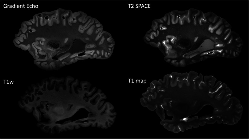 Figure 1
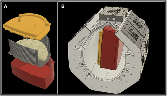 Figure 2
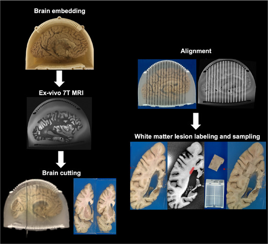 Figure 3
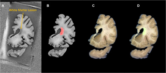 Figure 4
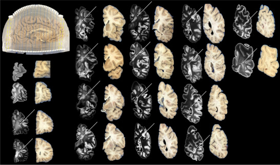 Figure 5
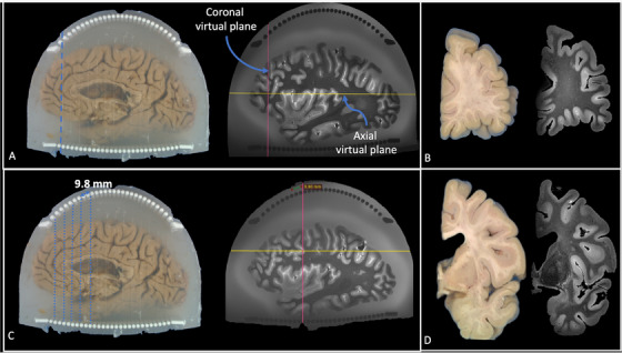 Figure 6
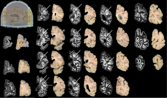 Figure 7
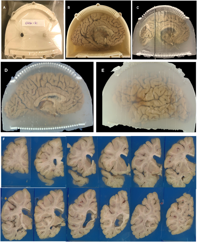 Figure 8
Peer Reviews
No public reviews on file for this paper yet. If you reviewed it on a platform where reviews are public (OpenReview, ICLR, NeurIPS, ICML), you can paste yours below so the community can read it here.
Videos
No videos yet. Explain this paper in a talk, walkthrough, or lecture? Add one.
Taxonomy
TopicsAdvanced Neuroimaging Techniques and Applications · Advanced MRI Techniques and Applications · Glioma Diagnosis and Treatment