Protocol for measuring cilium length using 3D confocal fluorescence microscopy, CiliaQ software, and a quality control pipeline
Daniel Burgdorf, Seniz Yüksel, Katharina Sieckmann, Jan N. Hansen, Dagmar Wachten, Nathalie Jurisch-Yaksi

TL;DR
This paper provides a detailed protocol for measuring cilium length using 3D confocal imaging and a custom software pipeline.
Contribution
The novel contribution is a complete workflow combining 3D imaging, CiliaQ software, and a Python-based analysis pipeline for accurate cilium length measurement.
Findings
A protocol for immunolabeling and 3D confocal imaging of cilia in cell culture is described.
CiliaQ plugin and Python pipeline enable segmentation, quantification, and statistical analysis of cilium morphology.
The method is applicable to various ciliated systems beyond cell culture, including tissues and organoids.
Abstract
Primary cilia are cellular antennas responding to various biological cues. Cilia undergo substantial changes in length, affecting their ability to sense extracellular signals and trigger downstream effector responses. Here, we present a protocol to image cilia in 2-dimensional cell culture using immunofluorescence staining and confocal microscopy. We describe steps to analyze cilium length using the CiliaQ plugin in Fiji/ImageJ and for using CiliaQ Explorer, a Python-based pipeline that plots CiliaQ-derived data and performs a statistical analysis after quality control. For complete details on the use and execution of this protocol, please refer to Hansen et al.1 •Protocol for immunolabeling and 3D confocal imaging of cilia in cell culture•Procedure to segment and quantify cilia morphology in 3D via the ImageJ plugin CiliaQ•Python pipeline to analyze CiliaQ data (plotting, statistics,…
Genes, proteins, chemicals, diseases, species, mutations and cell lines named across the full text — each resolved to its canonical identifier and authoritative record.
Click any figure to enlarge with its caption.
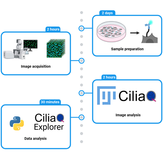 Figure 1
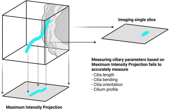 Figure 2
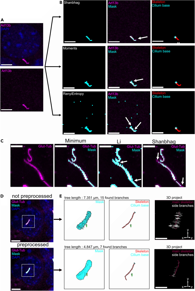 Figure 3
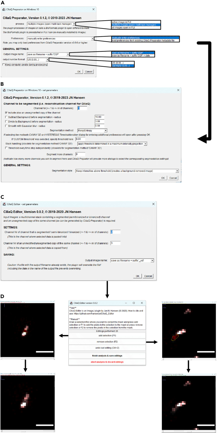 Figure 4
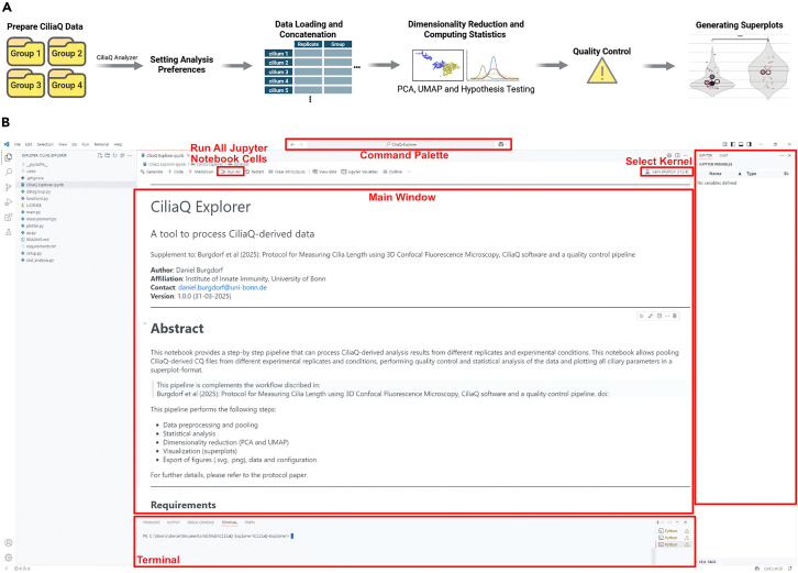 Figure 5
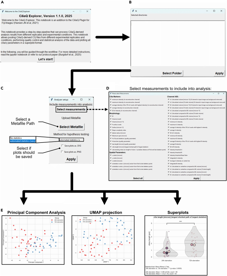 Figure 6
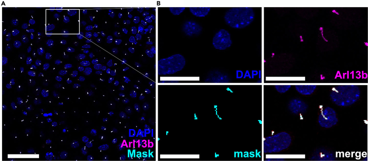 Figure 7
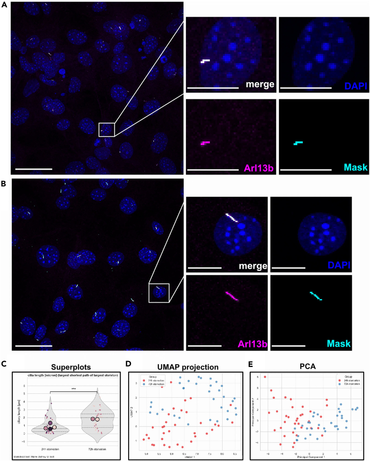 Figure 8
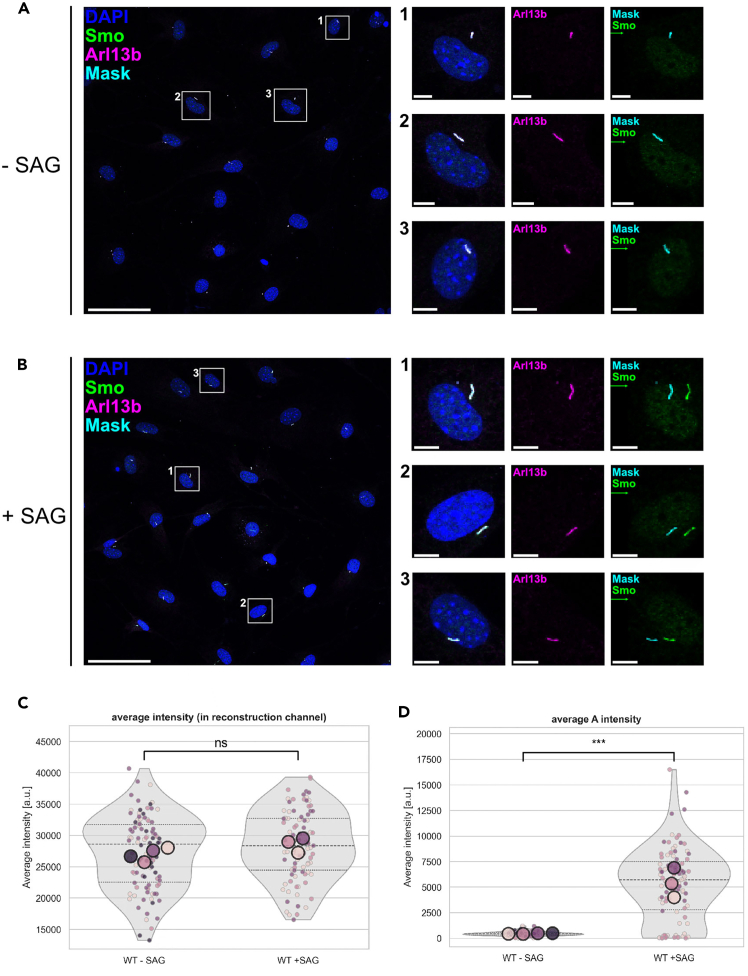 Figure 9
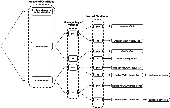 Figure 10
Peer Reviews
No public reviews on file for this paper yet. If you reviewed it on a platform where reviews are public (OpenReview, ICLR, NeurIPS, ICML), you can paste yours below so the community can read it here.
Videos
No videos yet. Explain this paper in a talk, walkthrough, or lecture? Add one.
Taxonomy
TopicsGenetic and Kidney Cyst Diseases · Pediatric Urology and Nephrology Studies · Fetal and Pediatric Neurological Disorders