Three-dimensional spatiotemporal analysis for the assessment of retinal capillary perfusion using a clinical OCT system
Yudan Chen, Jun Song, Hoyoung Jung, Tiffany Tse, Valerie Mok, Jennifer Tsang, Zaid Mammo, Myeong Jin Ju

TL;DR
This paper presents a new method to analyze retinal capillary perfusion using OCTA data, offering more accurate insights into early signs of vision-threatening diseases.
Contribution
The study introduces a novel protocol using unprocessed OCTA data and depth-resolved CoV analysis for retinal perfusion assessment.
Findings
The proposed method provides more accurate measurements of retinal perfusion heterogeneity compared to conventional CoV analysis.
Using unprocessed OCTA data enhances the reliability of perfusion analysis by incorporating depth-dependent signals.
The developed techniques can support future research on retinal vascular disorders.
Abstract
Growing evidence suggests that subtle changes in retinal microcirculation may precede structural damage in vision-threatening diseases. Among these, perfusion heterogeneity within the retinal capillary network has emerged as a promising biomarker for early detection and disease monitoring. Recent advances in optical coherence tomography (OCT) and OCT-based angiography (OCTA) have enabled high-resolution, three-dimensional imaging of retinal morphology and vasculature. However, commercial systems remain limited in their ability to accurately analyze retinal perfusion dynamics due to reliance on proprietary and undisclosed post-processing algorithms. This paper introduces an effective protocol for spatial and temporal analysis of capillary perfusion heterogeneity using unprocessed OCTA volume data acquired by a commercial retinal imaging system. The proposed method employs a novel…
Genes, proteins, chemicals, diseases, species, mutations and cell lines named across the full text — each resolved to its canonical identifier and authoritative record.
Click any figure to enlarge with its caption.
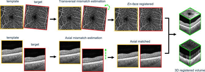 Figure 1
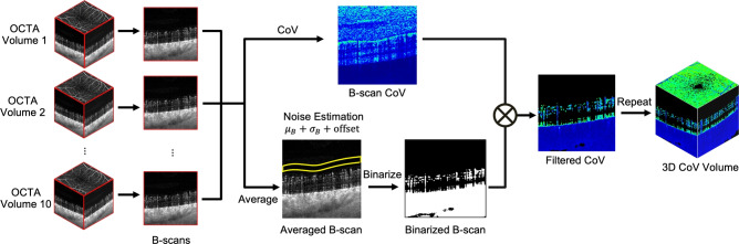 Figure 2
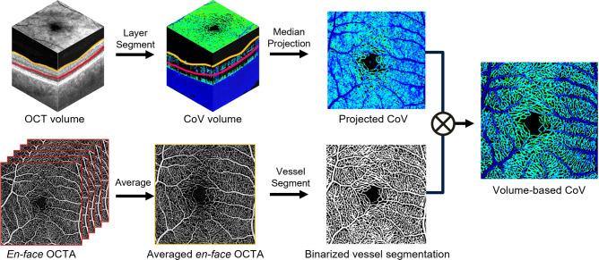 Figure 3
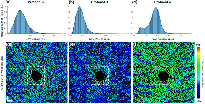 Figure 4
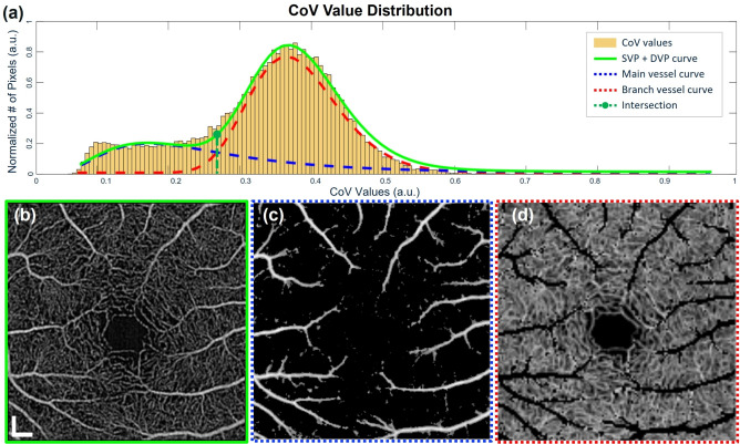 Figure 5
 Figure 6
 Figure 7
 Figure 8
Peer Reviews
No public reviews on file for this paper yet. If you reviewed it on a platform where reviews are public (OpenReview, ICLR, NeurIPS, ICML), you can paste yours below so the community can read it here.
Videos
No videos yet. Explain this paper in a talk, walkthrough, or lecture? Add one.
Taxonomy
TopicsRetinal Imaging and Analysis · Optical Coherence Tomography Applications · Retinal Diseases and Treatments