Multiparametric MRI to stratify risk factors for hemorrhagic complications in inoperable glioblastomas following stereotactic needle biopsy
Matia Martucci, Claudia Tocilă-Mătășel, Luigi Ruscelli, Giuseppe Varcasia, Giammaria Marziali, Francesco Schimperna, Giovanni Pentassuglia, Amato Infante, Quintino Giorgio D’Alessandris, Alessandro Olivi, Simona Gaudino

TL;DR
This study uses MRI to identify risk factors for bleeding after brain biopsies in inoperable glioblastomas, finding that tumor location is the strongest predictor.
Contribution
The study introduces a multiparametric MRI approach to predict hemorrhagic complications after stereotactic needle biopsy in glioblastomas.
Findings
Lesion location was the strongest predictor of post-biopsy hemorrhage, with deep lesions showing the highest frequency.
rCBVmax showed a significant linear association with hemorrhage area but not with hemorrhage incidence.
Grade 3 ITSS lesions were linked to more extensive bleeding, while peritumoral edema showed no correlation.
Abstract
Histological confirmation of glioblastoma (GB) is essential for therapeutic planning, even in inoperable cases where stereotactic needle biopsy (STNB) is the only option. However, post-procedural bleeding remains a known risk. This study aimed to evaluate the association between MRI features of GB and hemorrhagic complications following STNB. This retrospective, single-center study included 78 patients with IDH-wildtype GB (mean age: 61 years; 33 females) who underwent pre-biopsy MRI (including SWI and DSC-perfusion) and post-biopsy CT within 72 h. Lesions were anatomically classified into four groups based on their location: cortical/superficial grey matter (sGM n = 12), subependymal white matter (sWM; n = 36), deep nuclei/thalamus (n = 26), or brainstem (n = 4). Hemorrhage incidence and area were correlated with lesion location, intratumoral susceptibility signal (ITSS) grade,…
Genes, proteins, chemicals, diseases, species, mutations and cell lines named across the full text — each resolved to its canonical identifier and authoritative record.
Click any figure to enlarge with its caption.
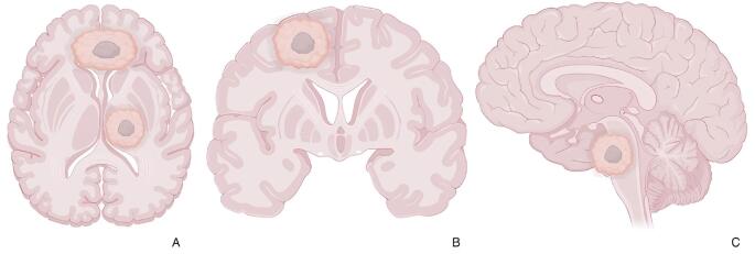 Figure 1
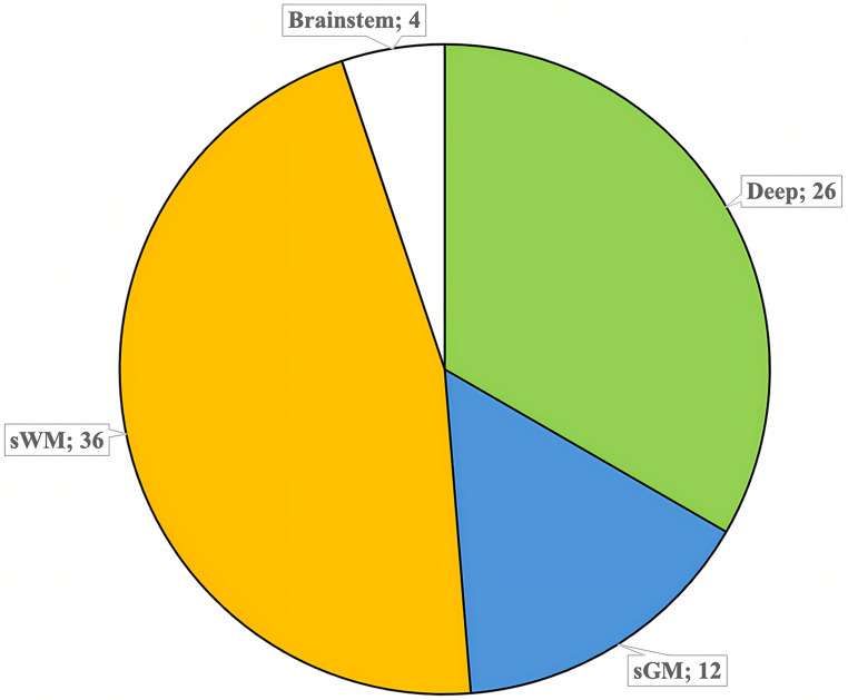 Figure 2
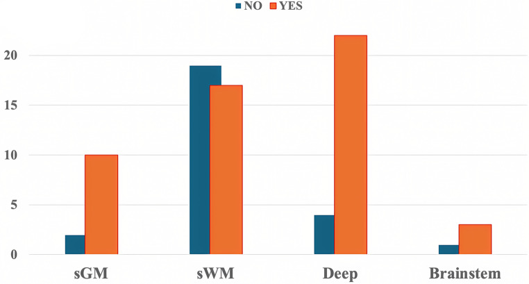 Figure 3
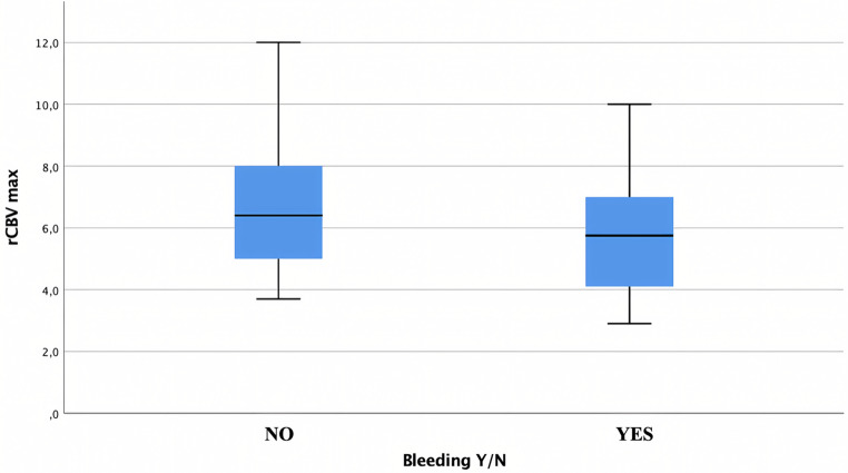 Figure 4
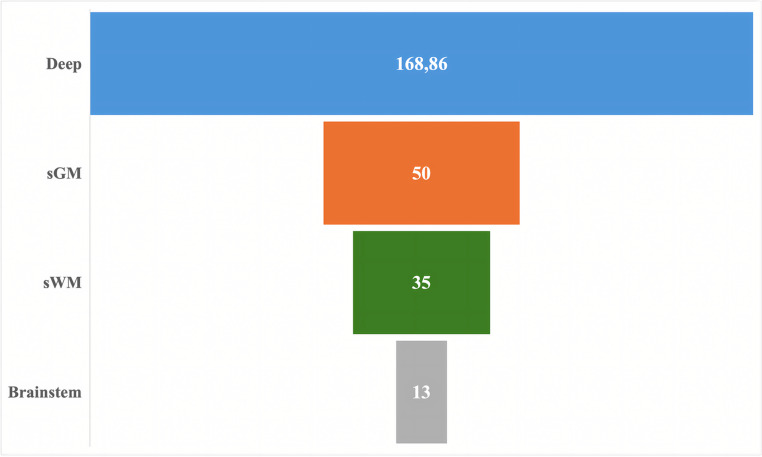 Figure 5
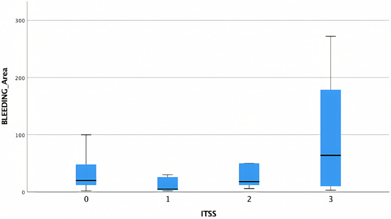 Figure 6
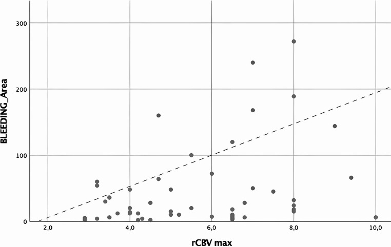 Figure 7
 Figure 8
Peer Reviews
No public reviews on file for this paper yet. If you reviewed it on a platform where reviews are public (OpenReview, ICLR, NeurIPS, ICML), you can paste yours below so the community can read it here.
Videos
No videos yet. Explain this paper in a talk, walkthrough, or lecture? Add one.
Taxonomy
TopicsGlioma Diagnosis and Treatment · MRI in cancer diagnosis · Radiomics and Machine Learning in Medical Imaging