Automatic diagnosis of type 2 diabetes mellitus with mild cognitive impairment using artificial intelligence based on routine T1-weighted MRI
Chang Li, Jun Zhang, Bo Xue, Yuwei Xia, Feng Shi, Xingyan Le, Junbang Feng, Peng Chen, Chuanming Li

TL;DR
This study uses AI and MRI scans to automatically diagnose type 2 diabetes patients with mild cognitive impairment, offering a precise and efficient diagnostic tool.
Contribution
The novel contribution is an AI-based model using structural MRI features and lab data to accurately detect early cognitive impairment in type 2 diabetes patients.
Findings
A Random Forest model achieved high accuracy (AUC 0.959 in training, 0.887 in testing) in diagnosing T2DM with MCI.
Eight features, including brain volume and lab metrics, were identified as potential biomarkers for early cognitive impairment.
The model enables objective and efficient differentiation of T2DM patients with and without MCI.
Abstract
Patients with type 2 diabetes mellitus (T2DM) exhibit a heightened susceptibility to developing dementia, especially those who already present with mild cognitive impairment (MCI). Nevertheless, the fundamental etiology remains elusive, underscoring the pressing need for an objective and precise diagnostic approach in clinical settings. This study investigates the utilization of machine learning algorithms in conjunction with high-resolution sagittal T1-weighted structural imaging to facilitate automated diagnosis of T2DM patients with MCI, differentiating them from both T2DM patients without MCI and healthy controls (HCs). Thirty patients with T2DM and MCI, thirty T2DM patients without MCI, and thirty matched healthy controls (HCs) were enrolled to identify independent biomarkers and develop a diagnostic model for early cognitive impairment in T2DM. Whole-brain structural…
Genes, proteins, chemicals, diseases, species, mutations and cell lines named across the full text — each resolved to its canonical identifier and authoritative record.
Click any figure to enlarge with its caption.
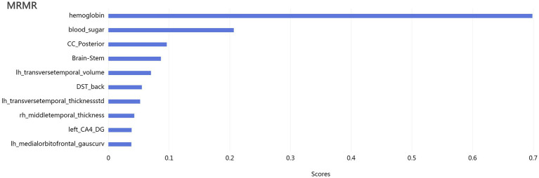 Figure 1
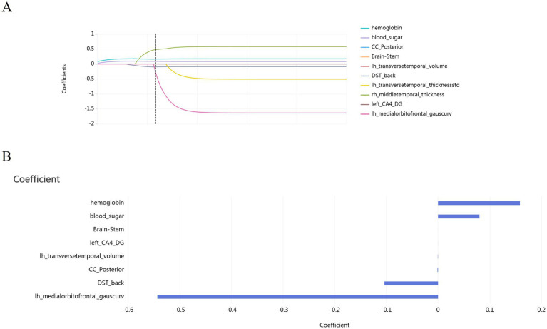 Figure 2
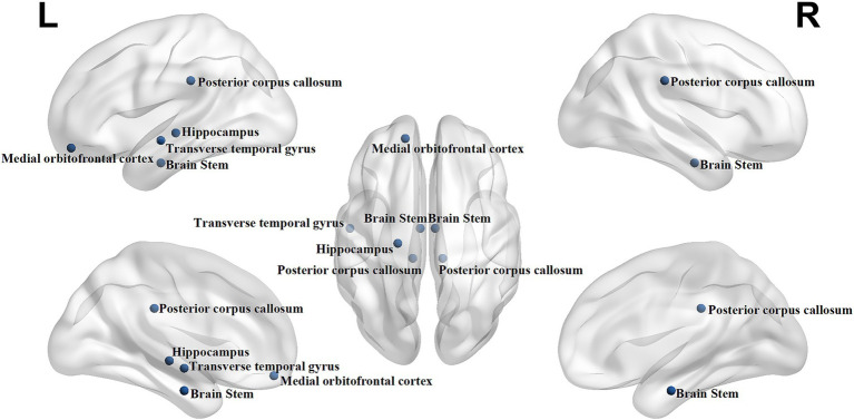 Figure 3
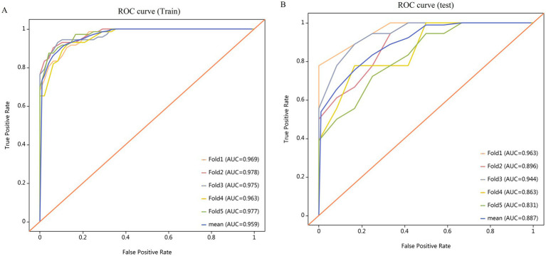 Figure 4
Peer Reviews
No public reviews on file for this paper yet. If you reviewed it on a platform where reviews are public (OpenReview, ICLR, NeurIPS, ICML), you can paste yours below so the community can read it here.
Videos
No videos yet. Explain this paper in a talk, walkthrough, or lecture? Add one.
Taxonomy
TopicsAdvanced MRI Techniques and Applications · Traditional Chinese Medicine Studies · Cerebrovascular and Carotid Artery Diseases