Revealing Erythema Nodosum Leprosum in a Leprosy Patient: A Case of Treatment Noncompliance
Bassem Alhariri, Memon Noor Illahi, Namaa Abubaker Suliman Elshaikh, Rashid Gamal Mohamed Omer, Noof AlQahtani, Muhammad Sharif

TL;DR
A leprosy patient developed severe inflammation after stopping treatment, highlighting the importance of following medical advice to avoid complications.
Contribution
This case highlights the clinical consequences of treatment noncompliance in leprosy patients, emphasizing the risk of Type-2 lepra reactions.
Findings
Noncompliance with MDT can trigger severe systemic inflammation resembling sepsis.
Early recognition and management of ENL are crucial to prevent complications.
Type-2 LR occurs predominantly in lepromatous leprosy patients during or after treatment.
Abstract
Lepra reactions (LR) are acute inflammatory conditions with immune mediators that are highly morbid. Patients with the lepromatous end of the leprosy spectrum (BL-LL) are the only ones who develop Type-2 LR. 90% of the time, it happens during or right after treatment, usually within 2 years. The emergence of Erythema Nodosum Leprosum (ENL) suggests a paradoxical immune reaction and hypersensitivity to leprosy bacteria that results in painful erythematous nodules and systemic symptoms. Here, we present a case of Type 2 LR in a 41-year-old Indian man diagnosed with lepromatous leprosy 2 years ago. He was treated with MDT regime for 12 months and was compliant with treatment till 1 month before presenting with fever, cough, red eyes, and painful erythematous nodules involving his trunk and extremities. This case underscores the severe systemic inflammatory response that can be triggered by…
Genes, proteins, chemicals, diseases, species, mutations and cell lines named across the full text — each resolved to its canonical identifier and authoritative record.
Click any figure to enlarge with its caption.
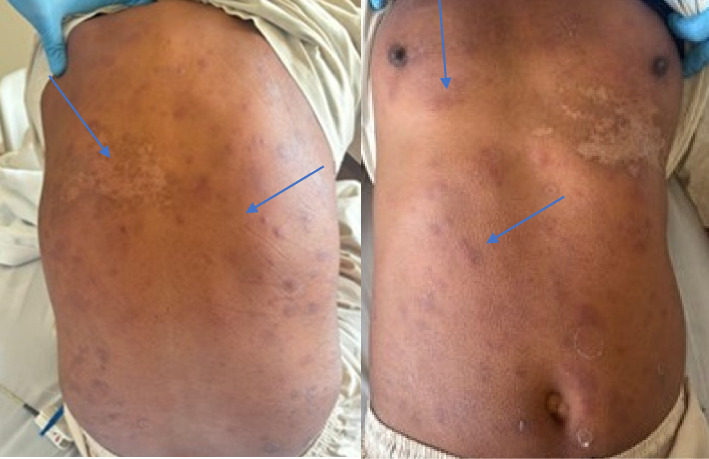 Figure 1
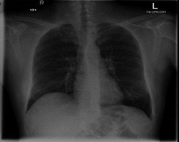 Figure 2
Peer Reviews
No public reviews on file for this paper yet. If you reviewed it on a platform where reviews are public (OpenReview, ICLR, NeurIPS, ICML), you can paste yours below so the community can read it here.
Videos
No videos yet. Explain this paper in a talk, walkthrough, or lecture? Add one.
Taxonomy
TopicsLeprosy Research and Treatment · Infectious Diseases and Tuberculosis · Mycobacterium research and diagnosis
1. Introduction
Leprosy, also known as Hansen's disease, is a chronic infectious disease caused by Mycobacterium leprae that primarily affects the skin, peripheral nervous system, upper respiratory tract, eyes, and testes [1]. According to the World Health Organization (WHO), more than 120 nations still have cases of this neglected tropical disease, with over 200,000 new cases reported annually [2]. In 2019, Brazil, India, and Indonesia reported over 10,000 new cases [3]. Lepra reactions (LR), which are acute inflammatory reactions that might complicate leprosy, are a major characteristic of the disease. The two main types of LR are Type 1 (reversal reaction) which typically occurs in tuberculoid (TL) and borderline tuberculoid leprosy (BT), and Type 2 also known as Erythema Nodosum Leprosum (ENL) [1, 4–6]. ENL is a delayed immune-complex-mediated response to high levels of circulating antimycobacterial antibodies, leading to systemic inflammation and multisystem involvement [5, 7]. Clinically, this multisystem disorder usually presents as erythematous, painful papules and nodules in the extremities of approximately 50% of individuals with lepromatous leprosy (LL) and, less frequently, in about 25% of those with borderline lepromatous leprosy (BL) [6]. Apart from skin nodules, patients may present with other systemic manifestations such as fever, iritis, arthritis, lymphadenitis, orchitis, and neuritis [1]. The diagnosis of ENL is mainly clinical, supported by the presence of leprosy and exclusion of other conditions. Despite the availability of effective treatments for leprosy, ENL has an important incidence and demands further understanding in terms of its pathophysiological and clinical presentation and therapeutic approaches.
2. Case Presentation
A 41-year-old Indian gentleman came in presenting with fever, cough, vomiting, eye redness, and generalized painful rash all over his body. Symptoms started 1 month ago. The patient was diagnosed with Hansen's disease LL (Type 2 LR) back in November 2022 during a visit in India. The diagnosis was based on clinical and histopathological findings. His baseline Bacteriological Index (BI) was reported to be 4+; however, records of the Morphological Index (MI) were not available. He was then started on the WHO multidrug treatment (MB-MDT) regimen for multibacillary leprosy (rifampicin, clofazimine, and dapsone) with a planned duration of 12 months; however, he stopped taking medications 1 month ago just before experiencing current symptoms. He has missed his scheduled follow-ups as he travelled back to his home country. The patient also complains of loss of appetite but denies any unintentional loss of weight. He has also been diagnosed with Type 2 diabetes mellitus, which is moderately controlled, and is on metformin. He is a nonsmoker and does not drink.
3. Physical Examination
According to the patient's vital signs on admission, the patient had borderline hypotension with systolic blood pressure of 99 mmHg. Other vitals were unremarkable.
On skin examination, there were multiple erythematous, raised, tender nodules all over his body and face, more notably on the back (Figure 1). Cardiovascular, respiratory, neurological, and abdominal examinations were insignificant (Table 1).
The clinical impression at the time of examination was sepsis versus an immune reaction. Significant laboratory findings include leucocytosis, elevated inflammatory markers, and hyponatremia (Table 2).
A chest X-ray and Echocardiography, which turned out unremarkable (Figure 2), were done on admission to rule out pneumonia and any cardiac pathology.
Blood samples were taken for blood culture which showed no growth.
3.1. In-Patient Management
Patients started on IV antibiotics (ceftriaxone and azithromycin) for empirical coverage of sepsis, steroids (intravenous methylprednisolone 40 mg every 8 h), insulin sliding scale, and IV fluids. Antibiotics were discontinued after 48 h once blood cultures were negative and the diagnosis of ENL was established.
Dermatology was consulted, and a biopsy was taken from the left upper back for pathology review. Later, the results of the biopsy showed unremarkable epidermis with dermal epithelioid histiocytes surrounding small cutaneous nerves, vessels, and adnexal structures. Macrophages are found in poorly circumscribed masses in the dermis with few lymphocytes. Slit-skin smears were not performed during this acute admission.
On the third day, the patient was discharged with referral to the infectious disease clinic for further follow up. Discharge plan was to continue oral prednisolone 60 mg daily with a taper plan of 10 mg weekly over 6 weeks taper off prednisolone at home. Patient was also advised to discontinue antibiotics as cultures were negative continue the oral antibiotics for 3 more days and continue with oral hypoglycaemic medication.
After 1 week, the patient was seen in the infectious disease clinic, where he was restarted on the WHO MB-MDT regimen for a full 12-month course alongside multidrug treatment regimen with thalidomide 100 mg twice daily for 2 more years as a steroid-sparing agent for long-term ENL control and stopped his current treatment regimen from India. Further follow-up was planned every other 4 weeks.
4. Discussion
Type 2 LR, referred to as ENL reaction, is an immune-complex-mediated inflammatory response that usually occurs in patients having LL with high load of leprosy bacilli. ENL may develop at any time of the disease course: before, during, or after initiation of treatment with MDT. According to the literature, 90% of cases acquired ENL after starting treatment, usually within 2 years [1, 4, 8]. The abrupt cessation of MDT, particularly rifampicin, likely led to a rapid change in the antigenic load and a surge in immune complex formation, precipitating this severe ENL reaction. The systemic inflammatory response in ENL, driven by a cytokine storm, can clinically mimic sepsis, as seen in this case with leukocytosis, markedly elevated CRP, and fever, explaining the initial diagnostic challenge.
The patient's comorbid type 2 diabetes mellitus may have contributed to an altered immune state, potentially facilitating both the initial infection and the severity of the inflammatory reaction upon treatment interruption.
When it comes to its pathophysiology, ENL appears to be a complex interaction between various aspects of the immune system. For the past years, theories have considered the role of neutrophils and immune-complex formation as a hallmark of ENL. Recent studies showed a multitude of factors such as proinflammatory cytokines, Treg cells, TLR-9, CCL-5, IFN-γ, and even possibly B cells, bringing humoral immunity into the picture [6, 9, 10]. Furthermore, genetic predisposition and environmental factors may influence the likelihood of developing ENL, suggesting that the pathophysiology is multifactorial and is still unclear [11].
Clinically, ENL presents as crops of multiple erythematous to dark brown, painful papules and nodules over the face commonly and predominantly on the extensor surface of the limbs [4]. Fever, malaise, arthritis, neuritis, vasculitis, uveitis, lymphadenopathy, and SIRS are other systemic signs of ENL [12]. Our patient was diagnosed with LL 2 years prior and ceased his treatment 1 month before presenting with this painful erythematous nodule involving his trunk and limbs, which is likely triggered by fluctuations in immune response following discontinuation of treatment. These nodules were preceded by fever, cough, and eye redness. This wide range of clinical presentations highlights the complex nature of ENL and the significance of maintaining a high level of suspicion of ENL in patients with LL. Patients with ENL may experience recurrent outbreaks that correspond with changes in their immunological status, often necessitating adjustments in treatment.
Regarding management of ENL, it is crucial to initiate appropriate anti-inflammatory treatment while addressing the underlying leprosy. The WHO's guidelines for treatment of ENL-complicated LL involve corticosteroids as the first-line treatment as they are effective in alleviating symptoms, reducing inflammation and preventing irreversible nerve damage [10, 13]; however, their use must be carefully monitored due to the potential fluctuations in immune function which can results in emergence of new nodules and other complications. Thalidomide has a considerable efficacy in men and women of nonreproductive potential; however, its use is limited due to its teratogenesis [9, 13]. In addition to these treatments, it is essential to re-initiate MDT for LL, as stopping treatment can lead to the progression of the disease and further complications. Our patient was on MDT till 1 month before the appearance of skin nodules. He was started on high dose of prednisolone and after 1 week, he was seen in the outpatient clinic, and all his lesions had subsided. He was restarted on MDT with thalidomide, and his steroid therapy was tapered. This rapid improvement in the patient symptoms highlighted the vital role of patient education, treatment adherence and regular follow up in enhancing the patient outcomes.
In this case, high-dose corticosteroids were initiated first to rapidly control the severe inflammation. Thalidomide was subsequently chosen for long-term ENL management due to its proven efficacy, its role as a steroid-sparing agent, and its suitability for this male patient, thus avoiding the risks associated with long-term high-dose steroid therapy. The decision to restart a full 12-month course of WHO MB-MDT was imperative to treat the underlying multibacillary disease, as the patient had not completed the standard regimen and was at high risk for relapse. Drug resistance testing was not performed due to lack of clinical suspicion (no prior MDT failure) and limited availability.
This case highlights that treatment nonadherence is a critical risk factor for severe ENL reactions. It underscores the necessity of patient education, structured follow-up programs, and a multidisciplinary approach to ensure compliance and prevent serious complications.
5. Conclusion
ENL remains a complex and critical complication of LL, reflecting the intricate relationship between the immune system and persistent infection. This case illustrates the importance of recognizing various systemic manifestations of ENL in patients with LL. A thorough grasp of the pathophysiology, clinical implications, and management strategies is crucial for enhancing the quality of life for patients with this complex condition. Additionally, the multidisciplinary approach, involving dermatologists, infectious disease specialists, and possibly ophthalmologists, is crucial in providing comprehensive care for this patient.
The reference list from the paper itself. Each links out to its DOI / PubMed record.
- 1Joanne M. Bargman K. L. S. No Ti Harrison’s Principles of Internal Medicine 2018 p. 2111
- 2Mungroo M. R. Khan N. A. Siddiqui R. Mycobacterium Leprae: Pathogenesis, Diagnosis, and Treatment Options Microbial Pathogenesis 2020 December 149p. 10447510.1016/j.micpath.2020.10447532931893 · doi ↗ · pubmed ↗
- 3https://www.who.int/news-room/fact-sheets/detail/leprosy
- 4Akinboro A. Erythema Nodosum Leprosum Associated With Lepromatous Leprosy: A Case Report and Review of Literature Leprosy Review 202192
- 5Jonas K. Type II Reaction Erythema Nodosum Leprosum: A Case Report Open Access 20248
- 6Bhat R. Vaidya T. What is New in the Pathogenesis and Management of Erythema Nodosum Leprosum Indian Dermatology Online Journal 2020114 p. 48210.4103/idoj.idoj_561_19PMC 741343532832433 · doi ↗ · pubmed ↗
- 7Polycarpou A. Walker S. L. Lockwood D. N. J. A Systematic Review of Immunological Studies of Erythema Nodosum Leprosum Frontiers in Immunology 2017 March 8p. 23310.3389/fimmu.2017.002332-s 2.0-85017277427 PMC 534688328348555 · doi ↗ · pubmed ↗
- 8Park L. Wallace C. E. Vasile G. Buckley C. A Case of Lepromatous Leprosy With Erythema Nodosum Leprosum Cureus 2023 January 151p. e 3384610.7759/cureus.33846 PMC 993494036819324 · doi ↗ · pubmed ↗