Contribution of UbrA, a ubiquitin ligase essential for Arg/N-degron pathway, to peptidase gene expression in Aspergillus oryzae
Waka Muromachi, Mao Ohba, Yasuaki Kawarasaki, Youhei Yamagata, Mizuki Tanaka

TL;DR
This study shows that UbrA, a protein involved in protein degradation, controls peptidase gene expression in the fungus Aspergillus oryzae, affecting enzyme production.
Contribution
The study reveals a novel role of UbrA in regulating peptidase and transporter gene expression in A. oryzae via the Arg/N-degron pathway.
Findings
Disruption of ubrA reduces acidic peptidase activity and gene expression but increases alkaline peptidase production.
UbrA regulates dipeptide/tripeptide transporter and peptidase genes independently of the PrtR transcription factor.
UbrA disruption reduces mRNA levels of dipeptidyl-peptidase and tripeptidyl-peptidase genes.
Abstract
The degradation of intracellular proteins by N-degron pathways depends on their N-terminal amino acids. In budding yeast, the Arg/N-degron pathway controls the expression of dipeptide/tripeptide transporter gene by degrading a transcriptional repressor. However, there is no detailed information on the N-degron pathway in filamentous fungi, and its role in regulating microbial nitrogen metabolism is unclear. Here, we demonstrated that the E3 ubiquitin ligase, UbrA, which is required for the Arg/N-degron pathway, regulates peptidase gene expression in the filamentous fungus Aspergillus oryzae. Using ubiquitin-fused green fluorescent protein as a reporter, we showed that the Arg/N-degron pathway in A. oryzae is similar to that in budding yeast. Disruption of ubrA significantly reduced the activities of acidic endopeptidase and carboxypeptidase in submerged culture using soy protein as the…
Genes, proteins, chemicals, diseases, species, mutations and cell lines named across the full text — each resolved to its canonical identifier and authoritative record.
Click any figure to enlarge with its caption.
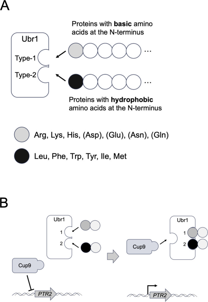 Fig 1
Fig 1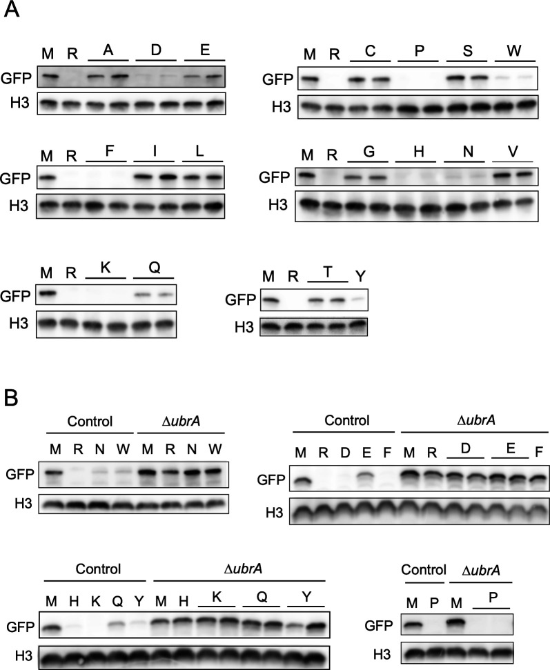 Fig 2
Fig 2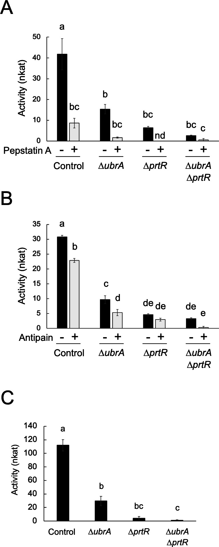 Fig 3
Fig 3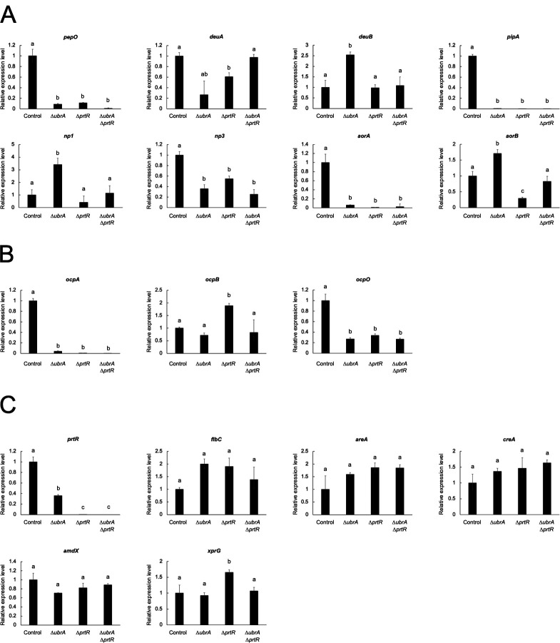 Fig 4
Fig 4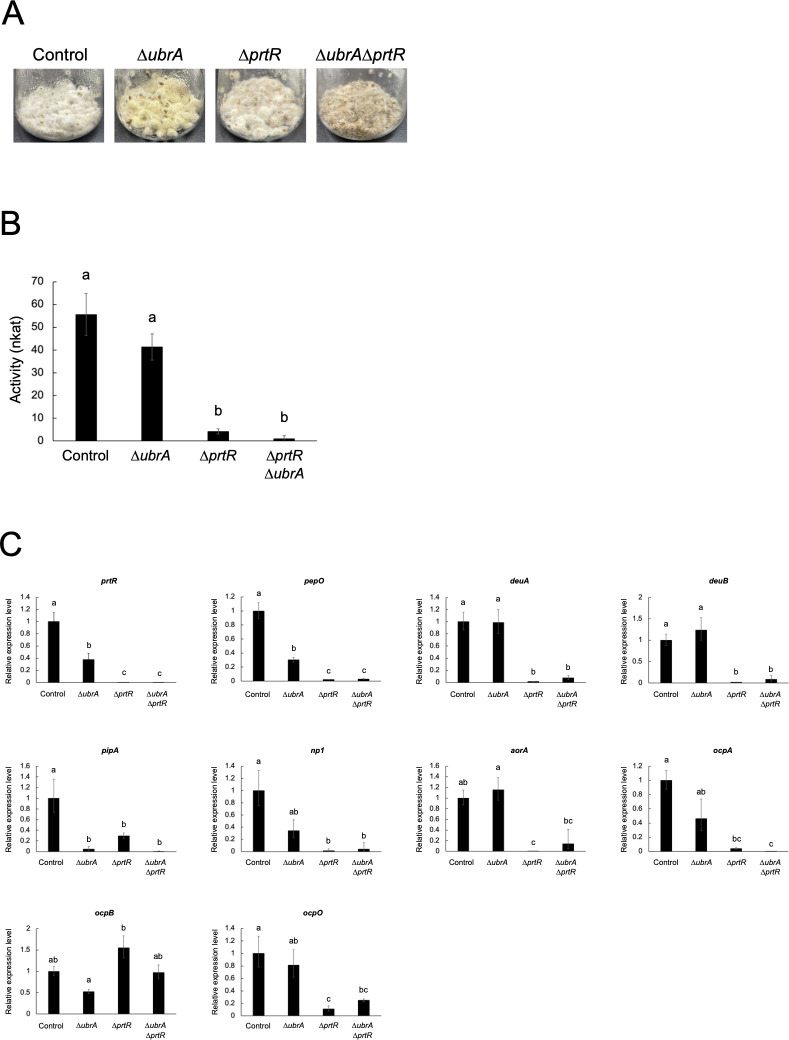 Fig 5
Fig 5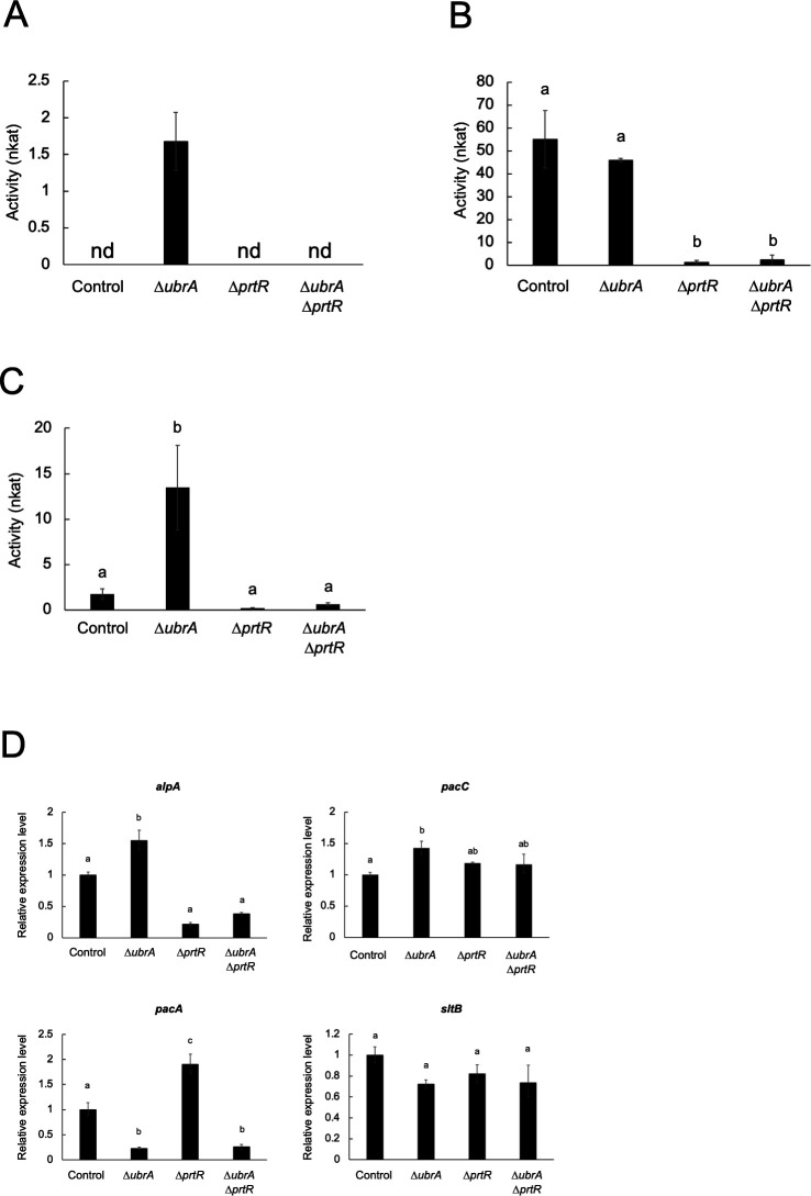 Fig 6
Fig 6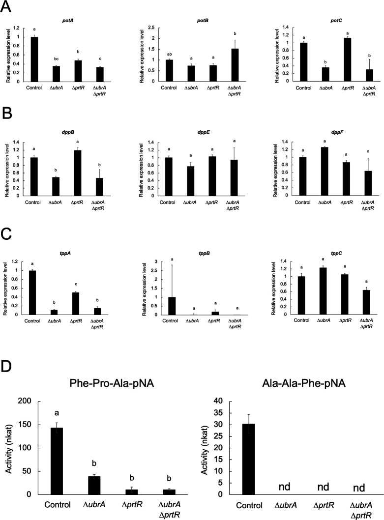 Fig 7
Fig 7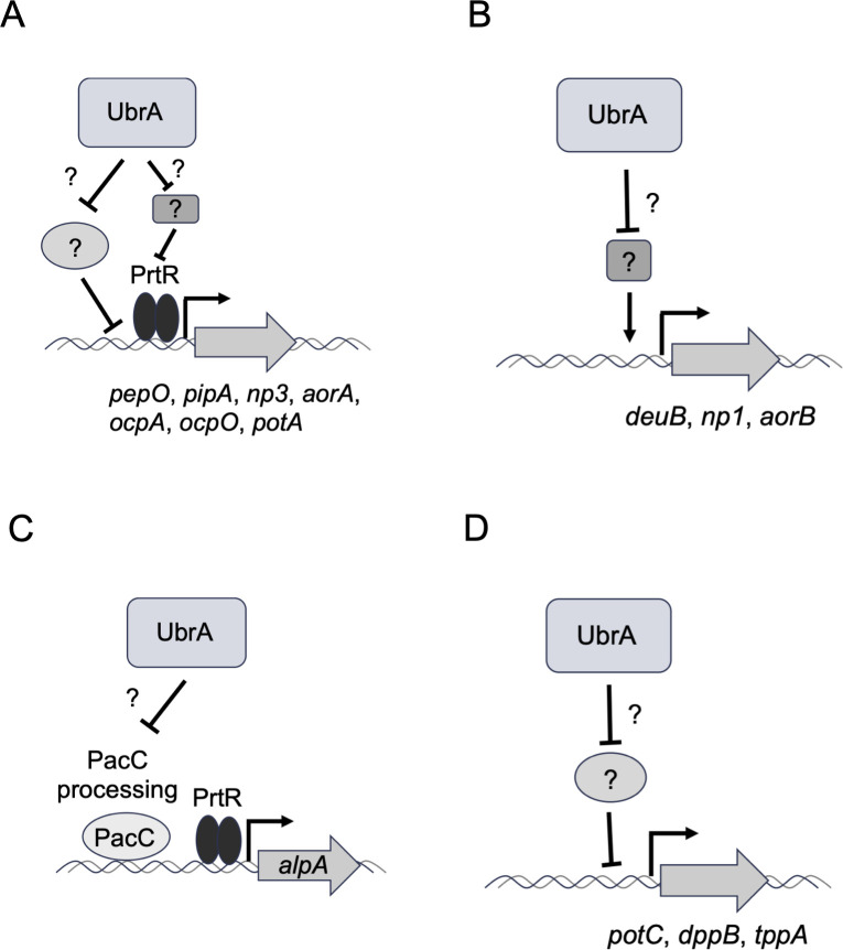 Fig 8
Fig 8| Strain | Mycelial semi-dry weight (g) | |
|---|---|---|
| 36 h | 48 h | |
| Control | 1.42 ± 0.09 | 1.66 ± 0.08 |
|
| 1.26 ± 0.01 | 1.59 ± 0.06 |
|
| 0.91 ± 0.09 | 1.66 ± 0.16 |
|
| 0.72 ± 0.02 | 1.35 ± 0.13 |
| Strain | pH |
|---|---|
| Control | 3.86 ± 0.05 |
|
| 3.91 ± 0.01 |
|
| 3.61 ± 0.04 |
|
| 4.46 ± 0.04 |
- —Institute for Fermentation, Osakahttp://dx.doi.org/10.13039/100007802
- —Noda Institute for Scientific Researchhttp://dx.doi.org/10.13039/100010157
- —Mayekawa Houonkai Foundationhttp://dx.doi.org/10.13039/100015645
- —Japan Society for the Promotion of Sciencehttp://dx.doi.org/10.13039/501100001691
- —Japan Society for the Promotion of Sciencehttp://dx.doi.org/10.13039/501100001691
Peer Reviews
No public reviews on file for this paper yet. If you reviewed it on a platform where reviews are public (OpenReview, ICLR, NeurIPS, ICML), you can paste yours below so the community can read it here.
Videos
No videos yet. Explain this paper in a talk, walkthrough, or lecture? Add one.
Taxonomy
TopicsPeptidase Inhibition and Analysis · Ubiquitin and proteasome pathways · Studies on Chitinases and Chitosanases
INTRODUCTION
Most proteins undergo post-translational processing, such as methionine removal by methionine aminopeptidase (MetAP), cleavage by endopeptidase, and deamidation or arginylation of N-terminal amino acids. The stability of intracellular proteins depends on their N-terminal amino acid residues. In eukaryotic cells, the recognition of N-terminal amino acids by ubiquitin ligases determines the stability of intracellular proteins. This system is called the N-degron pathway (or N-end rule) (1, 2). In Saccharomyces cerevisiae, the E3 ubiquitin ligase, Ubr1, directly recognizes the N-terminal amino acid residues at two distinct substrate-binding sites, type-1 and type-2 (3, 4). The type-1 site recognizes the basic amino acids arginine, lysine, and histidine. The type-2 site recognizes the hydrophobic amino acids leucine, phenylalanine, tryptophan, tyrosine, isoleucine, and unacetylated methionine (Fig. 1A). Proteins with N-terminal asparagine, glutamine, aspartate, and glutamate are also recognized by Ubr1 because the amido group of N-terminal asparagine and glutamine residues is removed by amidase, and arginine is conjugated to N-terminal aspartate and glutamate by arginyl-tRNA-protein transferase (1, 2). The proteins recognized by Ubr1 are ubiquitinated and rapidly degraded by the proteasome. This Ubr1-mediated degradation pathway is called the Arg/N-degron pathway. When the N-terminal methionine itself or N-terminal alanine, serine, threonine, valine, cysteine, and glycine exposed after methionine removal by MetAP are acetylated by the ribosome-anchored acetyltransferase complex, these acetylated N-terminal amino acids are recognized by the membrane-embedded E3 ubiquitin ligase, Doa10 (5). This acetylation-dependent degradation is known as the Ac/N-degron pathway. The degradation of proteins with an N-terminal proline recognized by the glucose-induced degradation-deficient (GID) ubiquitin ligase complex is called the Pro/N-degron pathway (6).
The Arg/N-degron recognition by Ubr1 in budding yeast. (A) Schematic diagram of substrate recognition by Ubr1. The type-1 site recognizes N-terminal basic amino acids, while the type-2 site recognizes N-terminal hydrophilic amino acids. Proteins with N-terminal asparagine, glutamine, aspartate, and glutamate are indirectly recognized by type-1 site because the amido group of N-terminal asparagine and glutamine residues is removed by amidase, and arginine is conjugated to N-terminal aspartate and glutamate by arginyl-tRNA-protein transferase. (B) Schematic diagram of a positive feedback mechanism for the effective uptake of dipeptide/tripeptides. When both the type-1 and type-2 sites of Ubr1 are occupied by dipeptides, a distinct substrate-binding site of Ubr1 recognizes the C-terminus-proximal region of Cup9, the transcriptional repressor of PTR2 encoding dipeptide/tripeptide transporter. The degradation of Cup9 then induces PTR2 expression and promotes dipeptide/tripeptide uptake.
The N-degron pathways regulate several cellular processes (2). One of the most well-studied processes is the regulation of dipeptide/tripeptide import by the Arg/N-end pathway in S. cerevisiae. The transcriptional expression of PTR2, which encodes a transmembrane dipeptide/tripeptide transporter, is suppressed by the transcriptional repressor, Cup9 (7). When both the type-1 and type-2 substrate-binding sites of Ubr1 are occupied by dipeptides, a distinct substrate-binding site of Ubr1 recognizes the C-terminus-proximal region of Cup9 (8, 9). The degradation of Cup9 then induces PTR2 expression and promotes dipeptide/tripeptide uptake (Fig. 1B). This is a positive feedback mechanism for the effective uptake of dipeptide/tripeptides as nutrient sources in response to environmental conditions (10).
The filamentous fungus Aspergillus oryzae secretes various hydrolytic enzymes for the degradation of raw materials and is used for the production of traditional Japanese fermented foods, such as sake, soy sauce, and miso (soybean paste) (11). A. oryzae has more peptidase genes than other closely related Aspergillus species, and these translational products have been used as industrial enzymes for various food-processing and pharmaceutical applications (12). On the other hand, although A. oryzae is used as a host for producing homologous and heterologous proteins (13, 14), degradation by peptidases is one of the major problems for efficient production (15). Therefore, controlling peptidase gene expression in A. oryzae is critical for various industrial applications. Although the expression of peptidase genes in filamentous fungi is regulated by several transcription factors, such as FlbC, AreA, CreA, AmdX, and XprG (16–21), the Zn(II)2_Cys_6-type transcription factor, PrtT, plays a central role in the regulation of extracellular peptidase gene expression (22–26). In A. oryzae, the expression of most extracellular peptidase genes is positively or negatively regulated by PrtR (ortholog of PrtT) in response to a nitrogen source (27). In filamentous fungi, the expression of hydrolytic genes is often regulated in concert with the expression of transporter genes that import the substrates of hydrolytic gene expression or the degradation products of polymers by hydrolytic enzymes (28, 29). We previously identified three dipeptide/tripeptide transporter genes, potA, potB, and potC, in A. oryzae and found that the expression of potA and potB is positively regulated by PrtR (30). This suggests that the gene expression of peptidases and dipeptide/tripeptide transporters is cooperatively regulated. The disruption of ubrA, the ortholog of yeast UBR1, reduces the expression levels of POT genes, especially potC (30). However, the role of UbrA in regulating peptidase gene expression remains unclear. Additionally, the N-degron pathway in filamentous fungi has not been studied in detail.
In this study, we investigated the presence of the N-degron pathway in A. oryzae and examined whether UbrA is involved in this pathway. Furthermore, to clarify whether UbrA participates in the regulation of peptidase gene expression, we analyzed the effects of ubrA disruption on peptidase production and the transcription of peptidase genes under both submerged and solid-state cultivation.
RESULTS
Involvement of UbrA in the N-degron pathway
To investigate the involvement of A. oryzae UbrA in the N-degron pathway, ubiquitin-fused green fluorescent protein (Ub-X-GFP) was used as a reporter substrate to determine the N-end rule. When this fusion protein is expressed in eukaryotic cells, ubiquitin is removed by deubiquitinating enzymes, resulting in the expression of X-GFP with the desired amino acid X at the N-terminus (31, 32). The mutant Ub-G76V-GFP, in which ubiquitin is not cleaved from GFP, is degraded by the proteasome independently of the N-degron pathway (31, 32). To verify whether Ub-X-GFP is available to examine the N-degron pathway in A. oryzae, Ub-Met (M)-GFP, Ub-Arg (R)-GFP, and Ub-G76V-GFP were expressed in the control and ∆ubrA strains. In the control strain, M-GFP was detected by western blotting using an anti-GFP antibody, whereas R-GFP, a typical substrate of the Arg/N-degron pathway in eukaryotes, was undetectable (Fig. S1). In contrast, R-GFP was detected equally with M-GFP in the ∆ubrA strain, and Ub-G76V-GFP was not detected in either strain (Fig. S1). This indicates that UbrA mediates the degradation of R-GFP, depending on the N-terminal arginine. In addition, the results suggest that Ub-X-GFP can be used to examine the N-degron pathway in A. oryzae. Using this system, GFPs containing other 18 amino acids at the N-terminus were expressed in the control strain. In addition to R-GFP, the signals of eight other GFPs, Asp (D)-GFP, Pro (P)-GFP, Trp (W)-GFP, Phe (F)-GFP, His (H)-GFP, Asn (N)-GFP, Lys (K)-GFP, and Tyr (Y)-GFP, were hardly detected (Fig. 2A). Although Glu (E)-GFP and Gln (Q)-GFP were detectable, their amounts were approximately half of that of M-GFP. The remaining eight X-GFP species were detected in similar or higher amounts than that of M-GFP. To determine whether reduced abundance X-GFPs were degraded in a UbrA-dependent manner, they were expressed in the ∆ubrA strain. The absence of P-GFP detection even in the ∆ubrA strain suggests that P-GFP is degraded independently of UbrA (Fig. 2B). In addition to R-GFP, D-GFP, W-GFP, F-GFP, H-GFP, N-GFP, K-GFP, Y-GFP, E-GFP, and Q-GFP, excluding P-GFP, were detected in similar amounts to that of M-GFP in the ∆ubrA strain (Fig. 2B). This result indicated that A. oryzae UbrA is essential for the Arg/N-degron pathway.
Effect of ubrA disruption on the abundance of X-GFPs. (A) Western blot analysis of all X-GFPs in the control strain. Approximately 2 × 10⁷ conidiospores of Ub-X-GFP expressing strains were grown at 30°C for 20 hours in liquid CD + 0.1% polypepton. Two independent transformants were cultured, except for the Ub-Y-GFP expression strain. The strains expressing Ub-M-GFP and Ub-R-GFP were used as positive and negative controls, respectively. After harvesting the mycelium, approximately 30 µg extracted intracellular protein was subjected to western blot analysis using anti-GFP antibody. Histone H3 (H3) was detected as loading control by anti-histone H3 antibody. (B) Western blot analysis of unstable X-GFPs in the ∆ubrA strain. The X-GFP and histone H3 were detected as described for panel A.
Involvement of UbrA in peptidase production in a submerged culture
The ∆ubrA strain described above has two auxotrophic mutations, niaD^−^ (deficient in the nitrate reductase gene) and sC**^−^** (deficient in the ATP sulfurylase gene), that can be used as selection markers for transformation (30). Since these auxotrophic mutations disable nitrate and sulfate metabolism and might affect peptidase expression, we regenerated the ∆ubrA strain without auxotrophic mutation (Fig. S2A through C). To investigate the relationship between UbrA and PrtR, the transcription factor regulating a broad range of extracellular peptidase genes, in peptidase production, a double disruption mutant of ubrA and prtR was also generated (Fig. S2A through C). The ∆ubrA and ∆ubrA∆prtR strains formed compact colonies on the agar medium (Fig. S3). To examine the involvement of UbrA in peptidase production, the activities of acidic endopeptidase in the culture supernatants of these disrupted strains were compared with those of control and ∆prtR strains. After 48 hours of cultivation in liquid medium with soy protein as the nitrogen source (CD/soy), the proteolytic activity using casein as the substrate at pH 3.0 was significantly reduced by approximately 70% and 85% by ubrA and prtR disruption, respectively (Fig. 3A and B). To determine the type of peptidase in the culture supernatant, inhibitor assays were performed using pepstatin A, which specifically inhibits aspartic proteases (APases), and antipain, which inhibits serine/cysteine proteases (Fig. 3A and B). The acidic endopeptidase activity of the control strain was significantly inhibited by 80% with pepstatin A and by 26% with antipain, respectively. In the ∆ubrA strain, the activity was inhibited by 89% with pepstatin A and was significantly inhibited by 45% with antipain. When comparing the activities inhibited by the inhibitors, disruption of ubrA reduced 59% of the pepstatin A-sensitive activity and 46% of the antipain-sensitive activity. This suggests that ubrA disruption reduces the production of both APases and serine/cysteine proteases. The semi-dry mycelia weight of the ∆ubrA strain after cultivation in CD/soy medium for 48 hours was comparable to those of the control and ∆prtR strains (Table 1), indicating that the decrease in acidic endopeptidase activity by ubrA disruption was not due to differences in growth. Moreover, ubrA and prtR disruption reduced acidic carboxypeptidase activity by approximately 75% and 95%, respectively (Fig. 3C). This activity was completely abolished by the double disruption of ubrA and prtR. These results suggest that the disruption of ubrA reduces the production of a wide range of acidic peptidases.
Effect of ubrA disruption on the acidic peptidase production in submerged culture. (A) Acid endopeptidase activity with or without pepstatin A. Approximately 2.5 × 10⁷ conidiospores of each strain were grown at 30°C for 48 hours in liquid CD/soy medium containing 0.6% soy protein as a nitrogen source. Activities of the culture supernatants were measured using 2% casein (pH 3.0) as the substrate with pepstatin A (+) or dimethyl sulfoxide (−), the solvent for pepstatin A. “nd” means not detected. Error bars indicate the standard error of three biological replicates. Statistical analysis was performed using the Tukey–Kramer method. Different lowercase letters indicate significant differences (P < 0.05). (B) Acid endopeptidase activity with or without antipain. Activities of the culture supernatants were measured with antipain (+) or water (−). Error bars indicate the standard error of three biological replicates. Statistical analysis was performed using the Tukey–Kramer method. Different lowercase letters indicate significant differences (P < 0.05). (C) Acid carboxypeptidase activity. Activities of the culture supernatants were measured using 1 mM Z-Glu-Tyr (pH 3.5) as the substrate. Error bars indicate the standard error of three biological replicates. Statistical analysis was performed using the Tukey–Kramer method. Different lowercase letters indicate significant differences (P < 0.05).
Involvement of UbrA in the transcriptional expression of peptidase genes in a submerged culture
To investigate the involvement of UbrA in the transcriptional expression of peptidase genes, the mRNA levels of well-characterized peptidase genes after cultivation in liquid CD/soy medium for 36 hours were examined by reverse transcription-quantitative PCR (RT-qPCR). The semi-dry mycelia weight at this cultivation time was comparable between the ∆ubrA and the control strains, whereas it was lower in the ∆prtR and the double disruption strains (Table 1). The mRNA levels of six endopeptidase genes (pepO, deuA, pipA, np3, aorA, and aorB) were significantly reduced following prtR disruption (Fig. 4A). The expression levels of all five genes, except aorB, were also reduced by ubrA disruption (Fig. 4A). Specifically, the expression levels of pepO, pipA, np3, and aorA encoding APase, glutamic endopeptidase, metalloendopeptidase, and aorsin A, respectively, were significantly reduced by ubrA disruption (Fig. 4A). In contrast, the expression levels of deuB, np1, and aorB encoding deuterolysin B, metalloendopeptidase, and aorsin B, respectively, were significantly increased by ubrA disruption, although they were unchanged or reduced by prtR disruption (Fig. 4A). The expression levels of two carboxypeptidase genes, ocpA and ocpO, were significantly reduced by the disruption of ubrA (Fig. 4B), suggesting that the decrease in carboxypeptidase activity caused by the disruption of ubrA is attributed to the reduced expression of these genes. The expression level of another carboxypeptidase gene, ocpB, was significantly increased by prtR disruption but was not altered by ubrA disruption. These results suggest that UbrA is involved in the regulation of the transcription of a wide range of peptidase genes through both cooperative and independent interactions with PrtR.
Effect of ubrA disruption on the transcriptional expression of peptidase genes in submerged culture. RT-qPCR analysis of endopeptidase (A), acid carboxypeptidase (B), and transcription factor genes (C) involved in the regulation of peptidase gene expression after cultivation in liquid CD/soy medium for 36 hours. Error bars indicate the range of SE determined by the standard deviations using ∆∆CT values of three biological replicates. Statistical analysis was performed using the Tukey–Kramer method. Different lowercase letters indicate significant differences (P < 0.05).
Next, we examined whether the expression levels of genes encoding transcription factors regulating peptidase gene expression were affected by ubrA disruption (Fig. 4C). The prtR expression level decreased significantly by approximately 65% after ubrA disruption. The mRNA levels of the other five transcription factors, AreA, CreA, AmdX, XprG, and FlbC, were not significantly affected by ubrA disruption. These results suggested that the disruption of ubrA specifically reduced prtR expression.
Involvement of UbrA in the transcriptional expression of peptidase genes in a solid-state culture
A. oryzae produces larger amounts of hydrolytic enzymes in solid state than in liquid cultures, particularly in solid-state culture using wheat bran as the substrate (33). To investigate the involvement of UbrA in peptidase production under solid-state cultivation, all strains were grown in solid-state culture using wheat bran as the substrate, and casein proteolytic activity at pH 3.0 in the crude enzyme extracts was measured. In the solid-state cultivation, the ∆ubrA strain covered wheat bran with yellow conidiaspores more than other strains at 48 hours, while the ∆ubrA∆prtR strain showed slightly slower mycelial growth than other strains (Fig. 5A). The proteolytic activity of casein was reduced by approximately 92% and 26% by prtR and ubrA disruption, respectively, although the latter reduction was not significant. (Fig. 5B). Most of the activity was abolished by the double disruption of prtR and ubrA. To investigate the effect of ubrA disruption on transcriptional expression, the mRNA levels of prtR and peptidase genes were examined (Fig. 5C). As in the submerged culture, disruption of ubrA significantly reduced prtR expression to approximately 40% of that in the control strain. Disruption of ubrA significantly reduced the expression level of pepO, while the expression levels of np1 and ocpA decreased, although not significantly. However, their levels were only slightly reduced compared with those caused by prtR disruption. In addition, the expression levels of dueA, deuB, aorA, and ocpO were significantly reduced by prtR disruption, whereas they remained mostly unchanged by ubrA disruption. Although there was no significant difference, the expression level of pipA was further reduced by ubrA disruption than by prtR disruption. These results suggest that disruption of ubrA affects the expression of some peptidase genes in solid-state culture, but that the effect of UbrA on peptidase gene expression is greater in submerged culture than in solid-state culture.
Effect of ubrA disruption on the expression of peptidases in solid-state culture. (A) Growth on wheat bran medium. Approximately 3 × 10⁶ of conidiospores of each strain were grown on wheat bran medium at 30°C for 2 days. (B) Acid endopeptidase activity. Approximately 3 × 10⁶ conidiospores of each strain were grown at 30°C for 36 hours in wheat bran medium. Activities of the crude enzyme extracts were measured using 2% casein (pH 3.0) as the substrate. Error bars indicate the standard deviations of three independent experiments. Statistical analysis was performed using the Tukey–Kramer method. Different lowercase letters indicate significant differences (P < 0.05). (C) RT-qPCR analysis of prtR and peptidase genes. Error bars indicate the range of SE determined by the standard deviations using ∆∆CT values of three biological replicates. Statistical analysis was performed using the Tukey–Kramer method. Different lowercase letters indicate significant differences (P < 0.05).
Involvement of UbrA in the expression of alkaline protease and alkaline-responsive genes
A. oryzae produces a chymotrypsin-type serine alkaline serine protease called oryzin (AlpA) (34, 35). To investigate the involvement of UbrA in alkaline protease production, proteolytic activity against casein at pH 9.0 was measured (Fig. 6A and B). In the control strain, no activity was detected when A. oryzae was cultured in the CD/soy liquid medium, whereas high activity was detected in the solid-state medium. When ubrA was disrupted, a low but distinct activity was detected when A. oryzae was cultured in CD/soy liquid medium, and the activity in solid-state culture was comparable to that of the control strain. The ∆prtR and ∆ubrA∆prtR strains had no detectable activity in CD/soy liquid culture and only slight activity in solid-state culture. To determine whether the detectable activity in the CD/soy liquid culture was derived from oryzin, we measured the degradation activity toward Suc-Leu-Leu-Val-Tyr-MCA, which is specifically degraded by a chymotrypsin-type serine protease. Compared with the control strain, the ∆ubrA strain showed approximately sevenfold more activity, whereas little activity was detected in the ∆prtR and ∆ubrA∆prtR strains (Fig. 6C). These results suggest that UbrA is not required for PrtR-dependent oryzin production in solid-state culture but has a role in repressing oryzin production in CD/soy liquid culture. To determine whether the induction of oryzin production by ubrA disruption depended on transcriptional expression, we examined the transcriptional expression level of alpA. The expression level of alpA increased significantly by approximately 1.5-fold in the ∆ubrA strain compared with the control strain (Fig. 6D). This suggests that UbrA is partially responsible for repressing alpA expression.
Effect of ubrA disruption on the alkaline peptidase expression. Alkaline endopeptidase activity in submerged (A) and solid-state (B) cultures. Activities of the culture supernatants after cultivation in CD/soy medium for 48 hours and crude enzyme extracts after cultivation in wheat bran medium for 36 hours were measured using 2% casein (pH 9.0) as the substrate. “nd” means not detected. Error bars indicate the standard deviations of three independent experiments. Statistical analysis was performed using the Tukey–Kramer method. Different lowercase letters indicate significant differences (P < 0.05). (C) Activity of chymotrypsin-type serine protease. Activities of the culture supernatants after cultivation in CD/soy medium for 48 hours were measured using Suc-Leu-Leu-Val-Tyr-MCA (pH 10.0) as the substrate. Error bars indicate the standard deviations of three independent experiments. Statistical analysis was performed using the Tukey–Kramer method. Different lowercase letters indicate significant differences (P < 0.05). (D) RT-qPCR analysis of alpA and alkaline-responsible factor genes. Error bars indicate the range of SE determined by the standard deviations using ∆∆CT values of three biological replicates. Statistical analysis was performed using the Tukey–Kramer method. Different lowercase letters indicate significant differences (P < 0.05).
The expression of alkaline protease alpA is induced under alkaline pH conditions (36). Although PrtR is required to induce alpA expression (22), pH-responsive transcription factor PacC also regulates alpA expression (36). Therefore, we examined the expression levels of pacC and other alkaline-responsive factors (Fig. 6D). The expression level of pacC increased significantly by approximately 1.5-fold by disruption of ubrA. In contrast, the expression level of the acidic phosphatase gene, pacA, in which expression is repressed by PacC (36), was significantly reduced by ubrA disruption. The expression levels of sltB, which is involved in the expression of cation/alkaline stress response genes and is induced by cation/alkaline stress (37, 38), were not increased by ubrA disruption. Moreover, the pH of the culture supernatant of each strain after cultivation in the CD/soy medium was approximately 4.0 (Table 2). These results suggest that disruption of ubrA induces the expression of alpA through PacC, independent of pH.
Involvement of UbrA in the gene expression of dipeptidyl and tripeptidyl peptidases
We previously identified three dipeptide/tripeptide transporter genes in A. oryzae. ubrA disruption reduced potA and potC expression levels but not potB when glycine or leucylglycine was used as the nitrogen source (30). Consistently, the expression levels of potA and potC, but not potB, were significantly reduced by the disruption of ubrA when cultured in CD/soy medium (Fig. 7A). Dipeptide/tripeptides are generated from polypeptides through cleavage by dipeptidyl peptidase (DPP) and tripeptidyl peptidase (TPP); A. oryzae produces three DPP and TPP (39–41). To investigate the involvement of UbrA in the generation of dipeptide/tripeptides, the expression levels of dpp and tpp were examined (Fig. 7B and C). The expression levels of dppB encoding Xaa-Prolyl DPP and tppA (sedB) were significantly reduced by ubrA disruption. Although there was no significant difference, the expression level of tppB was also reduced by ubrA disruption. In addition, TPP activities against Phe-Pro-Ala-pNA and Ala-Ala-Phe-pNA substrates were significantly reduced by the disruption of ubrA and prtR (Fig. 7D). Although the disruption of prtR also significantly reduced the expression levels of potA and tppA, the expression level of tppA was more significantly reduced by the disruption of ubrA compared with the disruption of prtR. Moreover, prtR disruption did not affect the expression of potC or dppB. These results suggest that UbrA regulates DPP and TPP gene expression, and not only of dipeptide/tripeptide transporter genes, independent of PrtR.
Effect of ubrA disruption on the expression of dipeptidyl-peptidase and tripeptidyl-peptidase genes. RT-qPCR analysis of dipeptide/tripeptide transporter (A), dipeptidyl peptidase (B), and tripeptidyl peptidase (C) genes after cultivation in CD/soy medium for 36 hours. Error bars indicate the range of SE determined by the standard deviations using ∆∆CT values of three biological replicates. Statistical analysis was performed using the Tukey–Kramer method. Different lowercase letters indicate significant differences (P < 0.05). (D) Tripeptidyl-peptidase activity. Activities of the culture supernatants were measured using Phe-Pro-Ala-pNA and Ala-Ala-Phe-pNA (pH 5.5) as the substrates. Error bars indicate the standard deviations of three independent experiments. Statistical analysis was performed using the Tukey–Kramer method. Different lowercase letters indicate significant differences (P < 0.05). “nd means not detected.
DISCUSSION
The Arg/N-degron pathway regulates various cellular functions (2). Notably, it is important for obtaining nitrogen sources in budding yeast because it regulates the expression of dipeptide/tripeptide transporter gene. However, its involvement in regulating nitrogen metabolism, other than dipeptide/tripeptide import in microorganisms, is unknown. In this study, we showed that UbrA regulates peptidase gene expression in A. oryzae. This suggests that the Arg/N-degron pathway broadly regulates the nitrogen metabolism in filamentous fungi.
Although the stabilization of R-GFP by disrupting the UBR1 ortholog in Fusarium verticillioides has been reported (42), there is no detailed information on the N-degron pathways in filamentous fungi. In this study, we revealed that GFPs with N-terminal amino acids recognized by S. cerevisiae Ubr1, except for leucine and isoleucine, were degraded in a UbrA-dependent manner in A. oryzae (Fig. 2A and B). This suggests that the Arg/N-degron pathways in S. cerevisiae and filamentous fungi are similar. GFP with N-terminal proline was not detected even in the ∆ubrA strain (Fig. 2B). In S. cerevisiae, several glycogenic enzymes are cleaved by MetAP, leading to a proline at the N-terminus, which is recognized by the GID ubiquitin ligase complex, resulting in ubiquitination and proteasome-dependent degradation in the presence of glucose (6). Since the Pro/N-degron pathway is highly conserved from yeast to humans (43), GFP with an N-terminal proline in A. oryzae was probably degraded in a GID ubiquitin ligase complex-dependent manner. Although GFPs with N-terminal leucine and isoleucine were not reduced in the control strain, whether UbrA recognizes these N-terminal amino acids requires further investigation.
Although disruption of ubrA reduces prtR expression, the molecular mechanism by which UbrA regulates peptidase expression is unknown (Fig. 8A through D). The expression levels of transcription factors involved in the regulation of peptidase genes other than PrtR were not affected by the disruption of ubrA (Fig. 4C). However, since the activity of transcription factors is often regulated by nuclear localization and post-translational modifications (44), further analysis is needed to determine whether UbrA is involved in the activation of these transcription factors, including PrtR. There is also the possibility that UbrA is involved in the regulation of other transcription factors (Fig. 8B). In S. cerevisiae, the binding of dipeptides to the UBR-box sites of Ubr1 allows the recognition of the transcriptional repressor, Cup9, at a distinct substrate-binding site of Ubr1, which induces the expression of PTR2 encoding a dipeptide/tripeptide transporter by promoting the degradation of Cup9 (7–10). Therefore, one hypothesis is that Ubr1 degrades transcriptional repressors to suppress prtR and peptidase gene expression (Fig. 8A and D). In filamentous fungi, NmrA is a corepressor of the GATA transcription factor, AreA, which regulates the expression of nitrogen metabolism genes under nitrogen limitation and starvation conditions (45, 46). It has been reported that NmrA is cleaved by three proteases, including the trypsin-like serine protease, PrmB, when AreA function is required (47, 48). Although the relationship between PrtR and NmrA remains unknown, UbrA may degrade intact NmrA or its cleaved products. Identifying the proteins recognized by UbrA is expected to provide novel insights into the regulatory mechanisms of peptidase gene expression.
The proposed schematic diagrams illustrating how UbrA is involved in the regulation of peptidase and dipeptide/tripeptide transporter gene expression. (A) UbrA is involved in the PrtR-dependent induction of peptidase gene expression through the degradation of putative factor(s) that repress prtR and/or peptidase gene expression. (B) UbrA is involved in the repression of peptidase gene expression through the degradation of putative factor(s) responsible for the activation of peptidase gene expression. (C) UbrA is involved in the repression of alpA expression through the degradation of PacC or factor(s) responsible for PacC processing. (D) UbrA contributes to the PrtR-independent induction of peptidase gene expression through the degradation of putative factor(s) that repress peptidase gene expression.
Unlike several genes encoding acidic and neutral peptidases, the expression of alpA in submerged culture was induced by ubrA disruption (Fig. 6D). Because ubrA disruption also increased pacC expression and markedly decreased the pacA expression (Fig. 6D), this induction of alpA expression is likely to be mediated by PacC. It is well known that PacC is activated after truncation mediated by the Pal pathway (49–53). In Aspergillus nidulans, the sensing of alkaline pH by the transmembrane protein, PalH, induces post-translational modification of the arrestin-related trafficking adaptor protein, PalF, followed by the recruitment of PalA to intact PacC (PacC^72^). The calpain-like protease, PalB, binds to the PalA-PacC complex and cleaves PacC. The C-terminus of the resultant N-terminus of PacC (PacC^53^) was further truncated by the proteasome to generate the active form of PacC (PacC^27^). Thus, intact or truncated PacC may be degraded in a UbrA-dependent manner (Fig. 8C). Because the pH of the culture supernatant of each strain used in this study after cultivation in CD/soy medium was approximately 4.0 (Table 2), we speculated that UbrA plays a role in the clearance of active PacC under conditions where PacC is nonfunctional. Therefore, it is necessary to examine whether intact or truncated PacC is subject to degradation in a UbrA-dependent manner. Furthermore, it will also be necessary to investigate the involvement of UbrA in the regulation of pH-responsive factors other than PacC, as well as the effects of ubrA disruption under conditions controlled at the pH that activates PacC.
In A. oryzae, the expression of some hydrolytic genes is induced specifically in solid-state culture, and this specific expression requires FlbC (16). This indicates that the regulatory mechanisms for hydrolytic enzyme gene expression in liquid and solid-state cultures are very different and are more complex in solid-state cultures. The effect of ubrA disruption on peptidase gene expression was more limited in solid-state culture than in submerged culture (Fig. 5C). This suggests that the contribution of UbrA to the regulation of complex gene expression in solid-state culture was not significant. In addition, ubrA disruption promoted the formation of conidiospores in solid-state cultures (Fig. 5A); therefore, the effect of this morphological change on the regulation of peptidase gene expression should be considered.
The regulation of PTR2 expression through the degradation of Cup9 is one of the most well-studied functions of Ubr1. This is a positive feedback mechanism in which PTR2 expression is induced by dipeptides imported through Ptr2 (10). Similar to PTR2 in budding yeast, the expression of potA and potC in A. oryzae is regulated in a UbrA-dependent manner (Fig. 7A). In this study, we showed that UbrA is also involved in the expression of DPP and TPP genes in A. oryzae (Fig. 7B and C). UbrA-dependent expression of the DPP and TPP genes likely facilitates the positive feedback of dipeptide/tripeptide transporter gene expression in an environment where proteins are available as a nutrient source. Because the Cup9 ortholog is not conserved in the genomes of filamentous fungi, the identification of transcriptional repressors of the POT, DPP, and TPP genes is necessary to understand the dipeptide/tripeptide acquisition mechanism in filamentous fungi.
The significantly greater or distinct effects of ubrA disruption, compared with prtR disruption, on the expression levels of dueB, np1, aorB, potC, dppB, and tppA suggest that UbrA-mediated regulation is independent of PrtR (Fig. 8B and D). In this study, the expression levels of tppC, potB, potC, and all three DPP genes were unaffected by prtR disruption (Fig. 7A through C). In contrast, when A. oryzae was cultured in CD using casein as the nitrogen source, the expression of all three dpp genes was increased by prtR disruption (27). In Aspergillus fumigatus, prtT deletion reduced the expression of dppIV and sedB (tppA) but did not alter the expression of dppV when cultured in liquid medium containing bovine serum albumin (BSA) (24). In Penicillium oxalicum, prtT deletion enhances dppV expression when cultured in a liquid medium containing casein (54). These results suggest that PrtR (PrtT)-dependent gene expression varies with the culture conditions. As Ubr1 is activated by the binding of dipeptides, UbrA activation is also expected to be greatly affected by the peptides present in the medium. Therefore, the effects of ubrA disruption on peptidase gene expression under various culture conditions should be investigated in future studies.
A. oryzae genome contains approximately 130 peptidase genes, 31 of which are presumed to be secreted (27). In this study, we examined the gene expression of well-characterized 16 secretory peptidases. In the future, the role of UbrA in nitrogen metabolism will be clarified by investigating the effects of UbrA disruption on comprehensive gene expression, including the remaining peptidase genes, using RNA-seq analysis.
Peptidase production in filamentous fungi has been artificially manipulated through the disruption of peptidase genes, as well as the disruption or overexpression of transcription factors that regulate their expression (15, 27, 28). This study demonstrated the potential to manipulate peptidase expression by modulating the activation of UbrA. Furthermore, the considerable decrease in acidic peptidase production caused by ubrA disruption suggests that the ∆ubrA strain could serve as a useful host for heterologous protein production.
In conclusion, this study revealed that A. oryzae UbrA is essential for the Arg/N-degron pathway, positively regulates major acid peptidase genes, and negatively regulates alkaline peptidase gene. This study also revealed that UbrA regulates DPP and TPP gene expression in concert with the expression of dipeptide/tripeptide transporter genes. These results suggest that UbrA regulates the expression of various peptidase genes to facilitate positive feedback of dipeptide/tripeptide transporter genes. Further understanding of UbrA function will provide important information regarding nitrogen metabolism in filamentous fungi.
MATERIALS AND METHODS
Strains and media
The ∆ligD::ptrA (55) and ∆ubrA (30) strains were used as recipient strains to express Ub-X-GFPs. RIB40*∆ligD∆pyrG* (56) and RIB40*∆ligD∆prtR∆pyrG* (27) strains, which carry only a pyrG deficiency as an auxotrophic mutation, were used as host strains for ubrA disruption to examine peptidase expression. Deletion-control (56) and RIB40*∆prtR* (27) strains were used as control and ∆prtR strains, respectively, to examine peptidase expression. The A. oryzae strains used in this study are listed in Table S1. Escherichia coli DH5α was used to construct and propagate the plasmids. Czapek–Dox (CD) medium containing 0.6% NaNO_3_, 0.05% KCl, 0.2% KH_2_PO_4_, 0.05% MgSO_4_, trace amounts of FeSO_4_, ZnSO_4_, CuSO_4_, MnSO_4_, Na_2_B_4_O_7_, (NH_4_)6_Mo_7_O_24, and 1% glucose was used as the standard minimal medium for A. oryzae cultivation. L-methionine was supplemented at a final concentration of 0.003% to cultivate the sC-deficient strain. Hipolypepton N (Nihon Pharmaceutical Co., Ltd., Tokyo, Japan) was added to the liquid CD medium at a final concentration of 0.1% to promote growth when examining the N-end rule. Soy protein (0.6%; Fuji Oil Co., Ltd., Osaka, Japan) was used as the nitrogen source for analysis of peptidase production. To examine peptidase production in solid-state culture, wheat bran medium was prepared by adding 6 mL reverse osmosis water to 4.2 g wheat bran (Showa Sangyo Co., Ltd., Tokyo, Japan).
DNA fragments for expression of Ub-X-GFPs
Plasmid DNA for the expression of Ub-M-GFP, Ub-R-GFP, and Ub-G76V-GFP was constructed as follows: the DNA fragments encoding Ub-M-GFP, Ub-R-GFP, and Ub-G76V-GFP were amplified through PCR using the templates (pYES2-Ub-M-GFP, pYES2-Ub-R-GFP, and pYES2-Ub-G76V-GFP, respectively) (32) purchased from Addgene (Addgene plasmids #11952, #11953, and #11954) and the primers Ub-GFPsen MTS382 and Ub-GFPanti MTS383. The amplified DNA fragments were introduced between the thiA promoter and agdA terminator of NotI-digested pNthiA (57) using the SLiCE method (58). The resultant plasmids were designated as pNT-Ub-M-GFP, pNT-Ub-R-GFP, and pNT-Ub-G76V-GFP. These plasmids were digested with HpaI and integrated at the niaD locus via a single crossover in the ∆ligD::ptrA and ∆ubrA strains. The resultant transformants were designated as Ub-M-GFPsc, Ub-R-GFPsc, and Ub-G76V-GFPsc.
To analyze the Arg/N-degron pathway, DNA fragments for the expression of Ub-X-GFP were constructed as follows: DNA fragments of the partial niaD marker and the Ub-M-GFP or Ub-R-GFP expression cassette regions were amplified through PCR using pNT-Ub-M-GFP or pNT-Ub-R-GFP as templates and the primer sets niaDantiIFsC MTS471 + TagdA-niaDanti MTT576 and niaD5-TagdAanti MTT577 + niaD3-PthiAsen MTT578. The downstream region of niaD was amplified through PCR using the genomic DNA of A. oryzae RIB40 strain as a template and the primers PthiA-niaD3sen MTT579 and niaD3anti MTT580. These three PCR fragments were mixed, and a second round of PCR was performed with the primers niaDantiIFsC MTS471 and niaD3anti MTT580. The resulting PCR fragments were integrated at the niaD locus via a double crossover in the ∆ligD::ptrA and ∆ubrA strains. DNA fragments for the expression of other Ub-X-GFPs were amplified through fusion PCR. Two PCR fragments were amplified from the genomic DNA of the Ub-M-GFP-expressing strain using the primer sets niaDantiIFsC MTS471 + Ub-X-GFPanti and Ub-X-GFPsen + niaD3-PthiAsen MTT578 (X indicates the amino acid to be substituted with methionine). These two PCR fragments were mixed, and a second round of PCR was performed with the primers niaDantiIFsC MTS471 and niaD3anti MTT580. The resulting PCR fragments were integrated at the niaD locus via a double crossover in the ∆ligD::ptrA and ∆ubrA strains. The nucleotide sequences of all primers used in this study are presented in Table S2.
Construction of ubrA disruption strain used for examining peptidase production
The DNA fragment used for ubrA disruption was constructed as follows: the DNA fragments of ubrA 5ʹ flanking and 3ʹ coding regions for homologous recombination to the genomic DNA of host strains were amplified from A. oryzae genomic DNA via PCR with primer sets ubrAup_F1 + ubrAup_R2 and ubrAdown_F5 + ubrAdown_R6. Another ubrA 3ʹ coding region for loop-out after ubrA disruption was amplified from A. oryzae genomic DNA through PCR with primer sets ubrAdown_loopF3 and ubrAdown_loopR4. The A. nidulans pyrG marker fragment was amplified from the pUC/pyrG/niaD plasmid (59) through PCR using the primer sets AnpyrGsen and AnpyrGantiPstI. The four resulting amplified fragments were introduced into linearized pUC19 (Takara Bio Inc., Shiga, Japan) using NEBuilder HiFi DNA Assembly Master Mix (New England Biolabs Japan Inc., Tokyo, Japan). The resultant plasmid, pUC/∆ubrA/pyrGloop-out, was linearized via SmaI digestion and introduced into A. oryzae protoplast.
A. oryzae transformation
A. oryzae was transformed via protoplast polyethylene glycol method described by Gomi et al. (60). Yatalase (Ozeki Co., Hyogo, Japan) was used for the protoplast preparation.
pyrG marker recycling
Selection of ubrA disruption strains in which the pyrG marker was removed via loop-out was conducted according to Maruyama and Kitamoto (61). The ligD and pyrG were complemented in pyrG-removed strains, as previously described (56).
Southern blot analysis
Extraction of A. oryzae genomic DNA and Southern blotting were performed as previously described (56). The probe used to confirm ubrA disruption was amplified via PCR using the genomic DNA of A. oryzae RIB40 strain as a template and the primers ubrAprobeF15 and ubrAprobeR16.
Intracellular protein extraction for GFP detection
After culturing in liquid CD + 0.1% Hipolypepton N for 20 hours, mycelia were harvested using Miracloth (Merck KGaA, Darmstadt, Germany) and washed with distilled water. Mycelia were ground to a fine powder in liquid nitrogen using a mortar and pestle, suspended in 1× Laemmli sample buffer (62), and boiled for 3 minutes. After centrifugation at 20,400 × g for 10 minutes, the supernatant was collected as an intracellular protein extraction sample. Protein concentrations were quantified using Qubit 4 Fluorometer and Qubit protein assay kit (Thermo Fisher Scientific Inc., Waltham, MA, USA). Approximately 30 µg protein was subjected to western blot analysis.
Western blot analysis
The extracted intracellular proteins were separated by sodium dodecyl sulfate-polyacrylamide gel electrophoresis using AnykD Mini-Protean TGX gel (Bio-Rad Laboratories Inc., Hercules, CA, USA). The proteins were then transferred to a polyvinylidene difluoride membrane using the Trans-Blot Turbo Transfer System (Bio-Rad Laboratories Inc.). Horseradish peroxidase-conjugated anti-GFP (B-2) (Santa Cruz Biotechnology Inc., Dallas, TX, USA), anti-histone H3 (ab21054) (Abcam plc., Cambridge, UK), and anti-PGK1 (14) (Santa Cruz Biotechnology Inc.) antibodies were used for protein detection according to the manufacturer’s instructions. ImmunoStar LD (Fujifilm Wako Pure Chemical Corporation, Osaka, Japan) and Chemi-Lumi One L (Nacalai Tesque, Inc., Kyoto, Japan) were used as chemiluminescence reagents. ImageQuant LAS 500 (Cytiva, Tokyo, Japan) was used to detect chemiluminescent signals.
Measurement of enzymatic activities
The activities of acidic endopeptidases and carboxypeptidases after cultivation in CD/soy medium were measured as previously described (27). After removing moisture from the harvested mycelia with paper towels, their semi-dry weight was measured. To measure enzymatic activity in solid-state culture, approximately 3 × 10^6^ conidiospores of each strain were grown on wheat bran medium at 30°C for 36 hours. After cultivation, the molded wheat bran culture was suspended in 60 mL of deionized water and shaken for 180 minutes at 20°C to extract the secreted enzymatic proteins. Subsequently, the crude enzyme extract was obtained via filtration through Miracloth and dialyzed against a 10 mM acetate buffer (pH 5.0) at 4°C. The dialyzed crude enzyme extract was centrifuged at 15,000 × g for 30 minutes at 4°C and subjected to measurement of enzymatic activities.
The activity of alkaline endopeptidase was measured in 0.1 M Tris-HCl buffer (pH 9.0) with casein as the substrate. The specific activity of chymotrypsin-type serine protease was measured in 0.1 M boric acid buffer (pH 10.0) with Suc-Leu-Leu-Val-Tyr-MCA as the substrate. The amount of 7-amino-4-methylcoumarin (AMC) from Suc-Leu-Leu-Val-Tyr-MCA was determined as follows: 95 µL enzyme solution diluted with 1.3 mL 0.1 M boric acid buffer (pH 10.0) was preincubated at 30°C for 10 minutes, and then 5 µL 10 mM Suc-Leu-Leu-Val-Tyr-MCA preincubated at 30°C was added to the enzyme solution. The increase in emission at 440 nm with excitation at 360 nm was monitored every 10 seconds at 30°C using the Fluorescence Spectrophotometer F-2500 (Hitachi High-Technologies Corp., Tokyo, Japan). One katal of the chymotrypsin-type serine protease is defined as the enzyme amount that yields an equivalent of 1 mol AMC per second, using Z-Arg-Arg-MCA as the substrate under the conditions described above.
The activity of TPP was measured in 0.1 M citrate buffer (pH 5.5) with Phe-Pro-Ala-pNA and Ala-Ala-Phe-pNA as substrates. The amount of para-nitroaniline (pNA) from the substrates was determined as follows: 20 µL enzyme solution diluted with 178 µL 0.1 M citrate buffer (pH 5.5) was preincubated at 30°C for 10 minutes, and then 2 µL 10 mM Phe-Pro-Ala-pNA or Ala-Ala-Phe-pNA preincubated at 30°C was added to the enzyme solution. After 30 minutes of incubation at 30°C, the absorbance of the mixture at 405 nm was measured using the SpectraMax iD5 (Molecular Devices Japan, LLC., Tokyo, Japan). One katal of TPP is defined as the amount of enzyme that yields an equivalent of 1 mol pNA per second, using a standard curve obtained with known amounts of pNA.
Total RNA extraction, complementary DNA synthesis, and RT-qPCR analysis
After culturing in CD/soy medium for 36 hours, mycelia were harvested using Miracloth and washed with distilled water. After removing moisture from the harvested mycelia with paper towels, their semi-dry weight was measured. Total RNA was extracted as previously described (27, 56). Complementary DNA was synthesized using the PrimeScript FAST RT reagent kit with gDNA Eraser (Perfect Real Time) (Takara Bio Inc.). RT-qPCR analysis was performed using Mx3000P (Agilent Technologies Japan, Ltd., Tokyo, Japan). The actin gene (AO090701000065; actA) was used as a reference, and mRNA expression relative to the control strain was calculated using the comparative CT (∆∆CT) method. The standard deviation was calculated according to the ABI Prism 7700 Sequence Detection System User Bulletin #2 (Thermo Fisher Scientific).
The reference list from the paper itself. Each links out to its DOI / PubMed record.
- 1Varshavsky A. 2019. N-degron and C-degron pathways of protein degradation. Proc Natl Acad Sci USA 116:358–366. doi:10.1073/pnas.181659611630622213 PMC 6329975 · doi ↗ · pubmed ↗
- 2Varshavsky A. 2024. N-degron pathways. Proc Natl Acad Sci USA 121:e 2408697121. doi:10.1073/pnas.240869712139264755 PMC 11441550 · doi ↗ · pubmed ↗
- 3Pan M, Zheng Q, Wang T, Liang L, Mao J, Zuo C, Ding R, Ai H, Xie Y, Si D, Yu Y, Liu L, Zhao M. 2021. Structural insights into Ubr 1-mediated N-degron polyubiquitination. Nature 600:334–338. doi:10.1038/s 41586-021-04097-834789879 PMC 8798225 · doi ↗ · pubmed ↗
- 4Sherpa D, Chrustowicz J, Schulman BA. 2022. How the ends signal the end: regulation by E 3 ubiquitin ligases recognizing protein termini. Mol Cell 82:1424–1438. doi:10.1016/j.molcel.2022.02.00435247307 PMC 9098119 · doi ↗ · pubmed ↗
- 5Hwang CS, Shemorry A, Varshavsky A. 2010. N-terminal acetylation of cellular proteins creates specific degradation signals. Science 327:973–977. doi:10.1126/science.118314720110468 PMC 4259118 · doi ↗ · pubmed ↗
- 6Chen SJ, Wu X, Wadas B, Oh JH, Varshavsky A. 2017. An N-end rule pathway that recognizes proline and destroys gluconeogenic enzymes. Science 355:eaal 3655. doi:10.1126/science.aal 365528126757 PMC 5457285 · doi ↗ · pubmed ↗
- 7Byrd C, Turner GC, Varshavsky A 1. 1998. The N-end rule pathway controls the import of peptides through degradation of a transcriptional repressor. EMBO J 17:269–277. doi:10.1093/emboj/17.1.2699427760 PMC 1170377 · doi ↗ · pubmed ↗
- 8Du F, Navarro-Garcia F, Xia Z, Tasaki T, Varshavsky A. 2002. Pairs of dipeptides synergistically activate the binding of substrate by ubiquitin ligase through dissociation of its autoinhibitory domain. Proc Natl Acad Sci USA 99:14110–14115. doi:10.1073/pnas.17252739912391316 PMC 137845 · doi ↗ · pubmed ↗