Ectopic sebaceous glands of the esophagus presenting as sessile polyps
Kuan-Wei Liu, Sheng-Lei Yan

Abstract
Genes, proteins, chemicals, diseases, species, mutations and cell lines named across the full text — each resolved to its canonical identifier and authoritative record.
Click any figure to enlarge with its caption.
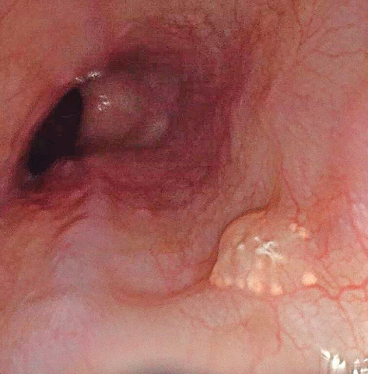 Fig. 1
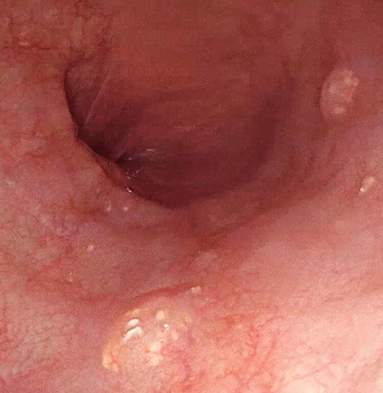 Fig. 2
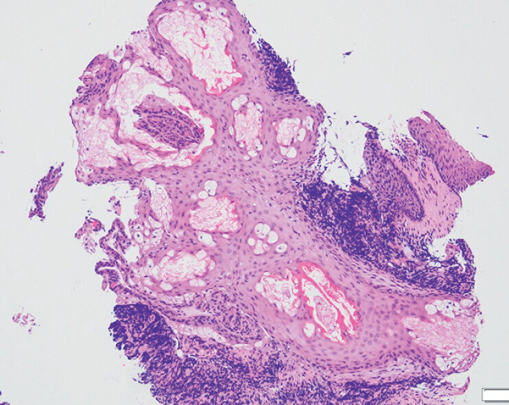 Fig. 3
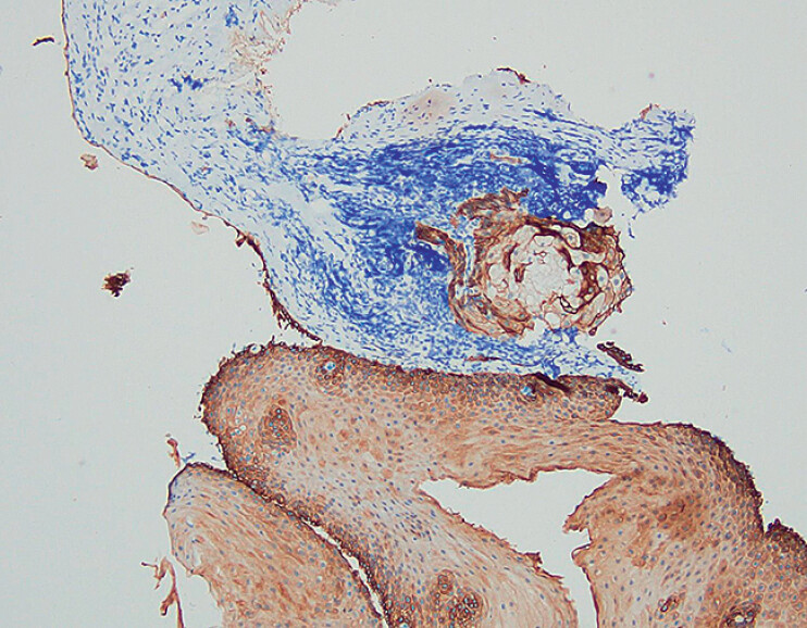 Fig. 4
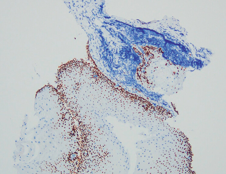 Fig. 5
Peer Reviews
No public reviews on file for this paper yet. If you reviewed it on a platform where reviews are public (OpenReview, ICLR, NeurIPS, ICML), you can paste yours below so the community can read it here.
Videos
No videos yet. Explain this paper in a talk, walkthrough, or lecture? Add one.
Taxonomy
TopicsNonmelanoma Skin Cancer Studies · Cancer and Skin Lesions · Tumors and Oncological Cases
Ectopic sebaceous glands (ESGs) of the esophagus are very rare lesions, typically discovered incidentally during endoscopic examinations 1 2 . In 1978, Ramakrishnan and Brinker reported the first case of esophageal ESGs identified via endoscopy 3 . Esophageal ESGs were found in 0.05% of asymptomatic subjects in a study involving a population undergoing gastric cancer screening 2 . Most reported patients with esophageal ESGs were either asymptomatic or presented with symptoms of gastroesophageal reflux disease (GERD 1 2 4 ). Endoscopically, esophageal ESGs may appear as yellowish patches, plaques, or elevated lesions of varying sizes 1 2 4 . Although esophageal ESGs can be found throughout the esophagus, they were most commonly located in the middle and lower thirds 2 4 . We report here a new case of esophageal ESGs that presented as multiple sessile polyps, with the diagnosis confirmed by histopathological examination.
A 44-year-old man presented to our institution with worsening symptoms of acid regurgitation following the consumption of a fatty meal and alcohol. His medical history was notable for alcoholic fatty liver disease and GERD. Upper endoscopy revealed a sessile polyp in the middle esophagus ( Video 1 and Fig. 1 ), measuring approximately 0.4 cm in length. The lesion appeared semitransparent, with multiple small whitish pellets along its border. Additional smaller sessile polyps with similar endoscopic features were identified in the lower esophagus ( Fig. 2 ). Due to the uncertain nature of the lesions, biopsy specimens were obtained. Histopathological examination revealed polygonal cells with small central nuclei and abundant clear, granular cytoplasm containing foam-like fat droplets, located within relatively normal squamous epithelium and lamina propria ( Fig. 3 ). Immunohistochemical staining showed positivity for CK ( Fig. 4 ) and p40 ( Fig. 5 ), while immunostains for mucin and CD20 were negative. These findings were consistent with a diagnosis of ESGs. The patient remained under follow-up at our institution following the upper endoscopy examination.
Endoscopic video showing a sessile polyp in the middle esophagus. The polyp appeared semitransparent, with small whitish pellets along its border. Similar sessile polyps were identified in the lower esophagus.Video 1
Endoscopic view showing a sessile polyp in the middle esophagus. The polyp appeared semitransparent, with multiple small whitish pellets along its border.
Smaller sessile polyps with similar endoscopic features were identified in the lower esophagus.
Photomicrograph showing polygonal cells with small central nuclei and abundant clear, granular cytoplasm containing foam-like fat droplets, located within relatively normal squamous epithelium and lamina propria (hematoxylin and eosin, magnification ×100).
Immunohistochemical staining revealed positivity for CK (magnification ×100).
Immunohistochemical staining revealed positivity for p40 (magnification ×100).
Endoscopy_UCTN_Code_CCL_1AB_2AC_3AH
The reference list from the paper itself. Each links out to its DOI / PubMed record.
- 1Hashimoto H Horiuchi H Miura S Clinicopathologic Characteristics of Esophageal Ectopic Sebaceous Glands: Chronological Changes and Immunohistochemical Analysis Int J Surg Pathol 20212937838410.1177/106689692095184432844680 · doi ↗ · pubmed ↗
- 2Park A Lee JH Park A Prevalence rate and clinical characteristics of esophageal ectopic sebaceous glands in asymptomatic health screen examinees Dis Esophagus 2017301510.1111/dote.1245326822541 · doi ↗ · pubmed ↗
- 3Harada A Tatsumi Y Masumoto T Ectopic sebaceous glands Gastrointest Endosc 2004609715229434 10.1016/s 0016-5107(04)01296-9 · doi ↗ · pubmed ↗
- 4Chen HF Lee HC Liao MK The clinical and endoscopic features of esophageal ectopic sebaceous glands Adv Dig Med 20207179187