Protocol to identify and sort antigen-specific B cells from Plasmodium-infected mice
Carolina Calôba, Allen M. Minns, Scott E. Lindner, Rahul Vijay

TL;DR
This paper describes a detailed protocol for isolating B cells specific to a malaria antigen in mice for advanced genetic and chromatin analysis.
Contribution
The protocol introduces a method using SpyCage reagents to target the MSP1-19 antigen in rodent malaria models for downstream single-cell sequencing.
Findings
Steps for preparing single-cell suspensions and enriching antigen-specific B cells are outlined.
Optimization of staining and sorting procedures is detailed for downstream RNA and ATAC sequencing.
The protocol facilitates isolation of live B cells for multi-omics analysis.
Abstract
Here, we detail a protocol to identify an antigen-specific B cell population using SpyCage reagents tailored to bind MSP1-19 antigen in a rodent malaria model. We describe steps for the preparation of splenic single-cell suspension, B cell enrichment, and staining to facilitate sorting of live cells for downstream applications such as single-cell RNA, V(D)J, and assay for transposase-accessible chromatin (ATAC) sequencing. For complete details on the use and execution of this protocol, please refer to Calôba et al.1 •Steps for preparing single-cell suspension and antigen-specific B cell enrichment•Instructions for optimizing staining of compensation/reference control beads•Guide to sort-purify antigen-specific B cells for downstream workflow Steps for preparing single-cell suspension and antigen-specific B cell enrichment Instructions for optimizing staining of…
Genes, proteins, chemicals, diseases, species, mutations and cell lines named across the full text — each resolved to its canonical identifier and authoritative record.
Click any figure to enlarge with its caption.
 Figure 1
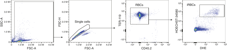 Figure 2
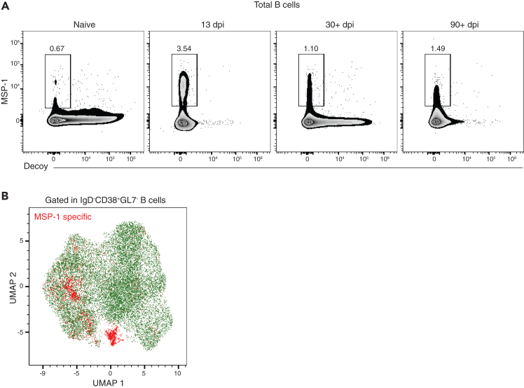 Figure 3
Peer Reviews
No public reviews on file for this paper yet. If you reviewed it on a platform where reviews are public (OpenReview, ICLR, NeurIPS, ICML), you can paste yours below so the community can read it here.
Videos
No videos yet. Explain this paper in a talk, walkthrough, or lecture? Add one.
Taxonomy
TopicsImmune Cell Function and Interaction · Monoclonal and Polyclonal Antibodies Research · Immunotherapy and Immune Responses
Before you begin
B cells play a critical role in protection against malaria by secreting antibodies specific to a wide range of Plasmodium antigens.2^,^3^,^4^,^5^,^6^,^7^,^8 The protocol below describes the specific steps for infection of mice with *Plasmodium yoelii-*17XNL Clone 1.1 (Py), spleen harvest, preparation of splenic single cell suspension, staining of MSP_1-19_-specific B cells, as well as sorting live cells. Stained cells can be either used for phenotypic characterization using flow cytometry or for transcriptomic and epigenetic analysis using single-cell sequencing techniques. While we used this methodology to characterize memory B cells from Plasmodium-infected mice, this approach can be broadly applied to identify non-Plasmodium antigens in mice, when the right combination of “SpyCage” reagents is used.
- 1.Prepare required buffers as described below.
- 2.If needed, perform a trial run of the experiment to ascertain the minimum quantity of antibodies and enrichment kits necessary, as the size of the spleen and, therefore, the number of cells will vary depending on the time point of harvest post infection.
Institutional permissions
C57BL/6 mice were purchased from The Jackson Laboratory, and Plasmodium yoelii clone 17xNL (Py) was obtained from the Malaria Research and Reference Reagent Resource center (MR4; BEI Resources, American Type Culture Collection). All experiments and procedures were approved by the Rosalind Franklin University of Medicine and Science Institutional Animal Care and Use Committee. Researchers who plan to execute this protocol must obtain approval from their institutional committees.
Innovation statement
This protocol is innovative in that it employs highly sensitive reagents to detect bona fide parasite-specific germinal center (GC) B cells and memory B cells (MBCs). These fluorescent protein–labeled SpyCage reagents are significantly brighter than conventional B cell tetramers, enabling confident identification of antigen-specific B cell populations, even when using a semi–high-throughput flow cytometry panel. Combined with a carefully optimized workflow-refined through multiple rounds of trial and error, this protocol enables high-resolution analysis of the phenotypic and molecular characteristics of antigen-specific MBCs during Plasmodium infection.
Plasmodium yoelii infection and assessment
Timing: variable
This step describes how to infect mice with Py and measure parasitemia by flow cytometry during the course of infection.9 Perform the following procedures in an ABSL2 facility.
-
3.Py preparation and infection.
-
a.Thaw Py stock stored in liquid nitrogen in a water bath at 37°C.
-
b.Dilute Py stock with sterile PBS to achieve a concentration of 5x10^6^ parasitized red blood cells (RBC) per mL.
-
c.Inject 200 μL (1x10^6^ parasitized red blood cells) of the diluted Py stock into each mouse either intravenously (i.v.) or retro-orbitally (r.o.). Note: Place the mouse under an infrared lamp (25–30 cm away) briefly (1–3 min) to facilitate vasodilation of the peripheral (tail) veins. Transfer the mouse into a mouse restrainer and sterilize the injection site (tail) using gauze soaked in 70% v/v ethanol before injection with a sterile 26-gauge syringe. Upon successful completion of the injection, slowly withdraw the needle and apply slight pressure to the injection site to prevent bleeding. Alternatively, perform r.o. injections in mice under controlled isoflurane anesthesia.
-
4.Monitor parasitemia (starting on day 5 post infection).
-
a.Aliquot 100 μL of PBS per well in a 96-well flat-bottom plate. Keep the plate on ice. The number of wells should be equal to the number of infected mice plus one extra well to be used as an unstained control.
-
b.Place a mouse into a mouse restrainer and snip the tip of the tail carefully using sharp and sterile scissors. Milk the tail gently from the base to the tip and collect blood using a 10 μL pipette set to 2 μL. Transfer the blood to the corresponding well containing 100 μL sterile PBS and mix by pipetting.
-
c.Add at least 1 μL of blood from one mouse per group to the well for the unstained control.
-
d.Add 100 μL of cold PBS and centrifuge at 200 xg for 5 minutes at 4°C.
-
e.Discard supernatant and pulse vortex the plate.
-
f.Prepare the staining mix with the following antibodies and dyes:
-
i.Hoechst-34580 (1:1000 dilution, BV570 on Cytek Aurora).
-
ii.Dyhydroethidium (DHE) (1:500 dilution, BB630 on Cytek Aurora).
-
iii.APC-conjugated anti-Ter119 (1:400).
-
iv.FITC-conjugated CD45.2 (1:200).CRITICAL: Preparation of the staining mix is done in PBS.
-
g.Add 100 μL of staining mix per well and incubate for 20 minutes at 4°C in the dark.
-
h.Wash wells using 100 μL of cold PBS and centrifuge at 200 xg for 5 minutes at 4°C.
-
i.Resuspend in 200 μL of cold PBS and acquire in a flow cytometer as soon as possible.
-
j.Data is analyzed using FlowJo software as illustrated in Figure 1.Figure 1. Assessment of parasitemia using flow cytometryGating strategy used to obtain the fraction of infected red blood cells (iRBC).
-
k.Repeat parasitemia measurements every other day to monitor the infection until the mice clear the infection or until the desired day of harvest.
Key resources table
REAGENT or RESOURCESOURCEIDENTIFIERAntibodiesAnti-mouse F4/80, PE/Cy7-conjugated (clone BM8) (1:250)BioLegendCat #: 123114; RRID:AB_893478Anti-mouse CD38, APC-conjugated (clone 90) (1:250)BioLegendCat #: 102712; RRID:AB_312932Anti-mouse IgD, Spark NIR 685-conjugated (clone 11-26c.2a)(1:500)BioLegendCat #: 405750; RRID:AB_2888693Anti-mouse CD3, APC/Fire 810-conjugated (clone 17A2) (1:200)BioLegendCat #: 100268; RRID:AB_2876392Anti-mouse B220, PerCP/Cy5.5-conjugated (clone RA3-6B2) (1:300)BioLegendCat #: 103235; RRID:AB_893354Anti-human/mouse GL7, eFluor 450-conjugated (clone GL-7)(1:300)InvitrogenCat #: 48-5902-82; RRID:AB_10870775Anti-mouse CD45.2, FITC-conjugated (clone 104) (1:200)BioLegendCat #: 109806; RRID:AB_313442Anti-mouse TER-119, APC-conjugated (clone TER-119) (1:400)BioLegendCat #: 116212; RRID:AB_313712FcBlock (CD16/32) (2 μg/sample)Prepared in the labN/AChemicals, peptides, and recombinant proteinsDihydroethidiumSigma-AldrichCat #: D70008Hoechst 34580Sigma-AldrichCat #: 63493MSP1-19 SpyCageCalôba et al.1N/APyUIS4 SpyCageCalôba et al.1N/AHEPESCorningCat #: 25-060-CIL-glutamine 200 mMGibcoCat #: 25030-081Penicillin/streptomycinGibcoCat #: 15140-122GentamicinGibcoCat #: 15710-064b-mercaptoethanolSigma-AldrichCat #: M3148-100MLKHCO_3_Fisher ChemicalCat #: P235-500NH_4_ClMallinckrodt ChemicalsCat #: 3384EDTA-Na_2_Sigma-AldrichCat #: ED2SSZombie Aqua fixable viability kitBioLegendCat #: 423102Critical commercial assaysMojoSort mouse pan B cell isolation kit IIBioLegendCat #:480088MojoSort streptavidin nanobeadsBioLegendCat #: 480016Protector RNase inhibitorSigma-AldrichCat #: 03335399001 Sigma-AldrichExperimental models: Organisms/strainsModel organism: mouse: C57BL/6 (WT), males or females, 6- to 8-weeks oldJackson LaboratoryStock: 00664Plasmodium yoelli 17XNL (clone 1.1)BEI ResourcesMRA-593Software and algorithmsSpectroFloCytek BiosciencesN/AFlowJo v.10.10.0BDhttps://www.flowjo.comBioRenderN/Ahttps://app.biorender.comOtherRPMI-1640 mediumSigma-AldrichCat #: R8758-500MLFetal bovine serum (FBS)GibcoCat #: 26140-079PBSCytivaCat #: SH30028.03
Materials and equipment
SC (Supplementum Complementum)ReagentFinal concentrationAmountRPMI-1640 MediumN/A200 mLHEPES (1 M)0.2 M100 mLL-Glutamine 200 mM40 mM100 mLPenicillin/Streptomycin2000 U/mL / 2000 μg/mL100 mLGentamicin0.2 mg/mL10 mLβ-Mercaptoethanol2 mM70 μL
- •Prepare SC stock to be used when preparing RP-1 medium.
- •Filter through 0.22 μM filter.
- •Aliquot 12.5 mL into 15 mL tubes for storage.
Note: Store SC at −20°C until use. RP-1 mediumReagentFinal concentrationAmountRPMI-1640 MediumN/A482.5 mLSC1X12.5 mL (1 aliquot)Fetal Bovine Serum1% v/v5 mL Note: Store RP-1 at 4°C for not more than a month. ACK Lysis BufferReagentFinal concentrationAmountddH_2_ON/A1 LKHCO_3_0.01 M1 gNH_4_Cl0.15 M8.29 gEDTA-Na_2_0.0001 M37.2 mg
- •Prepare ACK Lysis buffer stock to use for RBC lysis.
- •Adjust pH to 7.2–7.4 at room temperature with HCl before increasing the volume to 1 L.
- •Filter through 0.22 μM filter.
- •Aliquot 50 mL into 50 mL tubes for storage.
Note: Store ACK Lysis buffer at 25°C until use.
Step-by-step method details
Preparation of single-cell suspension from infected spleens
Timing: 1–1.5 h
This step outlines how spleens are processed into a single cell suspension, followed by red blood cell (RBC) lysis. Please note that these steps are performed on the day post infection that pertains to your experimental question. For our specific application, these steps were conducted on 38 days post infection (dpi).
- 1.Euthanize mice:
- a.Sacrifice mice using CO_2_ inhalation in a chamber until the animals become unconscious and cease to move.
- b.To confirm euthanasia, perform cervical dislocation.
- 2.Collect spleens into tubes containing 3 mL of RP-1 on ice.
- 3.Process the tissues:
- a.Transfer the spleen and RP-1 to a 10 cm petri dish containing a 180 μm wire mesh screen molded into a strainer.
- b.Using a syringe piston, push the spleens through the wire mesh strainer.
- c.Force 4 mL of fresh RP-1 through the wire mesh with the pipette gun set to high speed.
- d.Aspirate back the entire volume (7 mL) and wash the wire mesh again. Repeat this process at least 3 times.
- e.Transfer the cells into a 15 mL tube.
- f.After transferring the cells, repeat steps c to e. CRITICAL: The washes are important to guarantee a high yield of cells.
- 4.Lyse the RBCs:
- a.Centrifuge cells from step 3f at 300 xg for 5 minutes at 4°C.
- b.Dislodge the pellets by vortexing or by sliding the tubes against a tube rack.
- c.Add 1 mL of ACK lysis buffer. Mix it well using a P1000 pipette.
- d.Add an additional 2 mL of ACK lysis buffer and mix well.Note: The volume of ACK lysis buffer depends on the size of the spleen. For instance, for larger spleens from 15 dpi (Py) we usually add a total of 4 mL of ACK lysis buffer, while smaller spleens from a memory time point (post 35 dpi) will require 3 mL of ACK lysis buffer for optimal RBC lysis.
- e.Incubate for 2–3 minutes at 25°C.CRITICAL: Longer incubation times at 25°C may result in loss of immune cells. Incubation on ice may require longer times, which may also result in loss of immune cells.
- f.Add fresh RP-1 to a final volume of 14 mL to neutralize the ACK lysis buffer.
- g.Centrifuge cells at 300 xg for 5 minutes at 4°C.
- h.Resuspend in 5 mL of RP-1 and filter the cells through a 70-micron filter mesh into a new 15 mL tube. Keep the tubes on ice.
- i.Take a 10 μL aliquot and count the cells.
B cell enrichment and live/dead staining
Timing: 1–1.5 h
This step describes the steps involved in enriching B cells through negative selection using magnetic beads and performing live/dead stain before staining for antigen-specific B cells using SpyCage reagents. We followed the manufacturer’s protocol as outlined below.
- 5.Prepare cells for enrichment using MojoSort Pan B cell isolation kit.
- a.Centrifuge the single cell suspension from step 4 h at 300 xg for 5 minutes at 4°C.
- b.Discard supernatant and resuspend cells in RP-1 to a final concentration of 1x10^8^ cells/mL and transfer to a 5 mL polystyrene (FACS) tube.
- c.Add 100 μL of antibody mix/mL and mix with a P1000 pipette.
- d.Incubate for 15 minutes on ice.
- e.Add 100 μL of nanobeads/mL and mix with a P1000 pipette.
- f.Incubate for 15 minutes on ice.
- g.Add RP-1 to a final volume of 2.5 mL, mix with a P1000 pipette, and place the cells in the MojoSort magnet for 5 minutes.
- h.Without removing the tube from the magnet, hold the magnet and swiftly pour the liquid into a new 15 mL tube.CRITICAL: The transferred liquid is the enriched fraction; do not discard.
- i.Remove the tube from the magnet and add an additional 2.5 mL of fresh RP-1.
- j.Resuspend the beads and place the tube in the magnet for 5 minutes.
- k.Without removing the tube from the magnet, pour the liquid into the 15 mL tube (from step 5h) already containing 2.5 mL and discard the beads. Keep on ice.
- l.Take an aliquot of cells to use as the unstained reference control.
- m.Take a 10 μL aliquot and count the cells.Note: Other Pan B cell isolation kits should work besides the MojoSort Pan B cell enrichment kit.CRITICAL: If using any other B cell enrichment kits, be sure to use negative selection kits.
- n.Add ice-cold PBS to the tube from step 5k to a final volume of 15 mL.
- o.Centrifuge the cells at 300 xg for 5 minutes at 4°C.
- p.Resuspend cells in 15 mL of PBS and repeat step 5n.CRITICAL: Washing steps with PBS before proceeding to the next step (Live/Dead staining) are critical since the presence of serum will result in suboptimal Live/Dead staining.
- 6.Stain cells with Live/Dead staining.
- a.Resuspend cells in 1 mL of diluted Live/Dead dye.Note: Live/Dead dye (Zombie Aqua Fixable Viability Kit) should be diluted at 1:1000 in PBS.CRITICAL: Use PBS to dilute live/dead dye. Do not use media containing serum.
- b.Incubate for 20 minutes at 25°C in the dark.
- c.Add fresh RP-1media to a final volume of 15 mL to neutralize Live/Dead stain.
SpyCage staining and enrichment
Timing: 2.5–3 h
B cells are stained with SpyCage reagents along with other surface markers and the SpyCage^+^ cells are enriched through positive selection using magnetic beads.Note: The SpyCage reagent has been used in our prior studies to nanoparticle vaccine approaches in the context of SARS-CoV-2.10 SpyCage consists of the fusion of the SpyCatcher domain to the N-terminus of the engineered I3-01 protein, with two 6xHis tags present for protein purification.11^,^12 We created two variants of the SpyCage reagents for this study by appending additional sequences to the C-terminus of I3-01, either with mScarlet to create “RedCage” – or with mNeonGreen to create “GreenCage”. In this study, the RedCage was used as the antigen-specific experimental reagent by covalently loading it with 60 copies of the targeted immunodominant Py antigen MSP_1-19_, capable of detecting MSP_1-19_ specific B cells. The GreenCage was used as a decoy control and was covalently loaded with 60 copies of the liver-stage antigen PyUIS4, which is not expressed in the blood stage. The PyMSP_1-19_ (AA1619-1754) and PyUIS4 (AA80-224) antigens were expressed in E. coli as GST-fusion proteins bearing a C-terminal SpyTag to enable covalent bonding to SpyCage.13 The SpyCage scaffold is designed to be versatile and to accommodate a wide range of antigens for display. Antigens are produced separately, and loaded onto SpyCage simply by mixing in a compatible buffer (e.g. 1x PBS) and temperature (e.g. 4^o^C–37^o^C). Unbound antigens were purified away via standard column chromatography and/or dialysis approaches.
- 7.Stain cells with SpyCage reagent.
- a.Centrifuge cells (from step 6c) at 300 xg for 5 minutes at 4°C.
- b.Discard the supernatant and resuspend cells in 1 mL of RP-1 and add 2 μg of FcBlock (CD16/32) and 1.25 μg of the decoy “GreenCage” conjugated to PyUIS4 antigen. Pulse vortex the tubes 5 times gently.
- c.Incubate for 10 minutes at 25^o^C in the dark.
- d.Add 1.25 μg of the RedCage conjugated to PyMSP1(19) and gently vortex the tubes.
- e.Incubate for 30 minutes at 4^o^C in the dark.
- f.Add fresh RP-1 media to a final volume of 15 mL.
- g.Centrifuge at 300 xg for 5 minutes at 4^o^C.
- h.Discard the supernatant and resuspend cells in 2 mL of RP-1.
- i.Add 1 μg of biotinylated anti-scaffold antibody and gently vortex the tubes.
- j.Incubate for 30 minutes at 4^o^C in the dark.
- k.Add fresh RP-1 media to a final volume of 15 mL.
- 8.Enrich cells bound to the SpyCage scaffold.
- a.Centrifuge cells at 300 xg for 5 minutes at 4^o^C.
- b.Discard the supernatant and resuspend cells at 1x10^8^ cells/mL and add 100 μL of MojoSort streptavidin nanobeads/mL.
- c.Incubate for 15 minutes at 4^o^C in the dark.
- d.Add fresh RP-1 media to a final volume of 15mL.
- e.Centrifuge at 300 xg for 5 minutes at 4^o^C.
- f.Discard the supernatant and resuspend cells in 2.5 mL of RP-1 and transfer to a polystyrene (FACS) tube.
- g.Place the tube in the magnet and incubate for 5 minutes.
- h.Without removing the tube from the magnet, hold the magnet and swiftly pour the unlabeled fraction into another FACS tube.
- i.Remove the tube from the magnet and add 1 mL of RP1 into the tube to prevent cells from drying and keep it on ice. This is ‘labeled fraction 1’.Note: The labeled fraction 1 is enriched for SpyCage^+^ cells.
- j.Place the FACS tube from step 8h containing the unlabeled cells back in the magnet and incubate for 5 minutes.
- k.Without removing the tube from the magnet, hold the magnet and swiftly pour and discard the unlabeled fraction.
- l.Remove the tube from the magnet and add 1 mL of RP1 into the tube to prevent cells from drying, and combine with ‘labeled fraction 1’ from step 8i to a final volume of 2 mL.
Sorting of SpyCage+ B cells
Timing: 3–5 h
Enriched SpyCage^+^ B cells are stained with other relevant surface markers to sort-purify your population of interest.
- 9.Stain B cells for sorting.
- a.Centrifuge cells from step 8l at 300 xg for 5 minutes at 4^o^C.
- b.Resuspend in 500 μL of antibody cocktail mix. The antibody cocktail will contain the following antibodies resuspended in RP-1:
- i.B220 PerCP/Cy5.5 (1:300).
- ii.IgD Spark NIR (1:800).
- iii.GL7 eF450 (1:300).
- iv.CD38 APC (1:250).
- v.CD3 APC/Fire 810 (1:200).
- vi.F4/80 PE/Cy7 (1:250).Note: One may add more markers depending on the populations of interest.CRITICAL: Remember not to add any markers tagged with Spark YG593 or FITC or any fluorophores whose emission spectra peaks at YG2 or B2 on a Cytek instrument. These channels have already been used for the two SpyCage scaffolds. If using a different instrument, be sure to identify the respective channels.Optional: If one of the downstream applications involves CITE-seq, CITE-seq antibodies should be added during this step at 1:1000 dilution or as recommended by the manufacturer.
- c.Incubate for 30 minutes at 4^o^C in the dark.
- d.Add 3 mL of RP-1 to wash.
- e.Centrifuge at 300 xg for 5 minutes at 4^o^C.Note: If transcriptomic analysis is one of the downstream applications, add Protector RNase Inhibitor to fresh RP-1 media at this point. This media will be used to resuspend the cells for sorting and should contain 1 U/mL of Protector RNase Inhibitor. The media in the collection tubes should contain 0.5 U/mL of Protector RNase Inhibitor. For other applications, fresh RP-1 media will be sufficient.
- f.Discard the supernatant and resuspend cells at 1x10^7^ cells/mL of the appropriate media. Keep on ice.
- g.Add 500 μL of the appropriate media to the collection tubes. Vortex the tubes to coat their walls. Keep on ice.
- h.Proceed to cell sorting.Note: It is ideal to use a sorting nozzle with a pore size greater than 70 μm. In this protocol, we used a 100 μm nozzle. It is also important to gate on CD3^−^F4/80^−^ cells before proceeding with B cell gates.CRITICAL: While preparing single stained (compensation or reference) controls to be used in the cell sorter, use compensation beads such as UltraComp eBeads (Thermo Fisher) or similar. It is also critical that when beads are prepared with SpyCage reagents, first add biotinylated anti-aldolase antibody and then the respective SpyCage scaffolds. For all other markers in the staining panels, stain as usual.
- i.Proceed to the downstream workflow as desired.
Expected outcomes
This protocol enriches and stains for MSP_1-19_-specific B cells, which can be analyzed by flow cytometry or flow sorted and used for other downstream applications such as sequencing. We have also used this protocol in combination with transcription factor staining. A successful SpyCage staining will show a clear single positive MSP-1 population (Figure 2A). As expected, MSP_1-19_-specific B cells will be higher in frequency during the effector phase of infection and will contract during the memory time point. It is important to note that the frequency of MSP_1-19_-specific B cells is usually low (below 5%) even after the enrichment step. Clustering of MSP_1-19_-specific memory B cells can clearly detect the stained population among the other B cell populations, as shown in Figure 2B. Please refer to Calôba et al. for a more detailed high-dimensional analysis of MSP_1-19_-specific memory B cells.Figure 2MSP-1 staining(A) Representative plots of total B cells stained using SpyCage reagents on different time points of Py infection.(B) UMAP of memory B cells stained with SpyCage reagents and analyzed by flow cytometry on 38 dpi.
Quantification and statistical analysis
FlowJo software (v.10.10.0, BD) was used for analysis of flow cytometry data.
Limitations
Given the low relative frequency of MSP_1-19_-specific B cells, the success of this protocol critically depends on the efficient isolation of SpyCage-specific B cells using magnetic beads. It is important to note that, even after enrichment, the isolated cells will still contain other B cell subsets. However, SpyCage-specific B cells will be substantially enriched, allowing for their visualization and, if needed, further purification by cell sorting. For downstream applications such as sequencing, samples can be pooled from at least 3 mice to achieve the amount of cells needed.
This protocol has been validated only in mice infected with Plasmodium yoelii. Although there may be sequence similarities in the MSP_1-19_ antigen across different Plasmodium species,14 we have not yet assessed the reliability of this antigen for identifying and isolating antigen-specific B cells in mice infected with other strains.
Troubleshooting
Problem 1
Unable to visualize/identify MSP_1-19_ specific B cells.
Potential solution
The above-mentioned issue may arise due to suboptimal reference/compensation bead staining (step 9). As mentioned elsewhere in the protocol, staining with individual SpyCage reagents is a two-step process. The first step uses a fluorescently labeled scaffold, followed by the use of an antibody that is specific to the scaffold. Thus, when staining beads for reference/compensation control to be used on a flow cytometer, reverse the staining sequence - stain with the anti-aldolase antibody first, followed by the aldolase scaffold. This order is important because the beads will be unable to bind directly to the fluorescently labeled scaffold without being bound to the anti-aldolase antibody, leading to a complete absence of positively stained population.
Problem 2
Very few MSP_1-19_ specific B cells are detected.
Potential solution
Suboptimal RBC lysis (step 4). Depending on the time point of infection, one may have to add more ACK lysis buffer (up to 5 ml) and incubate for longer periods (up to 4 minutes) to get effective lysis of RBCs. Please be sure to incubate cells with RBC lysis buffer (ACK lysis buffer) at room temperature.
Problem 3
Dimmer fluorescence signal from SpyCage reagents.
Potential solution
Adjusting the autofluorescence extraction threshold prior to unmixing and identifying/sorting cells of interest (in a spectral flow cytometer).
Problem 4
Lower quality of RNA or cDNA library.
Potential solution
Ensuring that RNase inhibitor (step 9) added to the cell suspension is not expired or denatured.
Problem 5
MSP-1^+^ cells do not show a bimodal staining pattern but rather shows a spread.
Potential solution
This can be minimized by adding recommended amount of Fc block (step 7) as well as ensuring that F4/80 (step 9h) is used a dump gate before proceeding with B cell gates.
Resource availability
Lead contact
Requests for further information should be directed to and will be fulfilled by the lead contact, Rahul Vijay, [email protected].
Technical contact
Requests for further information should be directed to corresponding authors Scott E. Lindner ([email protected]) and Rahul Vijay ([email protected]).
Materials availability
Please contact Scott E. Lindner ([email protected]) for inquiries related to SpyCage, PyMSP1(19), and PyUIS4 proteins used in this study.
Data and code availability
This paper did not generate any new datasets or code.
Acknowledgments
This work was supported by 1R01GM125907 to S.E.L., the RFUMS startup funds, Schweppe Scholar Award 1R01AI182217 to R.V., and the AAI 2024 Careers in Immunology Fellowship to C.C. and R.V.
Author contributions
R.V. conceived the study. C.C. and R.V. designed, executed, and analyzed experiments and wrote the manuscript. S.E.L. conceived the SpyCage scaffolds. A.M.M. and S.E.L. generated and provided SpyCage, PyMSP1(19), and PyUIS4 proteins.
Declaration of interests
We wish to disclose the following two intellectual property claims related to this protocol: Lindner, SE & Hafenstein, S. US Patent Application 16/494,502 “Versatile Display of Proteins” and Lindner, SE., Hafenstein, S., & Butler, N. PCT/US2020/033785 “Specific Selection of Immune Cells Using Versatile Displace Scaffolds.”
The reference list from the paper itself. Each links out to its DOI / PubMed record.
- 1Caloba C.Sturtz A.J.Lyons T.A.John L.Ramachandran A.Minns A.M.Cannon A.M.Whalley J.P.Watts T.H.Kaplan M.H.Systemic 4-1BB stimulation augments extrafollicular memory B cell formation and recall responses during Plasmodium infection Cell Rep.44202511552810.1016/j.celrep.2025.115528 PMC 1207978340215168 · doi ↗ · pubmed ↗
- 2Cohen S.Mc GREGORI.A.Carrington S.Gamma-globulin and acquired immunity to human malaria Nature 192196173373710.1038/192733 a 013880318 · doi ↗ · pubmed ↗
- 3Julien J.P.Wardemann H.Antibodies against Plasmodium falciparum malaria at the molecular level Nat. Rev. Immunol.19201976177510.1038/s 41577-019-0209-531462718 · doi ↗ · pubmed ↗
- 4Blackman M.J.Heidrich H.G.Donachie S.Mc Bride J.S.Holder A.A.A single fragment of a malaria merozoite surface protein remains on the parasite during red cell invasion and is the target of invasion-inhibiting antibodies J. Exp. Med.172199037938210.1084/jem.172.1.3791694225 PMC 2188181 · doi ↗ · pubmed ↗
- 5Bouharoun-Tayoun H.Oeuvray C.Lunel F.Druilhe P.Mechanisms underlying the monocyte-mediated antibody-dependent killing of Plasmodium falciparum asexual blood stages J. Exp. Med.182199540941810.1084/jem.182.2.4097629503 PMC 2192140 · doi ↗ · pubmed ↗
- 6Bull P.C.Lowe B.S.Kortok M.Molyneux C.S.Newbold C.I.Marsh K.Parasite antigens on the infected red cell surface are targets for naturally acquired immunity to malaria Nat. Med.4199835836010.1038/nm 0398-3589500614 PMC 3836255 · doi ↗ · pubmed ↗
- 7Jafarshad A.Dziegiel M.H.Lundquist R.Nielsen L.K.Singh S.Druilhe P.L.A novel antibody-dependent cellular cytotoxicity mechanism involved in defense against malaria requires costimulation of monocytes Fcgamma RII and Fcgamma RIIIJ. Immunol.17820073099310610.4049/jimmunol.178.5.309917312157 · doi ↗ · pubmed ↗
- 8Mc Intosh R.S.Shi J.Jennings R.M.Chappel J.C.de Koning-Ward T.F.Smith T.Green J.van Egmond M.Leusen J.H.W.Lazarou M.The importance of human Fcgamma RI in mediating protection to malaria P Lo S Pathog.32007 e 7210.1371/journal.ppat.0030072 PMC 186895417511516 · doi ↗ · pubmed ↗