Long-read sequencing unmasks a cryptic three-way translocation resulting in an ETV6::PDGFRB fusion
Joseph Tripodi, Douglas Tremblay, Daiva Ahire, Vesna Najfeld

TL;DR
Long-read sequencing identified a complex three-way translocation causing a gene fusion in a rare blood cancer, enabling accurate diagnosis and effective treatment.
Contribution
Demonstrates the power of long-read sequencing to resolve cryptic genomic rearrangements in PDGFRB-rearranged neoplasms.
Findings
Long-read sequencing revealed a three-way translocation involving chromosomes 5, 12, and 20.
The ETV6::PDGFRB fusion was precisely characterized with base pair resolution.
The patient responded well to imatinib therapy after accurate diagnosis.
Abstract
Myeloid/Lymphoid Neoplasms (MLN) with eosinophilia and PDGFRB rearrangements are rare but distinct hematologic malignancies driven by the constitutive activation of the PDGFRB tyrosine kinase through gene fusions. These neoplasms are sensitive to tyrosine kinase inhibitors (TKIs) such as imatinib, which often leads to rapid and durable molecular remissions. However, diagnostic challenges frequently arise from cryptic rearrangements, necessitating comprehensive molecular approaches. A 37-year-old male patient initially presented with pancytopenia and a splenic infarct; subsequent bone marrow findings were suggestive of a myeloid/lymphoid neoplasm. Initial conventional cytogenetic analysis and fluorescence in situ hybridization (FISH) identified a PDGFRB gene rearrangement but were unable to fully resolve the structural complexity of the underlying genomic alteration. Long-read…
Genes, proteins, chemicals, diseases, species, mutations and cell lines named across the full text — each resolved to its canonical identifier and authoritative record.
Click any figure to enlarge with its caption.
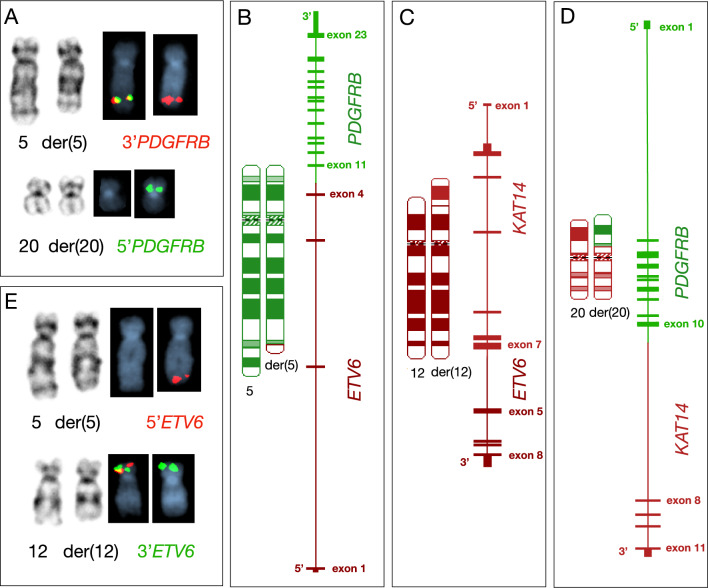 Figure 1
Peer Reviews
No public reviews on file for this paper yet. If you reviewed it on a platform where reviews are public (OpenReview, ICLR, NeurIPS, ICML), you can paste yours below so the community can read it here.
Videos
No videos yet. Explain this paper in a talk, walkthrough, or lecture? Add one.
Taxonomy
TopicsEosinophilic Disorders and Syndromes · Chronic Myeloid Leukemia Treatments · Cardiac tumors and thrombi
Background
Myeloid/lymphoid neoplasms (MLN) with eosinophilia and tyrosine kinase gene fusions are hematologic malignancies driven by rearrangements which lead to the kinase domain being constitutively activated, thereby resulting in cell signaling dysregulation that promotes proliferation and survival [1]. These neoplasms are characterized by the expression of fusion genes involving specific receptor tyrosine kinases, primarily PDGFRA, PDGFRB, or FGFR1. MLN with eosinophilia and PDGFRB rearrangements involve gene fusions with up to 40 known partner genes, with the most common of these rearrangements being ETV6::PDGFRB fusion resulting from a t(5;12)(q32;p13) translocation [2–4]. The ETV6*::*PDGFRB oncoprotein leads to ligand-independent dimerization and activation of the PDGFRB kinase domain, promoting uncontrolled cell proliferation and survival through downstream signaling pathways such as RAS/MAPK and PI3K/AKT [5]. Clinically, these neoplasms present frequently with marked peripheral eosinophilia and may manifest with myeloproliferative features, lymphoid involvement, and/or progression to acute leukemia. Unlike other eosinophilic disorders, PDGFRB-rearranged neoplasms are highly sensitive to tyrosine kinase inhibitors (TKIs) such as imatinib, often achieving rapid and durable molecular remissions [6]. However, due to the diversity of fusion partners and breakpoint locations, comprehensive molecular diagnostics—via karyotyping, fluorescence in situ hybridization (FISH), or RNA sequencing—are critical for accurate detection and therapeutic decision-making [7]. We report a case in which conventional cytogenetics and FISH studies detected a PDGFRB gene rearrangement, but long-read sequencing revealed its cryptic complexity, uncovering a three-way translocation that included the KAT14 gene on chromosome 20p as a partner gene. This case underscores the complementary role of long-read sequencing in surpassing the resolution of standard cytogenetic and FISH methods, enabling precise and comprehensive characterization of clinically significant rearrangements.
Case presentation
In March 2025, a 37-year-old male patient presented for an initial evaluation to our clinic for pancytopenia discovered in the setting of a recent emergency department admission for splenic infarct. Five years prior, he had abnormal pre-operative blood work for knee arthroscopy and was referred to a hematologist but did not follow up. Upon arrival at our emergency department, the patient was experiencing several episodes of abdominal pain and was diagnosed with a splenic infarct with splenomegaly (measuring 15.6 cm in coronal oblique dimension) and hepatomegaly (measuring 16.8 cm in the craniocaudal dimension). Complete blood tests at that time showed a white blood cell count of 21.6 × 10^9^/L with 37.9% neutrophils, 21.0% lymphocytes, 10.9% monocytes, 10.1% myelocytes, 8.4% band cells, 5.0% eosinophils, 3.4% promyelocytes, 1.7% metamyelocytes, and 0.8% basophils. The hemoglobin was 9.5 g/dL and platelet count was 22 × 10^9^/L. Spleen was palpable to 8 cm below the left costal margin. Upon further review of systems, he reported a nearly 20-pound unintentional weight loss over the last 8 months accompanied by early satiety and bloating as well as nightly drenching night sweats. A bone marrow biopsy was performed and demonstrated a hypercellular marrow (> 95%), trilineage hematopoiesis, myeloid hyperplasia with eosinophilia, and hypolobated atypical megakaryocytes. CD34 + blasts involved less than 2% of marrow cellularity and MF-2 + bone marrow fibrosis. A next-generation sequencing panel targeting 433 genes associated with hematologic malignancies was performed and did not identify any pathogenic mutations.
Due to a limited bone marrow aspirate as a result of marrow fibrosis, a peripheral blood (PB) sample was submitted for cytogenomic evaluation due to suspicion of a myeloid/lymphoid neoplasm and tyrosine kinase gene fusions. FISH analysis was performed using break-apart probes for PDGFRA, FGFR1, and PDGFRB (Abbott Molecular, Des Plaines, IL, USA) to assess for gene rearrangements. A PDGFRB rearrangement was detected in 84% of interphase nuclei, and was reported the following day. Two days later, conventional karyotyping revealed an apparently balanced translocation between chromosomes 5q and 20p, described as 46,XY,t(5;20)(q32;p11.2) in all 20 evaluated metaphases. Metaphase FISH confirmed this finding, demonstrating the 5’ region of PDGFRB translocated to the derivative chromosome 20p, while the 3’ region remained on the derivative chromosome 5, consistent with a balanced translocation (Fig. 1A).Fig. 1. Cytogenetic and Molecular Characterization of a Complex Three-Way Translocation Involving ETV6, PDGFRB, and KAT14. A Gene mapping using a PDGFRB break-apart FISH probe, with the 3′ region labeled in red and the 5′ region labeled in green, demonstrates a translocation involving chromosomes 5 and 20. B–D Chromosome ideograms illustrating a complex three-way translocation resulting in fusion genes: ETV6::PDGFRB, KAT14::ETV6, and PDGFRB::KAT14, located on derivative chromosomes 5, 12, and 20, respectively. E Gene mapping using an ETV6 break-apart probe, with the 5′ region labeled in red and the 3′ region in green, shows a translocation involving chromosomes 5 and 12
To further characterize this rearrangement and identify the partner gene on chromosome 20p, we performed long-read sequencing using Oxford Nanopore technology. Nanopore long-read sequencing works by passing single-stranded DNA or RNA molecules through a nanopore embedded in a membrane. As each nucleotide passes through the pore, changes in the electrical current are measured. These current disruptions are then decoded in real-time to determine the nucleotide sequence, yielding long, continuous reads without the need for prior amplification that span large genomic regions, including breakpoints. Whole-genome sequencing was performed achieving an average read depth of 40x. Structural variant analysis was conducted using Sniffles2, and final results were available within 72 h of sequencing initiation. While FISH helped guide the analysis toward PDGFRB, the complex three-way translocation involving chromosomes 5, 12, and 20—affecting the PDGFRB, ETV6, and KAT14 genes, respectively—could only be fully resolved by this novel platform. As shown in Fig. 1B-D, long read sequencing identified precise breakpoints within PDGFRB intron 10, ETV6 intron 4, and KAT14 intron 7. These breakpoints resulted in the formation of an ETV6::PDGFRB fusion gene on derivative chromosome 5, a KAT14::ETV6 fusion on derivative chromosome 12, and a PDGFRB::KAT14 fusion on derivative chromosome 20. Subsequent metaphase FISH was performed using an ETV6 break-apart probe (Abbott Molecular, Des Plaines, IL, USA) and showed the 5’ region of ETV6 on the derivative chromosome 5 and the 3’ region on the derivative chromosome 12, confirming the presence of an ETV6::PDGFRB fusion gene resulting from the complex rearrangement (Fig. 1E). The final karyotype was revised as seq[GRCh38] t(5;20;12)(q32;p11.2;p13) NC_000005.10:g.150129216_qterdelins[NC_000012.12:g.11856659_pter] NC_000012.12:g.11856659_pterdelins[NC_000020.11:g. 18165932_per] NC_000020.11:g.18165932_pterdelin[NC_000005.10:g.150129216_qter]. Shortly after diagnosis, he was started on imatinib 400mg daily and has attained clinical and hematological improvement.
Discussion and conclusion
Recently, Nanopore long-read sequencing (Oxford Nanopore Technologies, Oxford, United Kingdom) has emerged as a powerful tool for comprehensive genomic characterization of hematologic malignancies which enables real-time analysis within a single assay, providing meaningful results in as little as 15 min [8]. Long-read sequencing provides a major advancement in the detection of large chromosomal abnormalities and structural variants (SVs). Unlike short-read platforms, which often struggle to resolve SVs in repetitive or breakpoint-rich regions, long-read technologies generate extended, contiguous reads that span entire variants—including complex rearrangements, large insertions or deletions, and translocations. While base-calling accuracy for single nucleotide variants (SNVs) remains somewhat limited compared to short-read approaches, the ability of long-read sequencing to accurately resolve complex or cryptic structural alterations represents a significant advantage over conventional cytogenetic and sequencing methods. Moreover, long-read sequencing does not rely on prior knowledge of specific fusion events, enabling the unbiased identification of novel gene fusions that may be missed by targeted methods.
Here we report a cryptic ETV6::PDGFRB rearrangement that was identified using long-read sequencing. Although initial conventional cytogenetic and FISH analyses suggested a t(5;20) translocation involving PDGFRB and a novel partner gene on chromosome 20, it failed to fully resolve the structural complexity of the rearrangement. Long-read sequencing revealed a cryptic three-way translocation—t(5;20;12) leading to an ETV6::PDGFRB gene rearrangement. To our knowledge, only four other complex chromosomal rearrangements involving PDGFRB have been reported in the literature: [1] t(1;5;11), resulting in a GPIAP1::PDGFRB fusion; [2] t(4;5;5)(q23;q31;q33), involving a PRKG2::PDGFRB fusion; [3] t(1;12;5;12)(p36;p13;q33;q24), generating an ETV6::PDGFRB fusion; and [4] t(5;7;7)(q33.2;q32;q11.2), involving PDGFRB with an unidentified partner gene [9–11]. None of these cases utilized long-read sequencing; instead, these studies relied on multiple complementary methodologies—such as cytogenetics, FISH, and other molecular techniques—to infer the complexity of the rearrangements. While three of the four cases demonstrated clinical response to imatinib therapy and were initially diagnosed with a chronic myeloid neoplasm, one patient [t(5;7;7)] was diagnosed with AML and was treated with HiDAC consolidation chemotherapy along with imatinib until successful allogeneic stem cell transplant [10].
While conventional cytogenetics and FISH confirmed a PDGFRB gene rearrangement—enabling prompt initiation of TKI therapy—the presence of an unusual three-way translocation and unresolved partner chromosome warranted further investigation for several reasons. First, identifying the precise fusion partner can yield important insights into disease biology and potential mechanisms of treatment resistance, particularly when the partner gene is atypical or novel [12, 13]. Second, characterizing complex rearrangements contributes to the broader understanding of structural heterogeneity in PDGFRB-rearranged neoplasms. Third, recent studies have highlighted cases of “chromosomal mimicry,” in which chromosome morphology and FISH patterns appear consistent with known structural variants, yet the expected gene-level fusion is absent on molecular analysis [14]. This underscores the importance of definitive molecular confirmation in select cases.
In conclusion, long-read sequencing represents a powerful complementary technology to existing cytogenomic and RNA-based sequencing methods. Its ability to resolve complex structural rearrangements, including cryptic and multi-chromosomal fusions, makes it particularly valuable in genomically challenging cases.