Emerging Antifungal Resistance in Dermatophytosis: A Clinicomycological Study From North India
Anushka Lohariwala, Sanjeev Gupta, Narinder Kaur, Aneet Mahendra

TL;DR
This study from North India finds rising antifungal resistance in skin fungal infections, with common drugs like fluconazole and terbinafine showing reduced effectiveness.
Contribution
The study provides new clinicomycological data on antifungal resistance patterns in dermatophytosis in a North Indian population.
Findings
Trichophyton mentagrophytes was the most common fungal species isolated.
High resistance rates were observed for fluconazole (85.6%) and terbinafine (60%).
Recurrent infections were common despite mild severity scores.
Abstract
Introduction Dermatophytosis is a common superficial fungal infection affecting the skin, hair, and nails and is increasingly being recognized as a public health challenge. In recent years, there has been a noticeable increase in chronic and recurrent cases, possibly due to changing patterns of antifungal resistance, misuse of medications, and host-related factors. This study aimed to assess the demographic and clinical characteristics of patients with dermatophytosis, evaluate treatment history and recurrence, and analyze antifungal resistance patterns. Material and methods A total of 130 patients clinically diagnosed with dermatophytosis and attending the Dermatology Outpatient Department were included in this observational study. After obtaining informed consent, detailed data on demographic factors, clinical presentation using the Dermatophytosis Severity Score (DSS), treatment…
Genes, proteins, chemicals, diseases, species, mutations and cell lines named across the full text — each resolved to its canonical identifier and authoritative record.
Click any figure to enlarge with its caption.
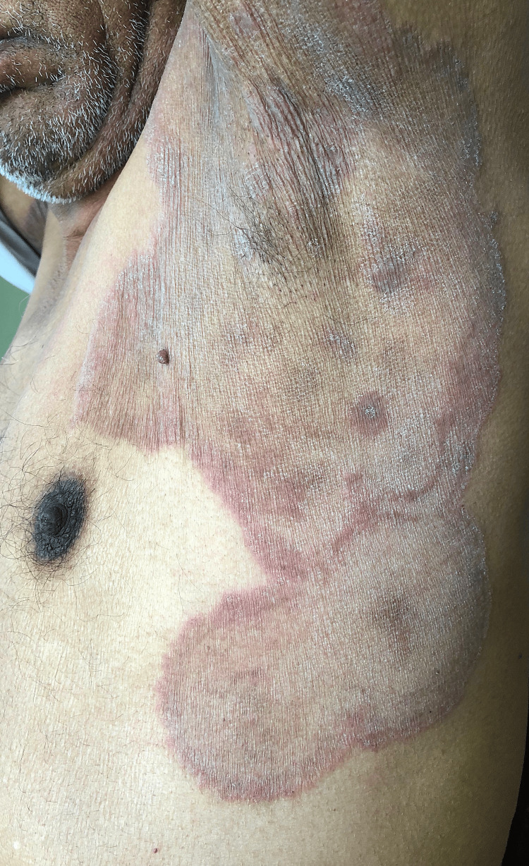 Figure 1
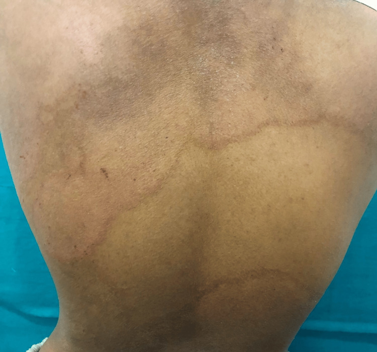 Figure 2
| Variable | Domain | Frequency | Percentage |
| Age | 15-30 years | 27 | 30.0% |
| 31-45 years | 38 | 42.2% | |
| 46-60 years | 18 | 20.0% | |
| 61-75 years | 7 | 7.8% | |
| Gender distribution | Males | 52 | 57.8% |
| Females | 38 | 42.2% | |
| Marital status | Married | 69 | 76.7% |
| Unmarried | 21 | 23.3% | |
| Locality | Urban | 18 | 20.0% |
| Rural | 72 | 80.0% | |
| Total | 90 | 100.0% |
| Diagnostic methods | Frequency | Percent | |
| KOH mount | Positive | 115 | 88.5% |
| Negative | 15 | 11.5% | |
| Sabouraud dextrose agar culture media of KOH mount positive cases | T. mentagrophytes | 87 | 75.7% |
| T. rubrum | 3 | 2.6% | |
|
| 5 | 4.3% | |
| No growth | 20 | 17.4% | |
| Total | 115 | 100.0% | |
| Drug | Frequency | Percent | |
| Fluconazole | Resistant | 77 | 85.6% |
| Sensitive | 13 | 14.4% | |
| Terbinafine | Resistant | 54 | 60.0% |
| Sensitive | 36 | 40.0% | |
| Itraconazole | Resistant | 0 | 0.0% |
| Sensitive | 90 | 100.0% | |
| Ketoconazole | Resistant | 0 | 0.0% |
| Sensitive | 90 | 100.0% | |
| Griseofulvin | Resistant | 0 | 0.0% |
| Sensitive | 90 | 100.0% | |
| Total | 90 | 100.0% | |
Peer Reviews
No public reviews on file for this paper yet. If you reviewed it on a platform where reviews are public (OpenReview, ICLR, NeurIPS, ICML), you can paste yours below so the community can read it here.
Videos
No videos yet. Explain this paper in a talk, walkthrough, or lecture? Add one.
Taxonomy
TopicsNail Diseases and Treatments · Plant Pathogens and Fungal Diseases · Fungal Biology and Applications
Introduction
Dermatophytosis refers to superficial fungal infections of keratinized tissues - skin, hair, and nails - caused by dermatophytes belonging to the genera Trichophyton, Epidermophyton, and Microsporum [1]. These fungi are classified into anthropophilic, zoophilic, and geophilic species based on their ecological niche. Clinical manifestations, including pruritus, erythema, and scaling, often cause significant physical discomfort and psychosocial stress [1]. Chronic dermatophytosis refers to infection persisting for more than six months to one year despite adequate treatment. Recurrent dermatophytosis is characterized by reoccurrence of lesions within a few weeks (usually less than six) after completion of treatment. Recalcitrant dermatophytosis refers to lack of clinical cure even after taking proper treatment including proper systemic antifungals for the recommended duration.
In India, the prevalence of dermatophytosis has risen sharply, accompanied by an alarming increase in chronic, recurrent, and treatment-resistant cases. Host-related risk factors such as diabetes mellitus, occlusive clothing, poor hygiene, and immunosuppression contribute to infection susceptibility. Host defense against dermatophytosis includes the skin’s physical and chemical barriers, ultraviolet exposure, and innate immunity involving keratinocytes, dendritic cells, and neutrophils. Pattern recognition receptors such as toll-like receptors (TLRs) and C-type lectin receptors (CLRs) detect fungal pattern recognition receptors (PAMPs), triggering cytokines (interleukin [IL]-6, IL-10, IL-17, TNF-α) and adaptive responses [2]. Cell-mediated immunity, especially Th1 and Th17, plays a central role in fungal clearance through IFN-γ and IL-17 [3]. In contrast, Th2 responses (IL-4, IL-5, IL-13) promote chronicity via IgE and IgG4 [4]. IL-10 suppresses Th1 but regulates immunity. Acute dermatophytosis involves delayed hypersensitivity, while chronic cases are linked to inadequate cellular responses and Th2 dominance and sometimes associated with allergic conditions such as asthma [5].
Standard treatments include topical and systemic antifungals such as terbinafine, itraconazole, and griseofulvin. However, widespread antifungal resistance has emerged, particularly to terbinafine, due to point mutations in the squalene epoxidase (SQLE) gene. A newly recognized species, Trichophyton indotineae, associated with genotype VIII of the T. mentagrophytes/*interdigitale *complex, has become endemic in India and is linked to high-level terbinafine resistance [6].
Given this backdrop, the present study evaluated the clinico-microbiological profile of dermatophytosis cases. Objectives include assessing clinical patterns, performing potassium hydroxide (KOH) microscopy and fungal culture, and determining the minimum inhibitory concentration (MIC) of antifungals through broth microdilution, with the goal of guiding effective, evidence-based treatment strategies.
Materials and methods
Study design
A descriptive, cross-sectional study design was adopted.
Study area
The study included clinically diagnosed cases of dermatophytosis and patients with high clinical suspicion of dermatophytosis but negative KOH smear. These patients presented to the Dermatology Outpatient Clinic at Maharishi Markandeshwar Institute of Medical Sciences and Research (MMIMSR), Mullana, Ambala, India. The study was conducted over a 12-month period from March 2024 to March 2025.
Study population and sample size
The study included 130 patients, of either gender, aged above five years. Cases of isolated Tinea capitis and Tinea unguium (involvement of hair or nails only) and samples showing growth of non-dermatophyte fungi on culture were excluded.
Ethical considerations
Institutional Ethics Committee approval was obtained prior to the commencement of the study (IEC-2940). Patients were enrolled after receiving an in-depth explanation about the nature of the study, and informed consent was obtained, reassuring confidentiality of the information shared.
Study measures
A structured proforma was used to collect data on demographic details, clinical history, previous treatments, hygiene practices, and risk factors. The Dermatophytosis Severity Score (DSS) was calculated based on clinical presentation [7]. Skin scrapings were taken from the active margins of lesions after cleaning with 70% alcohol. Samples underwent KOH mount microscopy and were cultured on Sabouraud dextrose agar (SDA) containing chloramphenicol and cycloheximide. Species identification was confirmed using colony morphology, lactophenol cotton blue (LPCB) mount, dermatophyte test medium (DTM), and urease test. Antifungal susceptibility testing was performed using the CLSI M38-A2 broth microdilution method to determine MICs for oral antifungal agents.
Statistical analysis
All samples were analyzed using Microsoft Excel (Microsoft Corp., Redmond, WA) and SPSS software Version 21 (IBM Corp., Armonk, NY). Proportions and percentages were used as appropriate, depending on the nature of the data obtained for the study. Sensitivity for the antifungal drugs was calculated.
Results
This study examined 130 patients who met the inclusion and exclusion criteria: 115 patients were KOH positive and 15 were KOH negative. Due to high clinical suspicion, 15 KOH-negative samples (part of inclusion criteria) were also processed for fungal culture, and they came out to be negative. Out of 115 KOH-positive samples, 90 samples had dermatophyte growth on fungal culture, which were further tabulated and processed for antifungal susceptibility testing, and the rest of the samples were discarded.
The most affected age group was 31-45 years (38; 42.2%), followed by 15-30 years (27; 30%). Males (52; 57.8%) were more frequently affected than females (38; 42.2%) (Table 1). A significant 72 (80%) patients were from rural areas. Homemakers (32; 35.6%) and students (19; 21.1%) made up the largest occupational groups, and a majority of patients were uneducated (58; 64.4%).
Most patients (40; 44.4%) had a disease duration of one to two years, while 83 (92.2%) had recurrent infections and seven (7.8%) had recalcitrant infections. A large number (79; 87.8%) had mild DSS between 1 and 10. Despite this, recurrent infections were common, even in patients with mild DSS.
Tinea corporis (79; 87.8%) (Figures 1, 2) was the most common clinical type, followed by Tinea cruris (51; 56.7%) and *Tinea faciei *(20; 22.2%). Tinea unguium, though rare (5; 5.6%), was associated with higher DSS. Patients had multiple sites involved, which could lead to generalized tinea. Only 11 (12.2%) patients had a positive family history, mostly with mild disease.
Clinical picture of an extensive Tinea corporis showing multiple erythematous annular scaly plaques with central clearing over the axilla and trunk.
Clinical picture of extensive Tinea corporis showing erythematous annular scaly plaques over the back of the trunk.
Mild DSS was more common in those with antifungal history, but moderate-to-severe cases were linked to topical steroid misuse (35; 38.9%) and combination creams (10; 11.1%). Alarmingly, 23 (25.6%) patients were unaware of the contents of the creams used, and many sought treatment from pharmacists or unqualified practitioners (35; 38.9%), while only eight (8.9%) patients consulted dermatologists.
Poor hygiene practices had a strong association with disease severity. Reusing unwashed clothes, infrequent bathing with soap, and changing clothes less frequently were all linked to higher DSS.
Diagnosis was primarily done by KOH mount, which was positive in 115 (88.5%) cases. Fungal culture confirmed growth in 95 patients, with T. mentagrophytes being the predominant species (87; 75.7%). *Trichophyton rubrum *was rare (3; 2.6%), confirming a shift in the causative organism pattern (Table 2).
Antifungal susceptibility testing revealed high resistance to fluconazole (77; 85.6%) and terbinafine (54; 60%), while no resistance was observed to itraconazole, ketoconazole, and griseofulvin (Table 3).
Discussion
Dermatophyte infections remain one of the most common fungal infections seen in dermatology outpatient settings worldwide, with India representing a significant burden due to its climatic conditions and socioeconomic factors. Over recent decades, there has been a noticeable rise in the prevalence, chronicity, and therapeutic challenges posed by dermatophytosis in India. This evolving pattern, characterized by prolonged disease duration, frequent recurrences, and mounting antifungal resistance, signals a shift from what was traditionally considered a simple superficial fungal infection to a chronic dermatological challenge with significant morbidity [8].
Out of 130 patients who met the inclusion and exclusion criteria, 115 patients were KOH positive and 15 were KOH negative. Due to high clinical suspicion, 15 KOH-negative samples (part of inclusion criteria) were also processed for fungal culture, and they came out to be negative. Out of 115 KOH-positive samples, 90 samples had dermatophyte growth on fungal culture, which were processed further for antifungal susceptibility testing, and the rest of the samples were discarded. The present study, including 90 patients with chronic superficial dermatophytosis, provides insight into the demographic, clinical, mycological, and therapeutic aspects of this evolving epidemic in India. The predominance of patients aged 31-45 years (42.2%) followed by those aged 15-30 years (30%) is consistent with other Indian studies such as that by Khurana et al. [9], who documented a peak incidence in the 21-40 years. This trend underscores the vulnerability of the economically productive and socially active segment of the population. Increased outdoor activities, sweating, and close interpersonal contact likely contribute to higher transmission and persistence in this group.
Male predominance (57.8%) aligns with findings reported by Patro et al. [10] and others. The higher incidence among males may be multifactorial, involving greater occupational exposure, increased physical activity, and possibly sociocultural factors influencing healthcare-seeking behavior. Notably, 76.7% of patients in our study were married, suggesting that close family contact and shared personal items such as towels or bedding might facilitate intra-familial transmission. This is compounded by the fact that 80% of patients hailed from rural areas where environmental factors such as humidity, poor sanitation, and limited healthcare access likely exacerbate fungal persistence and spread. Similarly, Kumar et al. [11] reported a significant rural predominance in dermatophytosis cases, highlighting socioenvironmental determinants as key contributors.
Occupationally, homemakers (35.6%) and students (21.1%) formed major proportions. The community environments, such as hostels, and repetitive exposure to fungal reservoirs in households may increase susceptibility in these groups. Farmers and unskilled workers also had notable representation, reflecting the role of prolonged exposure to soil, moisture, and unhygienic working conditions favoring dermatophyte colonization.
Educational status emerged as an important correlate of disease dynamics. A majority (64.4%) of patients were uneducated, paralleling findings by Verma et al. [12] and Shen et al. [13], who linked lower education levels with delayed diagnosis, inadequate treatment adherence, and poor preventive practices. Conversely, those with higher education were less prone to recurrence, likely due to better awareness of hygiene and early medical intervention.
Chronicity was evident, with 44.4% of patients suffering from disease for one to two years and 36.7% for less than a year. Importantly, 92.2% had recurrent dermatophytosis, while 7.8% were recalcitrant cases. This high recurrence rate is in line with national trends, indicating an epidemic-like increase in chronic and resistant infections, particularly involving T. indotineae and T. mentagrophytes. Verma et al. [12] and Sentamilselvi et al. [14] similarly documented prolonged disease courses and high relapse rates despite treatment. The predominance of disease duration ≤2 years in recalcitrant cases suggests that antifungal resistance and chronicity can manifest early, especially with improper treatment or steroid misuse, as also noted by Rudramurthy et al. [15].
Clinical severity, assessed by the DSS, was predominantly mild to moderate in recurrent cases, indicating that even less extensive disease can relapse repeatedly, possibly due to inadequate therapy, patient non-compliance, or persistent environmental reservoirs. Recalcitrant cases, though fewer, also primarily had low DSS, highlighting that clinical severity does not always correlate with resistance or chronicity. This reinforces the utility of DSS as a clinical tool to guide management, as advocated by Bhat et al. [7].
*Tinea corporis *(87.8%) and Tinea cruris (56.7%) were the common clinical forms observed, consistent with regional epidemiology. Tinea unguium, though infrequent (5.6%), was associated with higher severity scores, possibly reflecting more chronic, difficult-to-treat infections. Similarly, *Tinea faciei *cases demonstrated moderate-to-severe involvement in a notable fraction, underscoring the potential for extensive disease in certain clinical variants.
Family history was positive in only 12.2% of patients, and it did not significantly influence disease severity. This suggests that environmental exposures, treatment patterns, and personal hygiene play larger roles in disease burden than hereditary or familial predisposition alone.
Drug history revealed that 96.7% of patients had received treatment, with many patients using multiple modalities. Steroid misuse was highly prevalent, with 38.9% patients using topical steroids, 17.8% using antifungal creams alone, and 11.1% using triple combination creams containing steroids, antifungal, and antibiotics. This widespread and irrational use of topical corticosteroids, often obtained over-the-counter or from unqualified practitioners (38.9%), is a well-documented driver of chronicity and resistance in Indian dermatophytosis. Verma et al. [12] and Singh et al. [16] have emphasized the epidemic nature of steroid misuse fueling persistent infections. Our findings that steroid users had higher DSS compared to antifungal-only users further confirm the deleterious impact of inappropriate steroid application.
Hygiene practices were also closely linked with disease severity. Reuse of unwashed clothes (reported by 85.6%) and irregular soap use correlated with higher DSS, highlighting the importance of proper hygiene in disease control. These findings support public health messaging promoting daily changing and washing of clothes and regular bathing with soap to reduce fungal transmission and severity.
For diagnosis, KOH mount proved highly effective with an 88.5% positivity rate, reaffirming its role as a rapid, cost-effective screening tool in resource-limited settings. Culture confirmed the diagnosis in 95 cases (82.6% of positives), with some culture negativity likely due to prior antifungal therapy or low fungal load, as noted by Kidd and Weldhagen [17]. The shift in predominant species from *T. rubrum *to *T. mentagrophytes *seen in this study (75.7% versus 2.6%) aligns with recent Indian data and highlights a changing epidemiological landscape. This shift has important clinical implications given the association of T. mentagrophytes with increased antifungal resistance.
Antifungal susceptibility testing revealed alarming resistance patterns. Fluconazole resistance was highest at 85.6%, paralleling reports by Ebert et al. [6]. Terbinafine resistance was noted in 60% of isolates. This is consistent with Rudramurthy et al.’s findings [15], who linked such resistance to mutations in the squalene epoxidase gene, particularly in T. mentagrophytes and T. interdigitale. In India, there is a rampant use of fluconazole by unqualified practitioners in subtherapeutic doses. As per verbal communication and records available with the patient, oral fluconazole was given in inappropriate doses and duration, which could have been the reason for resistance in addition to poor patient compliance.
Interestingly, no resistance was observed to itraconazole, ketoconazole, and griseofulvin in our study. This is encouraging and supports their continued use in resistant and chronic cases. Martinez-Rossi et al. [18] highlighted that while resistance to terbinafine and fluconazole is rising, itraconazole remains effective in many cases. The absence of resistance to griseofulvin and ketoconazole may be attributed to their reduced usage in recent years, which could have limited the selection pressure on dermatophytes.
These results emphasize the necessity for routine antifungal susceptibility testing in chronic or recurrent cases to optimize therapy and prevent further resistance development. The widespread misuse of topical steroids and OTC combination creams, particularly in rural areas, underscores the urgent need for regulatory enforcement, healthcare provider education, and public awareness campaigns. Emphasizing rational drug use, discouraging self-medication, and improving hygiene practices should form the cornerstone of intervention strategies.
Conclusions
The current study reflects the changing epidemiology and clinical profile of dermatophytosis in India, marked by a predominance of T. mentagrophytes complex and rising resistance to commonly used antifungals such as fluconazole and terbinafine. Chronicity, recurrence, and steroid misuse contribute significantly to the disease burden. Targeted public health efforts, region-specific treatment guidelines, and incorporation of antifungal susceptibility testing are critical to controlling this emerging dermatological epidemic. Further multicenter studies with larger cohorts and molecular analyses are warranted to better understand resistance mechanisms and effective management strategies.
The reference list from the paper itself. Each links out to its DOI / PubMed record.
- 1Conventional and natural compounds for the treatment of dermatophytosis Med Mycol Lopes AI Tavaria FK Pintado ME 7077205820203177315310.1093/mmy/myz 116 · doi ↗ · pubmed ↗
- 2Pathogenesis of dermatophytoses Indian J Dermatol Tainwala R Sharma Y 2592615620112177258310.4103/0019-5154.82476 PMC 3132899 · doi ↗ · pubmed ↗
- 3IL-17 signaling in host defense against Candida albicans Immunol Res Gaffen SL Hernández-Santos N Peterson AC 1811875020112171706910.1007/s 12026-011-8226-x PMC 3257840 · doi ↗ · pubmed ↗
- 4Trichophyton antigens associated with Ig E antibodies and delayed type hypersensitivity. Sequence homology to two families of serine proteinases J Biol Chem Woodfolk JA Wheatley LM Piyasena RV Benjamin DC Platts-Mills TA 29489294962731998979265510.1074/jbc.273.45.29489 · doi ↗ · pubmed ↗
- 5Immunopathogenesis of dermatophytoses and factors leading to recalcitrant infections Indian Dermatol Online J Sardana K Gupta A Mathachan SR 3893991220213421190410.4103/idoj.IDOJ_503_20PMC 8202482 · doi ↗ · pubmed ↗
- 6Alarming India-wide phenomenon of antifungal resistance in dermatophytes: a multicentre study Mycoses Ebert A Monod M Salamin K 7177286320203230115910.1111/myc.13091 · doi ↗ · pubmed ↗
- 7Dermatophytoses Severity Score - a novel point-of-care scoring tool to assess the severity of dermatophytosis Mycoses Bhat RM Madhumita M Jayaraman J 3543616620233656498610.1111/myc.13560 · doi ↗ · pubmed ↗
- 8The menace of dermatophytosis in India: the evidence that we need Indian J Dermatol Venereol Leprol Panda S Verma S 2812848320172836691510.4103/ijdvl.IJDVL_224_17 · doi ↗ · pubmed ↗