Severe chest and back pain due to extra-pulmonary emphysema above 5,000 m: a series of case reports
Zhixin Gan, Jialin Wu, Jingdu Tian, Xiaoli Han, Qian Yang, Lei Zhang, Xi Yang, Xiaobo Han

TL;DR
This paper presents case reports of young men with chest and back pain at high altitudes due to extra-pulmonary emphysema and shows that hyperbaric oxygen therapy can be effective.
Contribution
The study highlights the importance of recognizing extra-pulmonary emphysema at high altitudes and suggests hyperbaric oxygen therapy as a viable treatment.
Findings
Nine young male patients with extra-pulmonary emphysema showed significant improvement after hyperbaric oxygen therapy.
Seven out of nine patients were initially misdiagnosed, highlighting the need for better clinical recognition.
Hyperbaric oxygen therapy at 2.0 atmospheres absolute (ATA) was effective in treating the condition.
Abstract
Extra-pulmonary emphysema is characterized by the presence of gas within loose soft tissues outside the lungs. This condition is frequently mistaken for acute cardiovascular diseases, leading to potential misdiagnosis. Enhancing clinical recognition of extra-pulmonary emphysema is critical for improving patient outcomes at high altitude. Notably, hyperbaric oxygen therapy has shown promise as a treatment modality for this condition. To evaluate the feasibility of early detection and hyperbaric oxygen therapy for extra-pulmonary emphysema in high-altitude regions. A retrospective analysis was conducted on non-trauma patients presenting with chest and back pain in areas exceeding 5,000 meters in altitude from May to December 2024. Patients with a confirmed diagnosis of extra-pulmonary emphysema based on computed tomography (CT) imaging were included. Clinical data and treatment details…
Genes, proteins, chemicals, diseases, species, mutations and cell lines named across the full text — each resolved to its canonical identifier and authoritative record.
Click any figure to enlarge with its caption.
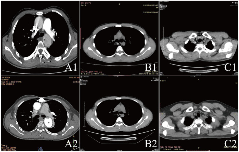 Figure 1
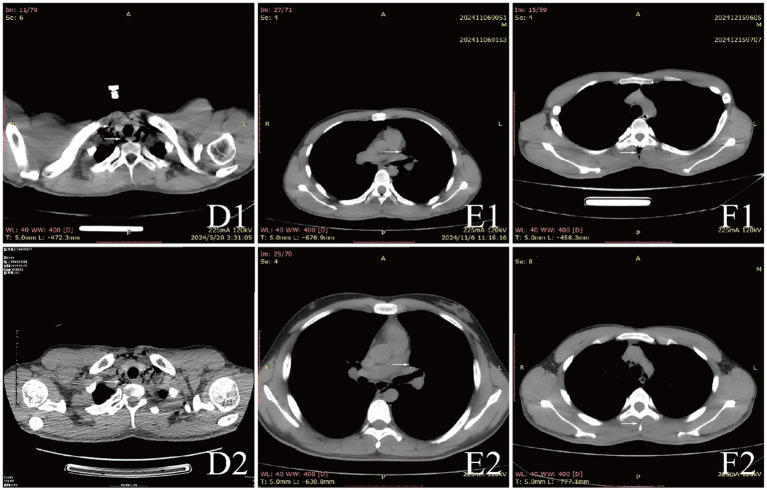 Figure 2
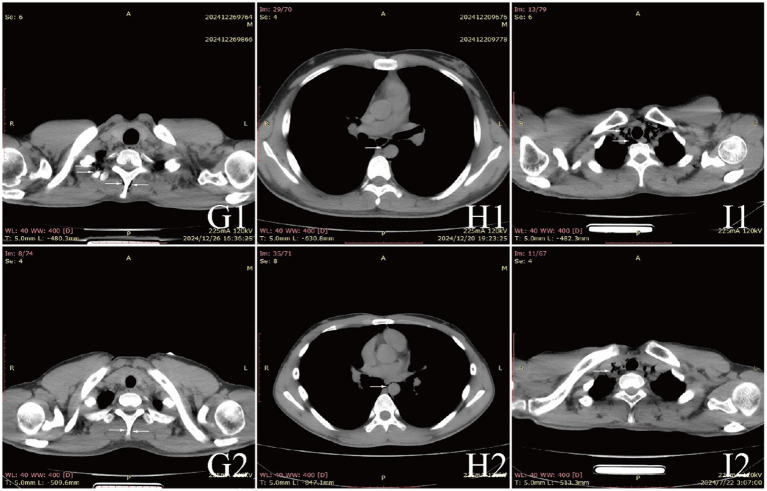 Figure 3
| No | Age | Sex | HA (m) | Time at HA (days) | Oxygen inhalation time (h) | Primary diagnosis | Final diagnosis | Comorbidities | Treatment | Outcome |
|---|---|---|---|---|---|---|---|---|---|---|
| 1 | 22 | Male | 5,100 | 30 | 0 | SE | SE、ME | Aortic Dissection、DVT | Oxygen Inhalation (Mask) + Blood Pressure Control (Sodium Nitroprusside) + Heart Rate Control (Esmolol) + Symptomatic Supportive Treatment + Transfer to Plain Area + Surgical Treatment (Artificial Aortic Arch Replacement) | Improvement |
| 2 | 23 | Male | 5,090 | 90 | 1 | SE | SE | Cerebral Edema、Tracheal Rupture | Oxygen Inhalation (Non-invasive Ventilator) + Sedation (Propofol) + Dehydration (Furosemide + Human Albumin) + Steroids (Dexamethasone) + Symptomatic Supportive Treatment + Hyperbaric Oxygen Therapy (2.0 ATA) | Improvement |
| 3 | 32 | Male | 5,100 | 94 | 4 | SE | SE | Tracheal Rupture | Oxygen Inhalation (Mask) + Symptomatic Supportive Treatment + Hyperbaric Oxygen Therapy (2.0 ATA) | Improvement |
| 4 | 21 | Male | 5,390 | 17 | 0 | ME | ME | Pericarditis | Oxygen Inhalation (Mask) + Anti-infection (Imipenem/cilastatin) + Symptomatic Supportive Treatment + Hyperbaric Oxygen Therapy (2.0 ATA) | Improvement |
| 5 | 20 | Male | 5,248 | 42 | 3 | ME | ME | ME | Oxygen Inhalation (Mask) + Symptomatic Supportive Treatment + Hyperbaric Oxygen Therapy (2.0 ATA) | Improvement |
| 6 | 22 | Male | 5,428 | 224 | 1 | SE | SE | Acute Appendicitis | Oxygen Inhalation (Mask) + Anti-infection (Levofloxacin) + Surgical Treatment + Symptomatic Supportive Treatment + Hyperbaric Oxygen Therapy (2.0 ATA) | Improvement |
| 7 | 33 | Male | 5,100 | 82 | 0 | SE | SE | Pneumonia、Pulmonary Edema、Pulmonary Emphysema | Oxygen Inhalation (Mask) + Dehydration (Furosemide) + Steroids (Dexamethasone) + Anti-infection (Levofloxacin) + Symptomatic Supportive Treatment + Hyperbaric Oxygen Therapy (2.0 ATA) | Improvement |
| 8 | 18 | Male | 5,100 | 229 | 4 | ME | ME | ME | Oxygen Inhalation (Nasal Cannula) + Symptomatic Supportive Treatment + Hyperbaric Oxygen Therapy (2.0 ATA) | Improvement |
| 9 | 21 | Male | 5,200 | 19 | 2 | ME | ME | ICN | Oxygen Inhalation (Nasal Cannula) + Pain Relief (Ibuprofen) + Symptomatic Supportive Treatment + Hyperbaric Oxygen Therapy (2.0 ATA) | Improvement |
| Variable | Mean or proportion |
|---|---|
| Age (years) | 23.56 ± 5.27 |
| Time at HA (days) | 82 ± 24.5 |
| Altitude (m) | 5195.11 ± 133.36 |
| Heart rate | 100.44 ± 17.89 |
| Systolic blood pressure | 133.56 ± 29.09 |
| Diastolic blood pressure | 83.67 ± 11.42 |
| Oxygen saturation | 81.33 ± 6.56 |
| Combined with pericarditis | 11.1% |
| Combined with aortic dissection | 11.1% |
| Combined with tracheal rupture | 22.2% |
| Combined with upper respiratory infection | 22.2% |
| Combined with pneumonia | 11.1% |
| Combined with acute appendicitis | 11.1% |
| Combined with cerebral edema | 11.1% |
| Pulmonary edema | 11.1% |
| Smoking | 33.3% |
| Alcohol consumption | 0 |
| Immunodeficiency | 0 |
| Surgical history | 0 |
| Symptoms | |
| Cough | 33.3% |
| Fever | 44.4% |
| Neck and back pain | 44.4% |
| Chest Pain | 33.3% |
| Variable | Case 1 | Case 2 | Case 3 | Case 4 | Case 5 | Case 6 | Case 7 | Case 8 | Case 9 |
|---|---|---|---|---|---|---|---|---|---|
| RBC (10^12/L) | 7.95 | 5.94 | 6.69 | 5.59 | 5.81 | 4.93 | 4.95 | 5.17 | 6.08 |
| HB (G/L) | 232 | 188 | 186 | 180 | 198 | 169 | 159 | 172 | 199 |
| WBC (10^9/L) | 12.9 | 6.26 | 8.5 | 8.13 | 8.04 | 14.06 | 6.98 | 9.8 | 7.6 |
| Neut count (10^9/L) | 11.74 | 3.82 | 4.44 | 4.87 | 5.43 | 12.05 | 5.72 | 6.78 | 5.22 |
| Neut (%) | 91 | 60.9 | 52.3 | 59.9 | 67.5 | 85.7 | 81.9 | 69.2 | 68.7 |
| PLT (10^9/L) | 119 | 198 | 350 | 243 | 160 | 287 | 159 | 335 | 177 |
| CK (U/L) | 58 | 125 | 199 | 80 | 261 | 95 | 94 | 90 | 75 |
| CK-MB (U/L) | 23.47 | 4.2 | 10.35 | 5.43 | 11.13 | 8.65 | 5.19 | 4.6 | 31.1 |
| LDH (U/L) | 347 | 244 | 339 | 221 | 233 | 272 | 126 | 312 | 159 |
| BUN (mmol/L) | 5.09 | 4.41 | 6.73 | 10.7 | 5.28 | 5.38 | 6.44 | 7.26 | 5.85 |
| UA (umol/L) | 443 | 345 | 478 | 473 | 454 | 357 | 457 | 170 | 483 |
| Cr (umol/L) | 63 | 100 | 63 | 106 | 85 | 83 | 95 | 81 | 94 |
| D-Dimer (mg/L) | 1.7 | 0.6 | 0.02 | 0.1 | 0.5 | 0.2 | 0.04 | 0.3 | 0.05 |
| GPT (U/L) | 30 | 17 | 41 | 20 | 25 | 42 | 21 | 24 | 13 |
| GOT (U/L) | 58 | 19 | 36 | 22 | 25 | 41 | 21 | 32 | 12 |
| TBil (umol/L) | 29.58 | 25.45 | 16.51 | 30.37 | 13.04 | 95.9 | 9.13 | 13.43 | 18.6 |
| DBIL (umol/L) | 4.08 | 15.77 | 2 | 5.89 | 3.51 | 13.19 | 2.21 | 3.12 | 4.49 |
| IBIL (umol/L) | 25.5 | 9.68 | 16.32 | 24.48 | 9.53 | 82.71 | 6.92 | 10.31 | 14.11 |
| TP (g/L) | 90.22 | 79.58 | 80.3 | 79.6 | 78.69 | 88.82 | 76.34 | 88.81 | 72.25 |
| ALB (g/L) | 54.62 | 54.38 | 49.69 | 51.9 | 44.3 | 52.31 | 48.71 | 48.42 | 45.94 |
| GLB (g/L) | 35.6 | 25.2 | 30.6 | 27.7 | 34.4 | 36.5 | 27.6 | 40.4 | 26.4 |
| TG (mmol/L) | 0.65 | 1.05 | 1.57 | 1.5 | 1.04 | 0.8 | 0.9 | 0.97 | 0.51 |
| TC (mmol/L) | 4.4 | 2.88 | 4.02 | 5.47 | 3 | 4.19 | 4.78 | 3.22 | 2.92 |
| K+ (mmol/L) | 5.84 | 4.55 | 5.3 | 5.52 | 5.68 | 5.76 | 4.68 | 6.7 | 4.46 |
| Na+ (mmol/L) | 136 | 139 | 138 | 136 | 137 | 131 | 152 | 124 | 144 |
- —Natural Science Foundation of Xinjiang
Peer Reviews
No public reviews on file for this paper yet. If you reviewed it on a platform where reviews are public (OpenReview, ICLR, NeurIPS, ICML), you can paste yours below so the community can read it here.
Videos
No videos yet. Explain this paper in a talk, walkthrough, or lecture? Add one.
Taxonomy
TopicsPneumothorax, Barotrauma, Emphysema · Chronic Obstructive Pulmonary Disease (COPD) Research · Pleural and Pulmonary Diseases
Introduction
Extra-pulmonary emphysema is defined as gas accumulation within soft tissues, particularly in areas such as the neck and chest. This condition can be classified into subcutaneous emphysema (SE) and mediastinal emphysema (ME), depending on the location of the gas infiltration. The etiology of extra-pulmonary emphysema is multifactorial, encompassing trauma from blunt or penetrating injuries, surgical interventions, and infections. Furthermore, any condition that creates a pressure differential between the alveoli and the surrounding vascular interstitial tissue can precipitate this phenomenon (1–3).
The pathophysiology of extra-pulmonary emphysema is explained by the Macklin effect, in which elevated intrathoracic pressure leads to alveolar rupture, permitting gas to escape into the perivascular interstitium and subsequently dissect into the mediastinum (4). Common triggers include violent coughing, breath-holding, strenuous exercise, acute asthma attacks, and traumatic injury. This mechanism is of particular relevance in high-altitude settings. Hypoxia can induce pulmonary vasoconstriction and endothelial injury, thereby facilitating the entry of gas into perivascular and peribronchial sheaths. Furthermore, the compensatory deep and labored breathing characteristic of high-altitude exposure exacerbates the pressure gradient between alveoli and vascular structures, increasing the risk of alveolar rupture (5). Physical exertion under such conditions can further raise intrathoracic pressure, promoting the development of extra-pulmonary emphysema.
Diagnosis of extra-pulmonary emphysema typically involves clinical assessment of swelling and crepitus over affected areas, while imaging studies are essential for evaluating deep tissue involvement (6). Treatment strategies for mild cases often include oxygen therapy, rest, and close monitoring. Most cases resolving spontaneously within a week. However, significant cases necessitate intervention, such as needle decompression or closed drainage (7). Notably, while hyperbaric oxygen therapy is generally contraindicated in severe cases of pneumothorax and mediastinal emphysema (8), it may lead to gas compression of the heart and major blood vessels, and even endanger life. Some studies suggest that its potential efficacy in treating extra-pulmonary emphysema when other serious conditions are ruled out (9).
At high altitude, the challenges posed by limited medical facilities and the potential for misdiagnosis of extra-pulmonary emphysema as cardiovascular emergencies underscore the importance of accurate and timely diagnosis. This case report details the clinical presentation and management of nine patients diagnosed with extra-pulmonary emphysema at an altitude of 5,000 meters, highlighting the need for heightened awareness and appropriate treatment strategies in such environments. However, due to the small sample size and the different etiologies in each patient, the specific mechanisms still need to be further investigated.
Case description
Basic clinical information
This study included nine male patients diagnosed with subcutaneous emphysema or mediastinal emphysema who visited a high altitude medical station (at 3,700 meters) from May to December 2024. They all developed the condition in areas above 5,000 meters and were transported down to the medical station for further treatment. The basic information of the nine patients are shown in Tables 1, 2. None of them had a history of chronic diseases, nor did they have a history of surgery or immunosuppression. Before the onset of illness, three patients had never received oxygen therapy, two patients received oxygen therapy for 1 h daily, one patient for 2 h, one patient for 3 h, and two patients for 4 h. The duration of residence at altitude varied, ranging from 17 to 229 days. Upon arrival at the medical facility, patients reported discomfort including neck pain, chest pain, and chest tightness. Initial physical examinations revealed crepitus in some patients, with CT confirming the diagnosis of extra-pulmonary emphysema. Only two patients were diagnosed with isolated extra-pulmonary emphysema among all the patients, while the others had definite etiologies.
Case 1 is 22 years old, he suddenly experienced severe neck pain while straining during a bowel movement. The pain persisted and radiated to the back. Six hours later, the patient was transferred to our department. His blood pressure was 202/103 mmHg in the right upper limb and 190/95 mmHg in another limb. The electrocardiogram showed “sinus rhythm, left axis deviation, left anterior fascicular block, and T-wave changes in the anterior leads.” Ultrasound revealed the formation of deep vein thrombosis. Non-contrast CT revealed emphysema in the muscles of the neck and back and subcutaneously, enhanced CT scan confirmed a thoracoabdominal aortic dissection. After stabilizing the blood pressure and heart rate, the patient was urgently transferred to a higher-level hospital for “aortic arch artificial vessel replacement.” Follow-up examinations indicated that the patient recovered well.
Case 2 and Case 3 are both young males. While engaging in heavy physical labor, both patients suddenly experienced chest pain. The patients were urgently transported to our medical facility and underwent chest CT, they were diagnosed with subcutaneous emphysema and tracheal rupture. Among them, case 2 also developed acute high altitude cerebral edema. After received symptomatic support and hyperbaric oxygen therapy (10, 11), the symptoms of these two patients significantly improved, and the extra-emphysema was also markedly absorbed. Follow-up showed that the tracheal rupture had also healed.
Case 4 is 21 years old. One week prior, he had a history of upper respiratory tract infection, and then He visited our hospital due to chest discomfort. The electrocardiogram suggested: “sinus rhythm, T-wave changes in the anterior leads,” and there was a slight increase in lymphocyte count. After underwent echocardiography and chest CT, he was diagnosed with mediastinal emphysema and pericarditis. In addition to antibiotic treatment, we also provided hyperbaric oxygen therapy (12, 13), and the patient eventually recovered well.
Case 6 is 22 years old, he experienced chest discomfort and abdominal pain after excessive alcohol consumption. After undergoing chest and abdominal CT, he was diagnosed with acute appendicitis and subcutaneous emphysema. Following an emergency “appendectomy,” he received hyperbaric oxygen therapy and had a good prognosis.
Case 7 is 33 years old, who developed symptoms of cough and chest pain after engaging in heavy physical labor outdoors. He was diagnosed with pneumonia and pulmonary edema through chest CT. Following this diagnosis, he received oxygen therapy, diuretics, antibiotic treatment, and hyperbaric oxygen therapy, his symptoms improved and he recovered well.
Case 5, Case 8, and Case 9 are all young males. All three patients presented with unexplained chest pain. Initially, primary healthcare institutions diagnosed them with cardiovascular emergencies and transferred them to our medical facility. After a series of examinations, including electrocardiogram, echocardiography, and chest and abdominal CT, Case 9 received a diagnosis of intercostal neuralgia, while Cases 5 and 8 were diagnosed with mediastinal emphysema. Follow-up after hyperbaric oxygen therapy showed significant resolution of mediastinal emphysema in all patients.
Laboratory results and auxiliary
Examinations affected by the hypoxic environment of high altitudes, all patients exhibited varying degrees of increased red blood cell count, hemoglobin concentration, and uric acid (UA). Case 6 and 7, who were diagnosed with acute appendicitis and pulmonary infection, respectively, showed increased percentages of neutrophils. Case 1, in the acute phase of aortic dissection, also had an elevated white blood cell count. Additionally, we observed that D-dimer in the blood is a good indicator of myocardial or vascular damage. It showed varying degrees of elevation in patients with aortic dissection, while this indicator remained normal in other patients, maintaining a high negative predictive value. It is noteworthy that the serum potassium (K^+^) levels of all patients were high, with five patients having levels above the normal range. The average serum K^+^ level for all patients was 5.39 ± 0.73 mmol/L, this may be due to the increased metabolism in high altitude areas (Table 3). The diagnosis of extra-pulmonary emphysema for all patients was confirmed through CT examination. Case 1, 2, 3, 6, and 7 showed subcutaneous emphysema, while the others showed mediastinal emphysema. Specific imaging manifestations are shown in Figure 1.
CT comparison images of extra-pulmonary emphysema treatment before and after for Case 1 to Case 3.
CT comparison images of extra-pulmonary emphysema treatment before and after for Case 4 to Case 6.
Diagnosis, treatment and prognosis
All patients were diagnosed with extra-pulmonary emphysema through CT scans, which showed gas infiltration in the mediastinum or subcutaneous tissue. The patient with aortic dissection was urgently transferred to a lower altitude area for “aortic arch artificial vessel replacement surgery.” Case 6 underwent “appendectomy” for treatment, and three patients received antibiotic therapy. All patients were given symptomatic and supportive treatment, including oxygen therapy. Six were given oxygen via a face mask, two received a nasal cannula, and one was assisted with non-invasive ventilation. In addition to case 1, they also received hyperbaric oxygen therapy at 2.0 ATA. Follow-up showed that all patients responded well to the treatment, their conditions improved, and the emphysema was largely absorbed (Figures 1–3).
CT comparison images of extra-pulmonary emphysema treatment before and after for Case 7 to Case 9.
Discussion
The occurrence of extra-pulmonary emphysema, particularly at high altitudes, represents a significant clinical challenge due to its rarity and potential for misdiagnosis (14–17). In our retrospective analysis of patients treated for this condition at altitudes exceeding 5,000 meters, we observed a concerning trend: many patients initially presented with symptoms that could easily be mistaken for cardiovascular emergencies, such as aortic dissection and pulmonary embolism. This underscores the critical need for heightened awareness and understanding of extra-pulmonary emphysema among healthcare providers, particularly in high-altitude settings where diagnostic resources may be limited. In the special environment of the plateau, medical resources are scarce, and most areas may only be equipped with X-ray examinations without conducting CT scans. This may lead to two types of misdiagnoses: either mistaking simple emphysema for cardiovascular emergencies such as pulmonary embolism or aortic dissection, or diagnosing emphysema solely based on X-ray examination while overlooking atypical critical illnesses. Both of these can result in excessive or insufficient treatment strategies. CT scans, through multiplanar imaging and 3D reconstruction analysis, can effectively avoid these situations. Through CT sections, we can observe vascular filling defects or intimal tears, thereby effectively obtaining diagnostic information for conditions such as pulmonary embolism and aortic dissection, which can guide subsequent medical procedures.
In our series, the majority of patients were young males with no significant medical histories, indicating that even previously healthy individuals can be susceptible to extra-pulmonary emphysema under extreme environmental stressors (18, 19). The clinical presentation varied, with symptoms such as neck pain, chest tightness, and crepitus upon examination. However, the diagnosis was often missed or delayed in primary care settings, reflecting a gap in knowledge regarding the manifestations of this condition at high altitudes. The initial misdiagnoses highlight a critical area for improvement in clinical practice, emphasizing the importance of considering extra-pulmonary emphysema in differential diagnoses for patients presenting with chest pain or dyspnea in such environments.
Furthermore, our findings support the efficacy of hyperbaric oxygen therapy as a treatment modality for extra-pulmonary emphysema. Eight out of nine patients showed improvement following this intervention, illustrating its potential in expediting recovery in high-altitude scenarios (20–22). Most cases of extra-pulmonary emphysema are asymptomatic or mildly symptomatic, and the standard treatment is typically the administration of pure oxygen under normal pressure. However, if hyperbaric oxygen therapy is administered, the volume of gas in the alveoli will change with the increase and decrease of ambient pressure during treatment. If the gas in bullae or air cysts cannot be normally expelled, it may lead to alveolar overexpansion and even rupture, thereby causing rare but serious complications such as pneumothorax and gas embolism. At the same time, the accumulation of a large amount of gas can affect the return flow of venous blood, leading to thrombotic diseases such as deep vein thrombosis or pulmonary embolism. In addition, if a patient with emphysema also has an untreated pneumothorax, this is an absolute contraindication for hyperbaric oxygen therapy, as the pneumothorax may rapidly worsen during treatment, leading to severe respiratory and circulatory dysfunction. Therefore, before undergoing hyperbaric oxygen therapy, patients with extra-pulmonary emphysema must undergo a comprehensive evaluation by a professional physician. During the treatment process, the general condition of the patient needs to be closely monitored to reduce the risk of pulmonary complications.
This analysis also draws attention to the limitations of medical facilities in high-altitude regions, where access to advanced imaging and definitive diagnostic capabilities may be restricted. The challenges of accurate diagnosis and timely intervention are compounded by the geographical isolation of many high-altitude communities, making early recognition of conditions such as extra-pulmonary emphysema crucial. Future studies should focus on developing standardized protocols for the diagnosis and management of extra-pulmonary emphysema in these settings, as well as exploring the long-term outcomes of patients treated at high altitudes.
In conclusion, our findings highlight the need for increased clinical awareness of extra-pulmonary emphysema, especially in high-altitude environments, where its presentation can mimic more serious cardiovascular conditions. Enhanced understanding and recognition of this condition may improve patient outcomes, as timely diagnosis and appropriate management strategies, such as hyperbaric oxygen therapy, can be life-saving (23–25). The lessons learned from our cases should inform future clinical practice and guide the development of emergency protocols for healthcare providers operating in high-altitude regions.
The limitations of this study are that the number of cases was small, and we do not have a control group. Meanwhile, after reviewing the relevant literature, we found that plasma sRAGE, CT images analyzed by deep learning, and blood-based transcriptomic and proteomic biomarkers can all serve as early warning indicators for the development of emphysema (26–28). Unfortunately, due to the limitations of our medical environment, we were unable to collect biological samples from the patients and thus could not conduct the relevant tests. In future work, we can pay more attention to research in this area.
Conclusion
In this study, a retrospective analysis of extra-pulmonary emphysema case data in extreme high altitude areas suggested that the diagnosis of extra-pulmonary emphysema should be emphasized in high altitude regions. After ruling out fatal diseases, administering hyperbaric oxygen therapy and symptomatic treatment is a reliable approach.
The reference list from the paper itself. Each links out to its DOI / PubMed record.
- 1Fosi S Giuricin V Girardi V Di Caprera E Costanzo E Di Trapano R. Subcutaneous emphysema, pneumomediastinum, pneumoretroperitoneum, and pneumoscrotum: unusual complications of acute perforated diverticulitis. Case Rep Radiol. (2014) 2014:1–5. doi: 10.1155/2014/431563 PMC 412722425136471 · doi ↗ · pubmed ↗
- 2Kukuruza K.Aboeed A. (2025). Subcutaneous emphysema (Archived). In: Stat Pearls [Internet]. Treasure Island (FL): Stat Pearls Publishing.31194349 · pubmed ↗
- 3Talwar A Rajeev A Rachapudi S Khan S Singh V Talwar A. Spontaneous pneumomediastinum: a comprehensive review of diagnosis and management. Intractable Rare Dis Res. (2024) 13:138–47. doi: 10.5582/irdr.2024.01020, PMID: 39220281 PMC 11350202 · doi ↗ · pubmed ↗
- 4Angelini M Belletti A Landoni G Zangrillo A De Cobelli F Palumbo D. Macklin effect: from pathophysiology to clinical implication. J Cardiothorac Vasc Anesth. (2024) 38:881–3. doi: 10.1053/j.jvca.2023.12.025, PMID: 38378321 · doi ↗ · pubmed ↗
- 5Sydykov A Mamazhakypov A Maripov A Kosanovic D Weissmann N Ghofrani H. Pulmonary hypertension in acute and chronic high altitude maladaptation disorders. Int J Environ Res Public Health. (2021) 18:1692. doi: 10.3390/ijerph 18041692, PMID: 33578749 PMC 7916528 · doi ↗ · pubmed ↗
- 6Medeiros B. J. D. C. (2018). Subcutaneous emphysema, a different way to diagnose. Rev Assoc Med Bras (1992). 64:159–163. doi: 10.1590/1806-9282.64.02.15929641666 · doi ↗ · pubmed ↗
- 7Iteen A. J.Bianchi W.Sharman T. (2025). Pneumomediastinum In: Stat Pearls [Internet]. Treasure Island (FL): Stat Pearls Publishing.32491372 · pubmed ↗
- 8Howell RS Criscitelli T Woods JS Gillette BM Gorenstein S. Hyperbaric oxygen therapy: indications, contraindications, and use at a tertiary care center: 1.3 www.Aornjournal.Org/content/cme [Journal Article]. AORN J. (2018) 107:442–53. doi: 10.1002/aorn.12097, PMID: 29595909 · doi ↗ · pubmed ↗