Bifocal β-hCG-secreting CNS Germinoma in a 13-Year-Old Boy: Clinical–Biochemical Pubertal Discordance and Long-Term Outcome
Corina Ramona Nicolescu, Sandrine Thouvenin Doulet, Lucie Bazus, Jean-Louis Stephan

TL;DR
A 13-year-old boy with a rare brain tumor showed signs of early puberty due to a hormone-secreting tumor, which was successfully treated with a combination of therapies.
Contribution
This case highlights the importance of β-hCG testing and neuroimaging in diagnosing rare CNS germinomas presenting with pubertal discordance.
Findings
β-hCG-secreting germinoma can induce gonadotropin-independent puberty in pediatric patients.
CSF sampling via EVD is a safe alternative when lumbar puncture is not feasible.
Coordinated oncologic and endocrine care leads to durable disease control in such cases.
Abstract
Intracranial germ cell tumors (GCTs) are rare in the pediatric population. They are classified as germinoma and nongerminomatous and may secrete specific proteins such as β subunit of human chorionic gonadotropin (β-hCG) and alpha-fetoprotein (AFP). When secreting β-hCG, they may induce gonadotropin-independent puberty (GIP), a clinical diagnostic clue that can precede neuroimaging findings. A 13-year-old boy presented with a first generalized tonic–clonic seizure after six months of headaches, vomiting, polyuria, and polydipsia. Examination showed pubertal penile length with peripubertal testes. Laboratory assessment revealed panhypopituitarism with suppressed gonadotropins and elevated testosterone. Brain magnetic resonance imaging (MRI) demonstrated bifocal lesions (pineal and suprasellar) with obstructive hydrocephalus. Cerebrospinal fluid (CSF) sample obtained via temporary…
Genes, proteins, chemicals, diseases, species, mutations and cell lines named across the full text — each resolved to its canonical identifier and authoritative record.
Click any figure to enlarge with its caption.
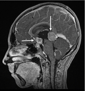 Figure 1
Peer Reviews
No public reviews on file for this paper yet. If you reviewed it on a platform where reviews are public (OpenReview, ICLR, NeurIPS, ICML), you can paste yours below so the community can read it here.
Videos
No videos yet. Explain this paper in a talk, walkthrough, or lecture? Add one.
Taxonomy
TopicsGlioma Diagnosis and Treatment · Testicular diseases and treatments · Neuroblastoma Research and Treatments
1. Introduction
Intracranial germ cell tumors (GCTs) are rare malignancies, accounting for 2%–3% of all primary central nervous system (CNS) tumors [1], predominantly affecting adolescents and young adults [2], with a male-to-female ratio of 2:1 [3]. They are commonly located in the midline region, may secrete β subunit of human chorionic gonadotropin (β-hCG), and are classified as germinomas or nongerminomatous GCTs [4].
A prolonged time to diagnosis is common, usually exceeding 6 months, although a recent study showed that delay in diagnosis does not adversely impact the outcome [5].
At presentation, the clinical features include endocrine dysfunction (most commonly, diabetes insipidus and pubertal disturbances), neurological symptoms (intracranial hypertension), and visual symptoms (Parinaud syndrome and visual field defects) [6].
Pubertal derangements vary by sex, age, and β-hCG secretion. Boys frequently show premature sexual development or discordant puberty [6, 7]. Girls predominantly present with hypogonadotropic hypogonadism [6], yet β-hCG-driven peripheral puberty has also been reported [8–12].
2. Case Presentation
A 13-year-old boy presented with a first generalized tonic–clonic seizure (5 min, loss of consciousness) following a 6-month history of progressive headaches, intermittent vomiting, polyuria, polydipsia, weight loss, and fatigue. He denied visual complaints. His past medical history was unremarkable. Family history was also noncontributory.
Clinical examination revealed an ill adolescent with a body weight of 29 kg (< 5^th^ percentile), height of 149 cm (10^th^ percentile), and body mass index (BMI) of 13.06 kg/m^2^ (< 3^rd^ percentile). His vital signs and systemic examination were otherwise normal. Genital examination showed an enlarged penis of 8 cm, bilateral testicular volume of 4 mL, and pubic hair without gynecomastia. The neurological examination was normal; funduscopic examination revealed bilateral papilledema.
Routine laboratory studies (full blood count, renal profile, liver function test, electrolytes, and serum calcium/phosphate) were within reference ranges (Table 1). Endocrine testing demonstrated complete anterior and posterior pituitary deficiency. Testosterone level was elevated, with suppressed gonadotropins (follicle-stimulating hormone [FSH] and luteinizing hormone [LH]) levels.
Brain magnetic resonance imaging (MRI) demonstrated two heterogeneous, contrast-enhancing lesions in the pineal (20 × 21 × 22 mm) and suprasellar (12 × 12 × 16 mm) regions, with moderate hydrocephalus (Figure 1). Spine MRI was normal.
Because papilledema and hydrocephalus contraindicated lumbar puncture, a temporary external ventricular drain (EVD) was placed and corticosteroids initiated, enabling safe cerebrospinal fluid (CSF) sampling. CSF cytology was negative for malignant cells, but β-hCG was elevated (Table 1). Serum β-hCG was 130 IU/L, and AFP was 4.3 ng/mL. A diagnosis of bifocal β-hCG-secreting CNS germinoma with secondary panhypopituitarism and raised intracranial pressure was established. Treatment approach aligned with contemporary consensus recommendations [4]: multimodal therapy comprising four cycles of ifosfamide, etoposide, and cisplatin, followed by complete neurosurgical resection and cranial radiotherapy (54 Gy). Early follow-up showed undetectable serum β-hCG and no residual disease (Table 1).
Hormone replacement therapy with hydrocortisone, desmopressin, and levothyroxine was progressively initiated within the first week after diagnosis. Growth hormone treatment was started 1 year after remission.
During surveillance, the patient experienced an intracranial relapse, as a contrast-enhancing lesion on the posterior aspect of the medulla oblongata. He was reinduced into remission with three cycles of gemcitabine, paclitaxel, and oxaliplatin, followed by high-dose etoposide and thiotepa with autologous hematopoietic stem-cell transplantation. Craniospinal irradiation was then delivered with dosimetric adaptation to previously irradiated proton fields (30 Gy craniospinal, 23 Gy cranial boost).
At 4 years of follow-up, MRI surveillance every 3 months has shown no recurrence.
Lifelong pituitary deficits were managed with growth hormone, hydrocortisone, levothyroxine, desmopressin, and testosterone.
At the time of writing, the patient is 18 years and 6 months old, with the final height of 162.7 cm (< 5^th^ percentile), and Tanner stage IV, with the testicular volume of 12 mL bilaterally. Final height was likely constrained by spinal proton irradiation and bilateral epiphysiodesis for severe genu valgum performed at the age of 16.
Key clinical lesson: The combination of neurological symptoms, pubertal dissociation (pubertal penile length with peripubertal testes), and panhypopituitarism with isolated high testosterone level was consistent with β-hCG-driven GIP due to CNS germinoma and should prompt targeted biochemical testing and neuroimaging.
3. Discussion
This case highlights a diagnostically valuable pattern in β-hCG-secreting CNS GCTs: discordant puberty features in male adolescents (pubertal penile length and peripubertal testicular volume) with panhypopituitarism and elevated testosterone level despite suppressed gonadotropins.
β-hCG germinoma secretion can precipitate premature sexual maturation, classically isosexual precocity in boys < 9 years and discordant puberty in older boys despite prepubertal gonadotropin levels. Two nonexclusive mechanisms are proposed, depending on two main tumor characteristics: location and the potential to produce β-hCG [13]. The anatomic pathway refers to the tumor location that may trigger early hypothalamic–pituitary activation (gonadotropin-dependent pubertal precocity) [14]. The biochemical pathway involves tumor-derived β-hCG, whose β subunit is homologous to LH and activates testicular LH receptors on Leydig cells, increasing testosterone synthesis independently of gonadotropin-releasing hormone (GnRH). In boys, GIP typically presents as a discrepancy between prepubertal testicular volume and pubertal penis length, prepubertal gonadotropins, and pubertal sex steroid levels.
As tumor detectability and β-hCG levels may fluctuate, GIP is considered a prodromal signal for GCTs; repeated hormonal testing and interval neuroimaging are warranted [15–17].
In girls, hypogonadotropic hypogonadism is common with suprasellar tumor location and hypothalamic–pituitary axis involvement, and β-hCG-mediated GIP is rare and physiologically complex. Three different biological hypotheses are discussed: (1) β-hCG could possess dual LH- and FSH-like activity, (2) tumor cells could produce a different substance with FSH-like activity, and (3) tumor cells might secrete β-hCG and simultaneously have aromatase activity, converting ovarian androgens to estrogens, thus inducing precocious pubertal development [10, 12]. Recently, a case report of suprasellar choriocarcinoma in a 7.5-year-old girl demonstrated the production of aromatase by tumor cells in immunohistological studies [8].
Peripheral β-hCG-producing tumors (choriocarcinoma of the liver, hepatoblastoma, and GCTs of the mediastinum) can similarly induce GIP in boys. In male adults with β-hCG-secreting tumors, gynecomastia was reported and might be secondary to the conversion of testosterone to estradiol via tumor cells aromatase synthesis [18–20]. Ovarian dysgerminoma with aromatase activity (and intratumoral estrogen synthesis) has been implicated in female precocious puberty [21–24].
Table 2 summarizes the characteristics of pediatric patients (boys and girls) with β-hCG-secreting intracranial GCTs (case reports compiled from PubMed, key words: β-hCG-producing tumor and early pubertal development).
In boys with intracranial β-hCG-producing tumors and sexual precocity, common findings included variable systemic symptoms/signs, a consistent pubertal discrepancy (small testes with penile enlargement), and similar biological profiles (suppressed and nonresponsive gonadotropin secretion and cerebral β-hCG secretion). Female GIP is an extremely rare event, and the relationship between precocious sexuality and CNS GCT is more complex and less explored.
4. Conclusion
We described an adolescent boy with bifocal β-hCG-secreting germinoma and overlapping symptoms, including neurological (pineal lesion) and endocrine (suprasellar lesion) dysfunction.
Discordant clinical and biochemical puberty (penile growth with small testes and high testosterone with suppressed gonadotropins) is a practical red flag for β-hCG-driven GIP in suspected CNS GCTs. Safe CSF sampling via EVD is appropriate when lumbar puncture is contraindicated by raised intracranial pressure.
Coordinated oncologic–endocrine care with structured MRI surveillance supports durable remission while managing endocrine sequelae.
The reference list from the paper itself. Each links out to its DOI / PubMed record.
- 1Echevarria M. E. Fangusaro J. Goldman S. Pediatric Central Nervous System Germ Cell Tumors: A Review The Oncologist 200813669069910.1634/theoncologist.2008-00372-s 2.0-4614911277618586924 · doi ↗ · pubmed ↗
- 2Villano J. L. Propp J. M. Porter K. R. Malignant Pineal Germ-Cell Tumors: An Analysis of Cases from Three Tumor Registries Neuro-Oncology 200810212113010.1215/15228517-2007-0542-s 2.0-4484914404218287340 PMC 2613814 · doi ↗ · pubmed ↗
- 3Jennings M. T. Gelman R. Hochberg F. Intracranial Germ-Cell Tumors: Natural History and Pathogenesis Journal of Neurosurgery 198563215516710.3171/jns.1985.63.2.01552-s 2.0-00220072182991485 · doi ↗ · pubmed ↗
- 4Frappaz D. Dhall G. Murray M. J. EANO, SNO and Euracan Consensus Review on the Current Management and Future Development of Intracranial Germ Cell Tumors in Adolescents and Young Adults Neuro-Oncology 202224451652710.1093/neuonc/noab 25234724065 PMC 8972311 · doi ↗ · pubmed ↗
- 5Partenope C. Pozzobon G. Weber G. Carceller F. Albanese A. Implications of Deferred Diagnosis of Paediatric Intracranial Germ Cell Tumours Pediatric Blood and Cancer 2023703 p. e 3016810.1002/pbc.3016836582128 · doi ↗ · pubmed ↗
- 6Partenope C. Pozzobon G. Weber G. Arya V. B. Carceller F. Albanese A. Endocrine Manifestations of Paediatric Intracranial Germ Cell Tumours: From Diagnosis to Long-Term Follow-Up Endocrine 202277354655510.1007/s 12020-022-03121-935767181 · doi ↗ · pubmed ↗
- 7Chen H. Ni M. Xu Y. Zhong L. Y. Precocious Puberty Due to Intracranial Germ Cell Tumors: A Case-Control Study Endocrine-Related Cancer 2022291058158810.1530/ERC-21-038135938923 · doi ↗ · pubmed ↗
- 8Shibata N. Nyuzuki H. Sasaki S. Ogawa Y. Okada M. Nagasaki K. Peripheral Precocious Puberty in a Girl With An Intracranial h CG-Producing Tumor: Case Report and Literature Review Endocrine Journal 202168121463146710.1507/endocrj.EJ 21-011734275973 · doi ↗ · pubmed ↗