Restenosis After Lacrimal Stent Intubation in a Patient With Indigo Carmine Positivity: A Case Report
Kosuke Aonuma, Hiroki Kaneko

TL;DR
A patient with a history of lacrimal intubation developed restenosis, and indigo carmine staining helped identify fibrotic tissue linked to the recurrence.
Contribution
This case report is the first to demonstrate indigo carmine's potential in detecting fibrotic mucosa after lacrimal stent intubation.
Findings
Indigo carmine staining revealed focal mucosal fibrosis after tube removal, correlating with later restenosis.
The patient developed restenosis six weeks post-tube removal, despite initial successful intubation.
The case suggests indigo carmine could help identify patients at risk for restenosis after lacrimal intubation.
Abstract
Nasolacrimal duct obstruction (NLDO) is commonly treated with lacrimal stent intubation, yet restenosis remains a frequent challenge. Indigo carmine staining has been reported in research settings as an adjunct in dacryoendoscopy for evaluating mucosal integrity, mainly after tube removal. However, its clinical application has not yet been established, and there are no reports of routine use in daily practice. We present the case of a 55-year-old woman who presented with epiphora, discharge, and itching of the left eye, with a history of lacrimal intubation 10 years earlier. At our clinic, dacryoendoscopy revealed right common canalicular obstruction and left NLDO with clinical features consistent with chronic dacryocystitis, for which bilateral lacrimal intubation was performed. Eight weeks later, the tubes were removed, and dacryoendoscopy was repeated. Indigo carmine (2 mg/0.5 mL)…
Genes, proteins, chemicals, diseases, species, mutations and cell lines named across the full text — each resolved to its canonical identifier and authoritative record.
Click any figure to enlarge with its caption.
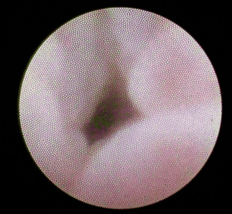 Figure 1
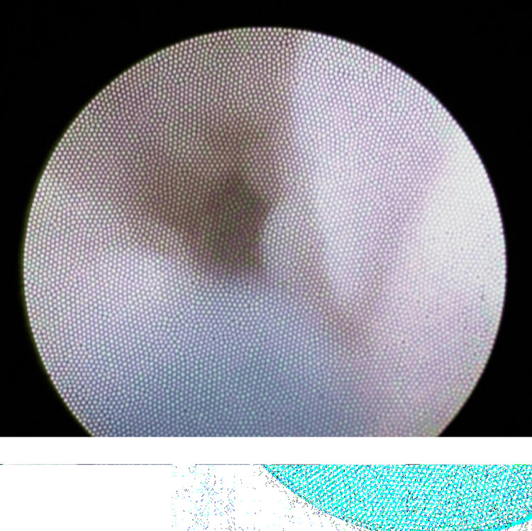 Figure 2
Peer Reviews
No public reviews on file for this paper yet. If you reviewed it on a platform where reviews are public (OpenReview, ICLR, NeurIPS, ICML), you can paste yours below so the community can read it here.
Videos
No videos yet. Explain this paper in a talk, walkthrough, or lecture? Add one.
Taxonomy
TopicsNasolacrimal Duct Obstruction Treatments · Sinusitis and nasal conditions · Salivary Gland Tumors Diagnosis and Treatment
Introduction
Nasolacrimal duct obstruction (NLDO) is a common condition that often leads to chronic epiphora, recurrent infections, and impaired quality of life. Lacrimal stent intubation is widely accepted as a first-line surgical option for adults with primary acquired nasolacrimal duct obstruction (PANDO), especially in cases without extensive fibrosis, because of its minimally invasive nature and relatively high success rate. However, restenosis after tube removal is not uncommon, particularly in patients with chronic dacryocystitis or advanced mucosal disease. Identifying patients at high risk of recurrence remains a major clinical challenge.
Indigo carmine staining has recently been introduced in research settings as an adjunctive method in dacryoendoscopy to evaluate lacrimal mucosa. Mimura et al. demonstrated that indigo carmine positivity correlates with histopathological findings of advanced fibrosis, including epithelial atrophy, goblet cell depletion, and subepithelial scarring, whereas negative staining was associated with inflammatory stages, suggesting a reversible process with preserved regenerative potential [1]. Despite these insights, indigo carmine has not yet been implemented in routine clinical practice, and, to date, no reports exist describing its widespread clinical use for postoperative evaluation in NLDO patients.
Here, we report a case of NLDO with chronic dacryocystitis in which indigo carmine staining, performed after lacrimal tube removal, revealed localized mucosal fibrosis. The patient subsequently developed restenosis of the left lacrimal drainage system. This case underscores both the potential and current limitations of indigo carmine staining in predicting postoperative outcomes.
Case presentation
A 55-year-old woman presented to a local clinic with complaints of ocular discharge, itching, and epiphora in the left eye. She had a history of lacrimal silicone tube intubation in the left eye performed at another hospital approximately 10 years earlier. There was no history of preservative-containing topical medications, chronic rhinosinusitis, or systemic inflammatory disease.
At the initial examination in our hospital, lacrimal irrigation with normal saline revealed reflux without purulent discharge in the right eye and reflux with purulent discharge in the left eye. Tear meniscus height was elevated bilaterally.
Three weeks after the initial visit, lacrimal silicone tube intubation was performed under dacryoendoscopic guidance. After local anesthesia around the puncta and lacrimal sac, punctal dilatation was performed with a dilator, and a dacryoendoscope was introduced into the lacrimal drainage system. In the right eye, a common canalicular obstruction was identified and successfully recanalized using direct endoscopic probing. In the left eye, mucoid and blood-stained secretions, suggestive of chronic inflammation, were observed in the lacrimal sac and nasolacrimal duct, with complete NLDO. The obstruction was relieved using a sheath-guided endoscopic probing technique. Silicone tubes were subsequently placed bilaterally under sheath guidance. The final diagnoses were common canalicular obstruction in the right eye and NLDO with clinical features consistent with chronic dacryocystitis in the left eye, although infection was not microbiologically confirmed. Postoperatively, the patient received topical corticosteroids and antibiotics four times daily. Lacrimal irrigation with 3 mL of saline was performed every two weeks to maintain patency.
Eight weeks after surgery, the tubes were removed, and dacryoendoscopic evaluation was repeated. Following lavage with distilled water, indigo carmine solution (2 mg/0.5 mL) was instilled through the irrigation channel of the dacryoendoscope and immediately washed out with distilled water to remove excess dye. Both lacrimal drainage systems were confirmed to be patent (Figure 1). The right side showed no staining, while the left nasolacrimal duct was mostly unstained except for a focal area of blue staining along the mucosa (Figure 2). This finding was explained to the patient as evidence of localized mucosal fibrosis, suggesting an increased risk of restenosis.
After tube removal, the left nasolacrimal duct remains patent.
Indigo carmine staining of the left nasolacrimal duct showing focal mucosal staining, indicating localized fibrosis.
Regular irrigation with saline was continued after tube removal. Although tear meniscus height initially remained stable, six weeks later, the left eye showed increased reflux during irrigation, indicating recurrent NLDO. Further intervention, such as dacryocystorhinostomy, was considered, and the patient was referred to a specialized tertiary facility.
Discussion
Lacrimal stent intubation is widely used for the treatment of NLDO. Nevertheless, recurrence remains a major challenge, particularly in patients with chronic dacryocystitis. Known risk factors include chronic inflammation, progressive mucosal fibrosis, and long-standing obstruction. Our case illustrates that even after technically successful intubation and initial patency, restenosis may still occur due to subtle mucosal changes.
Clinical studies have clarified the indications for silicone intubation. Mimura et al. reported that Nunchaku-style silicone tube intubation achieved excellent success in upper obstruction (94.6%) and in lower obstruction without dacryocystitis (82.9%), but outcomes were significantly worse when dacryocystitis was present (52.4%). In such cases, endonasal dacryocystorhinostomy achieved much higher success (95.5%) [2]. These findings indicate that while intubation is highly effective in early or uncomplicated PANDO, chronic inflammation and fibrosis substantially reduce long-term success.
Indigo carmine staining has been reported in experimental and research settings as an adjunct in dacryoendoscopy. Mimura et al. demonstrated that indigo carmine positivity correlates strongly with advanced fibrotic remodeling of the lacrimal mucosa, including epithelial atrophy and subepithelial scarring, while negative staining corresponds to earlier inflammatory stages with preserved regenerative potential [1]. Importantly, indigo carmine staining does not indicate infection or the presence of pus; rather, it reflects the degree of mucosal fibrosis and epithelial degeneration. Although promising, indigo carmine staining remains an experimental and exploratory technique, supported only by preliminary research. It has not been adopted into routine clinical practice, and no reports describe its widespread use. Our case, therefore, represents a rare example in which indigo carmine was applied after tube removal, with focal staining suggesting, but not definitively predicting, subsequent restenosis. This finding supports the hypothesis that indigo carmine positivity may identify mucosa at risk for fibrosis-related recurrence. However, as no histopathology or laboratory confirmation was performed in this case, the staining results should be regarded as hypothesis-generating rather than definitive proof of fibrosis. Rather than establishing a new standard of care, this case should be regarded as illustrative of indigo carmine’s potential role and as supportive of earlier research.
Recent evidence further highlights the evolving role of dacryoendoscopy as a platform technology. Wong et al. systematically reviewed 18 studies and confirmed that dacryoendoscopy achieves high treatment success (73.7-100%), with particularly excellent outcomes in congenital NLDO (97-100%) and favorable results in adults (73.7-96.9%). Reported complications were mostly minor, including edema, hemorrhage, and false passage formation, and severe events were rare [3]. In line with this, Kim and Lew found that dacryoendoscopy allowed real-time identification and correction of false passages, which occurred in 2.3% of PANDO cases, thereby improving safety and efficacy [4]. Beyond these clinical advantages, dacryoendoscopy has been increasingly recognized as a frontier technology. Since its introduction in the early 21st century, it has enabled direct visualization of intraluminal pathology and is now standardized, providing a reliable platform for integrating adjunctive techniques such as indigo carmine staining [5].
The duration of silicone tube intubation also plays a critical role in long-term outcomes. Zimmermann et al. demonstrated that removal earlier than three months was significantly associated with higher recurrence (success rate 38% vs. 61% for ≥3 months) [6]. In our case, the tube was removed after two months, and restenosis subsequently developed. This course is consistent with Zimmermann et al.’s findings and suggests that premature removal may compromise mucosal healing and promote fibrosis-related obstruction.
Histopathological investigations add further depth to this interpretation. Makselis et al. analyzed 275 lacrimal sac biopsies and found chronic nongranulomatous inflammation in over 70% of cases, while 1.1% harbored tumors such as adenoid cystic carcinoma, eccrine spiradenoma, and small B-cell lymphoma. Notably, clinical presentation alone did not distinguish inflammatory from neoplastic disease [7]. These results confirm that chronic inflammation and fibrosis dominate NLDO pathology, but rare malignant causes must also be considered. Ali further described PANDO as the outcome of a multifactorial cascade, progressing from recurrent inflammation through epithelial degeneration and goblet cell depletion to subepithelial fibrosis [8]. They also emphasized modifying factors such as anatomical narrowing, vascular changes, hormonal imbalance, microbial influences, and impaired local host defenses, with chronic dacryocystitis accelerating remodeling and worsening outcomes.
Taken together, our case highlights both the promise and the limitations of indigo carmine staining. Positive staining may serve as a prognostic marker for fibrosis-driven recurrence, complementing dacryoendoscopic visualization. Further prospective studies are required to validate its predictive value and establish standardized protocols for clinical use.
A limitation of this report is that dacryoendoscopic images obtained during the procedure were blurred, which is not uncommon in clinical practice. This limited the ability to fully document the intraluminal findings.
Conclusions
We report a case of NLDO with chronic dacryocystitis in which indigo carmine staining, performed after tube removal, revealed localized fibrosis of the nasolacrimal duct mucosa. The patient subsequently developed restenosis in the left lacrimal drainage system. This case suggests that indigo carmine staining may help identify high-risk mucosa prone to recurrence, although its clinical application remains unestablished. Future prospective studies are necessary to clarify its prognostic value and define its role in the surgical management of NLDO.
The reference list from the paper itself. Each links out to its DOI / PubMed record.
- 1Endoscopic evaluation of lacrimal mucosa with indigo carmine stain Ophthalmic Plast Reconstr Surg Mimura M Alameddine RM Korn BS Kikkawa DO Oku H Sato B Ikeda T 49543620203156791510.1097/IOP.0000000000001457 · doi ↗ · pubmed ↗
- 2Indications for and effects of Nunchaku-style silicone tube intubation for primary acquired lacrimal drainage obstruction Jpn J Ophthalmol Mimura M Ueki M Oku H Sato B Ikeda T 2662725920152591686410.1007/s 10384-015-0381-5 · doi ↗ · pubmed ↗
- 3Dacryoendoscopy in patients with lacrimal outflow obstruction: a systematic review Int Ophthalmol Wong NT Aljufairi FM Lai KK Chin JK Tham CC Pang CP Chong KK 904520254008535010.1007/s 10792-024-03388-z PMC 11909073 · doi ↗ · pubmed ↗
- 4The technique and its role of dacryoendoscopy in the management of the false passage of the lacrimal drainage system Sci Rep Kim M Lew H 224931220223657789510.1038/s 41598-022-27135-5PMC 9797485 · doi ↗ · pubmed ↗
- 5Dacryoendoscopy as a frontier technology for lacrimal drainage disorders Jpn J Ophthalmol Sugimoto M Inoue Y Shiraishi A 6616726920254077300110.1007/s 10384-025-01255-7PMC 12391239 · doi ↗ · pubmed ↗
- 6Nasolacrimal intubation in transcanalicular endoscopic dacryoplasty: a long-term follow-up study Sci Rep Zimmermann JA Esser EL MertéRL 75211320233716095010.1038/s 41598-023-34351-0PMC 10170144 · doi ↗ · pubmed ↗
- 7Acquired nasolacrimal duct obstruction: clinical and histological findings of 275 cases BMC Ophthalmol Makselis A Petroska D Kadziauskiene A 122220223498680810.1186/s 12886-021-02185-x PMC 8734260 · doi ↗ · pubmed ↗
- 8Etiopathogenesis of primary acquired nasolacrimal duct obstruction (PANDO)Prog Retin Eye Res Ali MJ 1011939620233739409310.1016/j.preteyeres.2023.101193 · doi ↗ · pubmed ↗