Cardiac adaptations in early equine pregnancy: heart rate elevation without heart rate variability alteration in Thai native crossbred mares
Sutheema Suwannarueang, Wanpitak Pongkan, Theerapong Pontaema, Wootichai Kenchaiwong, Pongphol Pongthaisong, Chayanon Chompoosan, Wichaporn Lerdweeraphon

TL;DR
This study finds that heart rate increases during early pregnancy in mares, but autonomic nervous system balance remains unchanged.
Contribution
The study provides new insights into cardiac adaptations during early equine pregnancy using HRV analysis.
Findings
Heart rate increases progressively in early and second trimesters of equine pregnancy.
No significant changes in HRV parameters or vasovagal tonus index were observed during early gestation.
Results suggest autonomic nervous system balance is maintained during the first and second trimesters.
Abstract
Pregnancy induces significant anatomical and physiological changes, many of which are regulated by the autonomic nervous system (ANS). Heart rate variability (HRV) is a well-established non-invasive tool for assessing ANS activity. While changes in heart rate (HR) and HRV during the third-trimester of equine pregnancy are documented, there is limited understanding of cardiac autonomic adaptations during the early stages of gestation. This study aimed to compare HR and time-domain HRV parameters between healthy non-pregnant mares and those in the first and second trimesters of pregnancy. A total of 45 Thai native crossbred mares were enrolled and divided into three groups: Non-pregnant (n = 5), first-trimester pregnant (0–114 days; n = 18), and second-trimester pregnant (115–226 days; n = 22). All mares were clinically healthy and free from cardiac abnormalities. Electrocardiographic…
Genes, proteins, chemicals, diseases, species, mutations and cell lines named across the full text — each resolved to its canonical identifier and authoritative record.
Click any figure to enlarge with its caption.
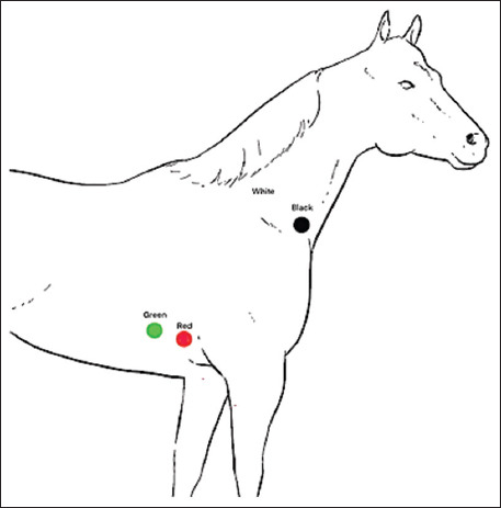 Figure 1
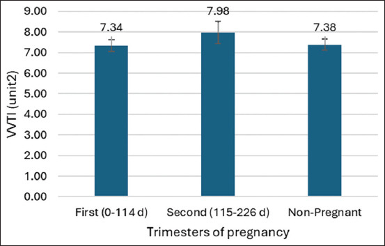 Figure 2
| Items | HR | SDNN | SDNN_idx | rMSSD | SDANN | pNN50 |
|---|---|---|---|---|---|---|
| Descriptive statistics | ||||||
| n | 45 | 45 | 45 | 45 | 45 | 45 |
| Mean | 47.2 | 168.5 | 154.5 | 87.7 | 44.6 | 18.4 |
| Standard deviation | 11.0 | 118.7 | 116.7 | 56.6 | 37.0 | 12.2 |
| Skewness | 1.7 | 1.4 | 1.5 | 1.4 | 1.8 | 0.6 |
| Coefficients variation | 23.3 | 70.5 | 75.5 | 64.6 | 82.9 | 63.9 |
| Standard error mean | 1.6 | 17.7 | 17.4 | 8.4 | 5.5 | 1.8 |
| Median | 45.0 | 124.0 | 110.0 | 76.0 | 29.5 | 16.0 |
| Mode | 42.0 | 121.0 | 38.0 | 36.0 | 60.0 | 13.0 |
| Maximum | 88.0 | 539.0 | 529.0 | 250.0 | 182.0 | 48.0 |
| Minimum | 33.0 | 35.0 | 34.0 | 30.0 | 4.0 | 1.0 |
| Tests for normality | ||||||
| Shapiro-Wilk (Pr < W) | 0.85 (<0.0001) | 0.85 (<0.0001) | 0.82 (<0.0001) | 0.82 (<0.0001) | 0.83 (<0.0001) | 0.94 (0.03) |
| HRV variables | Trimesters of pregnancy | n | Wilcoxon scores | Kruskal–Wallis test | ||
|---|---|---|---|---|---|---|
|
|
| |||||
| Sum of the scores | Mean score | Chi-square (df = 2) | Pr > Chi-square | |||
| HR | First (0–114 d) | 18 | 385.0 | 21.4ac | 6.84 | 0.032 |
| Second (115–226 d) | 22 | 596.5 | 27.1ab | |||
| Non-pregnant | 5 | 53.5 | 10.7c | |||
| SDNN | First (0–114 d) | 18 | 366.0 | 20.3 | 3.25 | 0.20 |
| Second (115–226 d) | 22 | 507.5 | 23.1 | |||
| Non-pregnant | 5 | 161.5 | 32.3 | |||
| SDNN_idx | First (0–114 d) | 18 | 363.0 | 20.2 | 3.15 | 0.21 |
| Second (115–226 d) | 22 | 512.5 | 23.3 | |||
| Non-pregnant | 5 | 159.5 | 31.9 | |||
| rMSSD | First (0–114 d) | 18 | 366.0 | 20.3 | 1.93 | 0.38 |
| Second (115–226 d) | 22 | 523.0 | 23.8 | |||
| Non-pregnant | 5 | 146.0 | 29.2 | |||
| SDANN | First (0–114 d) | 18 | 375.0 | 20.8 | 1.13 | 0.57 |
| Second (115–226 d) | 22 | 552.5 | 25.1 | |||
| Non-pregnant | 5 | 107.5 | 21.5 | |||
| pNN50 | First (0–114 d) | 18 | 462.5 | 25.7 | 1.34 | 0.51 |
| Second (115–226 d) | 22 | 459.5 | 20.9 | |||
| Non-pregnant | 5 | 113.0 | 22.6 | |||
| Trimesters of pregnancy | n | Wilcoxon scores | Kruskal-Wallis test | ||
|---|---|---|---|---|---|
|
|
| ||||
| Sum of the scores | Mean score | Chi-square (df = 2) | Pr > Chi-square | ||
| First (0–114 d) | 18 | 397 | 22.05 | ||
| Second (115–226 d) | 22 | 451 | 21.47 | ||
| Non-pregnant | 5 | 142 | 28.40 | 1.21 | 0.54 |
Peer Reviews
No public reviews on file for this paper yet. If you reviewed it on a platform where reviews are public (OpenReview, ICLR, NeurIPS, ICML), you can paste yours below so the community can read it here.
Videos
No videos yet. Explain this paper in a talk, walkthrough, or lecture? Add one.
Taxonomy
TopicsVeterinary Equine Medical Research · Reproductive Physiology in Livestock · Pharmacological Effects and Assays
INTRODUCTION
Pregnancy triggers a cascade of anatomical and physiological changes in the mare to support the developing fetus [1]. These changes begin shortly after conception and affect multiple organ systems. The autonomic nervous system (ANS) plays a pivotal role in orchestrating these adaptations, particularly through modulation of cardiac output (CO) and vascular resistance [2]. During early pregnancy, CO can increase by up to 20%, with noticeable changes occurring as early as 8 weeks of gestation [3]. Both heart rate (HR) and HR variability (HRV) are significantly altered during pregnancy [4], reflecting the influence of ANS adjustments. HRV serves as a non-invasive marker for evaluating the balance between sympathetic and parasympathetic nervous system activity [5].
In humans, the first-trimester is typically associated with an increase in HR and a reduction in HRV, indicating a shift toward greater sympathetic dominance and reduced parasympathetic activity [6]. However, comparable data on ANS function during early gestation in mares are limited. Understanding the trajectory of normal physiological changes in early pregnancy is essential for distinguishing healthy adaptation from pathological alterations in equine reproduction.
In horses, HRV is a widely accepted, non-invasive approach for assessing physiological and psychological states [7–10], as well as detecting arrhythmias [11]. HRV is defined as the variation in the intervals between successive heartbeats (RR or NN intervals), particularly those occurring between R waves. A specific time-domain index, vasovagal tonus index (VVTI), has proven useful in evaluating sympathovagal balance in horses and offers a simplified method for interpreting HRV in field conditions [12].
A recent study by Chompoosan et al. [13] has reported a decline in VVTI and an increase in HR in third-trimester pregnant mares compared with non-pregnant counterparts. Additionally, HR tends to rise before foaling, while HRV diminishes during parturition in smaller mare breeds [14]. Despite these observations, the pattern of HR and HRV changes during the early stages of pregnancy in mares remains unclear.
While numerous studies have explored the cardiovascular adaptations in pregnant mares during the late stages of gestation, especially in the third-trimester, there is a notable lack of data regarding these changes during early pregnancy. Previous research has demonstrated increased HR and decreased HRV in the final trimester, suggesting heightened sympathetic activity and reduced parasympathetic tone. However, these findings are limited to advanced pregnancy stages, and there is insufficient understanding of how early gestational changes, particularly during the first and second trimesters, affect autonomic regulation in mares. Furthermore, although the VVTI has been validated as a practical measure of autonomic tone in horses, its application in monitoring early pregnancy remains underexplored. This knowledge gap restricts veterinarians’ ability to distinguish between normal and pathological cardiovascular responses during the early stages of equine gestation, especially in field conditions where invasive diagnostics may not be feasible.
To address this gap, the present study aimed to investigate the effects of early pregnancy on HR and time-domain HRV parameters in Thai native crossbred mares. Specifically, the objective was to compare HR and HRV indices, including standard deviation of all NN intervals (SDNN), SDNN index, root mean square of successive differences (rMSSD), standard deviation of 5-min mean NN intervals (SDANN), percentage of successive NN intervals >50 ms (pNN50), and VVTI – among healthy non-pregnant mares and mares in their first and second trimesters of pregnancy. By evaluating these physiological markers, this study seeks to enhance understanding of ANS adaptations in early equine pregnancy and establish reference values that can support clinical monitoring, reproductive management, and welfare assessment in broodmares.
MATERIALS AND METHODS
Ethical approval
This study was approved by the Institutional Animal Ethics Committee of Mahasarakham University, Thailand (Approval number: IACUC-MSU-44/2024).
Study period and location
The study was conducted from October to November 2024 at the Husbandry Section of the 2^nd^ Livestock and Agriculture Division, Veterinary and Remount Department, located in Tha Phra Subdistrict, Mueang District, Khon Kaen Province, Thailand.
Animals and grouping
A total of 45 healthy Thai native crossbred mares were included, with an average age of 7.6 ± 2.5 years and body weight of 382.4 ± 71.0 kg. Pregnancy status and gestational age were determined using breeding history and transrectal ultrasonography. Based on gestational age, animals were allocated into three groups:
- Non-pregnant mares (n = 5)
- First-trimester pregnant mares (0–114 days; n = 18)
- Second-trimester pregnant mares (115–226 days; n = 22) [15].
All mares were free from cardiac abnormalities (e.g., murmurs, arrhythmias) and showed no pregnancy-related complications. The mares were housed under identical management conditions at a single facility (16°20’18.6”N, 102°48’23.5”E) and were fed Brachiaria mutica (para grass) ad libitum along with a 16% protein commercial concentrate (Charoen Pokphand Group, Bangkok, Thailand) at 1.6 kg/100 kg body weight. Water and mineral salts were also provided ad libitum. All recordings were performed between 2:00 and 3:00 p.m.
Experimental design and data collection
This study aimed to evaluate HR and time-domain HRV parameters, including SDNN, SDNN index, rMSSD, SDANN, pNN50, and VVTI. Before data collection, all mares underwent a physical examination and were allowed to stand quietly for 5-min with a halter to minimize excitement.
Electrocardiographic recording
Electrocardiogram (ECG) recordings were acquired using a Holter ECG system (SE-2012, EDAN Instruments Inc., China), which features a 3-channel display and settings of 25 mm/s paper speed and 10 mm/mV sensitivity. Mares were not sedated. Electrodes were attached using a modified base-apex lead system [16] as shown in Figure 1:
Electrode placement of the modified base-apex lead method [16].
- Green and red electrodes: 5^th^ intercostal space (right side)
- White and black electrodes: Right jugular furrow.
Each mare was recorded at rest for 15 min. The ECG data were saved to a secure digital card and transferred to a computer for analysis using Holter software.
HR and HRV analysis
HR and HRV parameters were automatically calculated from RR intervals using Holter software. Artifacts in the recordings were manually corrected before analysis. The following time-domain HRV metrics were computed:
- SDNN: Standard deviation of all RR intervals
- SDNN index: Mean of 5-min SDNN values
- rMSSD: Root mean square of successive RR differences
- SDANN: Standard deviation of 5-min mean RR intervals
- pNN50: Percentage of successive RR intervals >50 ms
- VVTI: Calculated using the first 20 consecutive RR intervals with the formula:
VVTI = NL [VAR (RR_1_–RR_20_)],
where NL is the natural logarithm and VAR represents variance in milliseconds [17].
Statistical analysis
The normality of HRV parameters was assessed using Q–Q plots and the Shapiro–Wilk test. Since most variables were not normally distributed, non-parametric methods (Kruskal–Wallis test) were applied. VVTI values were log-transformed based on the method by Pereira et al. [17] and analyzed using a generalized linear model. Significant differences were followed by post hoc comparisons using the Mann–Whitney U test. All analyses were performed using SAS University Edition (SAS/STAT, SAS Institute Inc., NC, USA). Data are presented as means, and statistical significance was set at p < 0.05.
RESULTS
Normality testing of HRV parameters
HRV parameters were assessed for normality using the Shapiro–Wilk test. The variables SDNN, SDNN index, rMSSD, SDANN, and pNN50 all exhibited high coefficients of variation (CV) (CV > 50%), indicating substantial variability among samples (Table 1). The Shapiro–Wilk test results showed that these parameters were not normally distributed (p < 0.05), necessitating the use of non-parametric statistical methods for further analysis.
Comparison of HR among groups
HR values significantly differed among the groups. The mean HR was significantly higher in second-trimester pregnant mares (51 ± 12.68 bpm) compared to first-trimester mares (46 ± 8.79 bpm) (p < 0.05). Additionally, first-trimester pregnant mares had a significantly higher HR than non-pregnant mares (39 ± 5.87 bpm) (p < 0.05) (Table 2). These findings suggest a progressive increase in maternal HR with advancing gestation.
Comparison of time-domain HRV parameters and VVTI
Despite the significant increase in HR, no significant differences were observed in time-domain HRV parameters – SDNN, SDNN index, rMSSD, SDANN, and pNN50 – across the three groups. Similarly, VVTI values did not significantly differ among non-pregnant, first-trimester, and second-trimester mares (Tables 2 and 3; Figure 2). These results suggest that while HR increases during early pregnancy, the autonomic balance, as reflected by HRV and VVTI, remains stable.
Comparison of the ls-means vasovagal tonus index parameters in each trimester of the pregnancy of mares using a generalized linear model.
DISCUSSION
Comparison with previous findings
A previous study by Chompoosan et al. [13] has reported an increase in HR and a reduction in VVTI in mares during the third-trimester of pregnancy (average 235 ± 35.8 days) compared to non-pregnant mares, suggesting heightened sympathetic activity in late gestation. However, evidence regarding HR and HRV changes during early pregnancy remains scarce. This study hypothesized that HR would increase and HRV would decrease in mares during the first and second trimesters (average 121.5 ± 47.9 days), compared to healthy non-pregnant mares. The primary finding confirmed an increase in HR during early pregnancy, but no significant changes were observed in time-domain HRV parameters.
HR elevation and physiological adaptation
HR was significantly higher in first-trimester mares than in non-pregnant mares and was highest in second-trimester mares. This pattern may be attributed to ongoing fetal development and the associated physiological demands during early gestation [18]. Cardiovascular changes are known to begin early in pregnancy, with CO increasing by approximately 20% by the 8^th^ week of gestation [3]. This increase is likely facilitated by a shift toward sympathetic dominance and reduced parasympathetic tone, allowing for enhanced uteroplacental perfusion [3, 19, 20]. In the current study involving medium-sized Thai native crossbred mares, second-trimester HR (50.4 ± 13.3 bpm) was higher than first-trimester HR (45.7 ± 8.8 bpm), aligning with findings in both horses [14, 21] and humans [4].
Stability of HRV parameters during early pregnancy
Although a clear increase in HR was observed, time-domain HRV parameters (SDNN, SDNN index, rMSSD, SDANN, and pNN50) did not differ significantly between pregnant and non-pregnant mares. This finding is consistent with previous studies by Nagel et al. [22] and Nagel et al. [23] reporting minimal HRV change during gestation in mares. While recent data have shown significantly reduced VVTI in third-trimester pregnant mares [13], our evaluation of VVTI during early pregnancy revealed no significant differences across groups. These results contrast with human studies, where reductions in HRV during both the first [24] and second trimesters [4] have been well documented.
Species-specific autonomic responses
The discrepancy between species may be explained by fundamental physiological differences. Hemodynamic and autonomic regulatory changes occur during early pregnancy, with increased sympathetic and decreased parasympathetic activity supporting fetal growth [25]. Horses, however, are known to have higher baseline vagal tone than many other species, including dogs, rabbits, mice, and humans, potentially contributing to the stability of HRV despite rising HR [26].
In equine physiology, HRV reductions are more commonly associated with stress [27], physical conditioning [28], or cardiovascular pathology [29], rather than normal pregnancy. The reduction in VVTI observed in third-trimester mares may be due to elevated fetal cortisol and sympathetic activation, reflecting prepartum stress responses [30, 31].
Implications and future directions
Given that VVTI is a simple yet effective time-domain measure of HRV, its use in third-trimester pregnancy monitoring appears promising. However, during early gestation, it may not be sensitive enough to detect subtle changes in autonomic tone. Hormonal markers – including progesterone, androgens, estrogens, and their metabolites – could complement HR and HRV assessments to better estimate gestational stages, especially when palpation or ultrasonography is not feasible [32].
Hematological and biochemical indicators such as albumin, blood urea nitrogen, and potassium also shift throughout pregnancy and may reflect metabolic adaptations across trimesters [33]. Future research should investigate the combined use of endocrine profiles, hematology, and advanced HRV analyses to refine monitoring strategies for pregnant mares.
Strengths, limitations, and methodological considerations
Although this study only employed time-domain HRV parameters, newer techniques such as frequency-domain and non-linear HRV analysis are gaining importance in equine research and will be considered in future studies by Physick-Sheard et al. [34] and Sanigavatee et al. [35]. The use of time-domain methods offered practical advantages for field-based ECG acquisition, particularly given the limited duration of recordings and the small number of non-pregnant control animals. The constrained sample size was due to the primary mission of the participating facility, to breed and maintain pregnant mares, which reduced access to non-pregnant subjects.
CONCLUSION
This study demonstrated that HR progressively increases during early pregnancy in Thai native crossbred mares, with significantly higher HR observed in first-trimester mares compared to non-pregnant mares and further elevation in the second-trimester. However, time-domain HRV parameters, including SDNN, SDNN index, rMSSD, SDANN, pNN50, and VVTI remained stable across all groups, suggesting that ANS balance is maintained during the early gestational period.
These findings provide important baseline data for the physiological cardiovascular adaptation in mares and support the use of HR monitoring as a simple and non-invasive indicator of early pregnancy progression. The use of a portable Holter ECG system in this study also highlights the practical feasibility of field-based cardiac assessments in equine practice.
A key strength of this research lies in its focus on early pregnancy stages, which have been underrepresented in equine cardiovascular studies. The inclusion of VVTI as a simple HRV measure further enhances its clinical applicability in routine reproductive monitoring.
However, the study is not without limitations. The relatively small number of non-pregnant mares, due to the breeding-focused nature of the study site, may limit broader generalization. Additionally, the analysis was confined to time-domain HRV parameters, without incorporating frequency-domain or non-linear measures, which could provide deeper insight into ANS dynamics.
Future research should consider integrating hormonal, hematological, and biochemical profiles alongside HR and HRV metrics to better predict gestational stages and parturition. Expanded use of advanced HRV analysis techniques and longitudinal monitoring throughout pregnancy would further clarify the trajectory of ANS modulation in mares.
In conclusion, while HR increases early in pregnancy as part of the cardiovascular adaptation, HRV remains unchanged, reflecting a stable autonomic environment in the first and second trimesters. These findings lay the groundwork for improved physiological monitoring and health assessment strategies in pregnant mares under field conditions.
AUTHORS’ CONTRIBUTIONS
WK: Designed the study and analyzed the data. SS, CC, PP, TP, WP, and WL: Recorded and analyzed the data. WL: Coordinated the study. WL, SS, WP, and WK: Drafted and revised the manuscript. All authors have read and approved the final version of the manuscript.
The reference list from the paper itself. Each links out to its DOI / PubMed record.
- 1Sanghavi M Rutherford J.D Cardiovascular physiology of pregnancy Circulation 201413012100310082522377110.1161/CIRCULATIONAHA.114.009029 · doi ↗ · pubmed ↗
- 2Mulder E.Gde Haas S Mohseni Z Schartmann N Abo Hasson F Alsadah Fvan Kuijk Svan Drongelen J Spaanderman M Ghossein-Doha C Cardiac output and peripheral vascular resistance during normotensive and hypertensive pregnancy - a systematic review and meta-analysis BJOG 2022129(5)6967073363040310.1111/1471-0528.16678 PMC 9291763 · doi ↗ · pubmed ↗
- 3Soma-Pillay P Nelson-Piercy C Tolppanen H Mebazaa A Physiological changes in pregnancy Cardiovasc. J. Afr 201627289942721385610.5830/CVJA-2016-021PMC 4928162 · doi ↗ · pubmed ↗
- 4Sarhaddi F Azimi I Axelin A Niela-Vilen H Liljeberg P Rahmani A.M Trends in heart rate and heart rate variability during pregnancy and the 3-month postpartum period:Continuous monitoring in a free-living context JMIR Mhealth Uhealth 2022106 e 334583565766710.2196/33458 PMC 9206203 · doi ↗ · pubmed ↗
- 5Duong H.T.H Tadesse G.A Nhat P.T.H Hao N.V Prince J Duong T.D Kien T.T Nhat L.T.H Tan L.V Pugh C Loan H.T Chau N.V.V Minh Yen L Zhu T Clifton D Thwaites L Heart rate variability as an indicator of autonomic nervous system disturbance in tetanus Am. J. Trop. Med. Hyg 202010224034073183347110.4269/ajtmh.19-0720 PMC 7008337 · doi ↗ · pubmed ↗
- 6Carpenter R.ED'Silva L.A Emery S.J Uzun O Rassi D Lewis M.J Changes in heart rate variability and QT variability during the first-trimester of pregnancy Physiol. Meas 20153635315452569010510.1088/0967-3334/36/3/531 · doi ↗ · pubmed ↗
- 7Huangsaksri O Sanigavatee K Poochipakorn C Wonghanchao T Yalong M Thongcham K Srirattanamongkol C Pornkittiwattanakul S Sittiananwong T Ithisariyanont B Pakpokai C Ninyeeruae S Chanda M Physiological stress responses in horses participating in novice endurance rides Heliyon 20241011 e 318743884592510.1016/j.heliyon.2024.e 31874 PMC 11153225 · doi ↗ · pubmed ↗
- 8Huangsaksri O Wonghanchao T Sanigavatee K Poochipakorn C Chanda M Heart rate and heart rate variability in horses undergoing hot and cold shoeing P Lo S One 2024196 e 03050313884325410.1371/journal.pone.0305031 PMC 11156413 · doi ↗ · pubmed ↗