First morphological description of the larval stages of three Microtendipes species (Diptera, Chironomidae) from South China with molecular confirmation

Abstract
Genes, proteins, chemicals, diseases, species, mutations and cell lines named across the full text — each resolved to its canonical identifier and authoritative record.
Click any figure to enlarge with its caption.
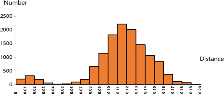 Figure 1
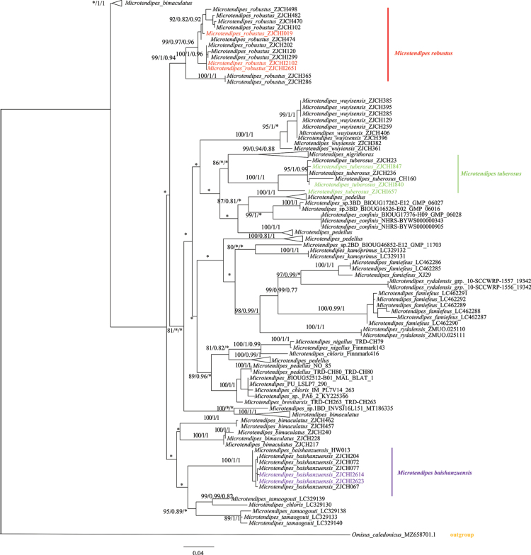 Figure 2
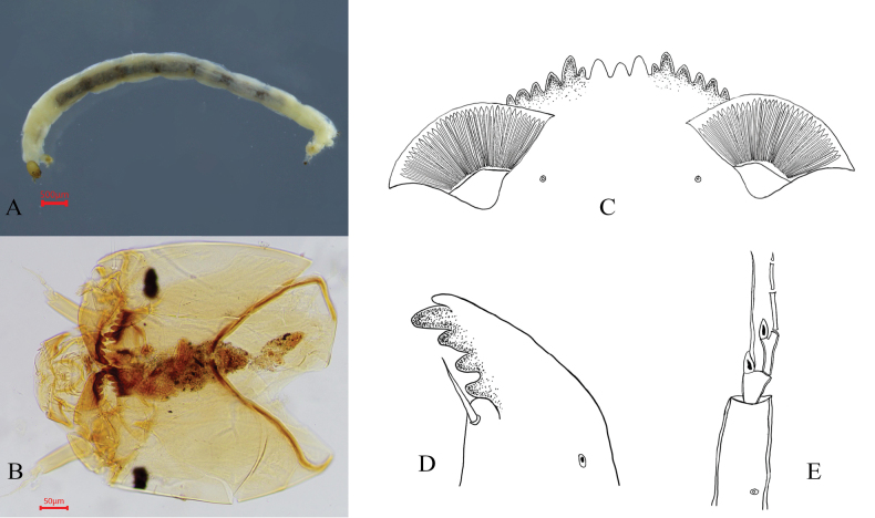 Figure 3
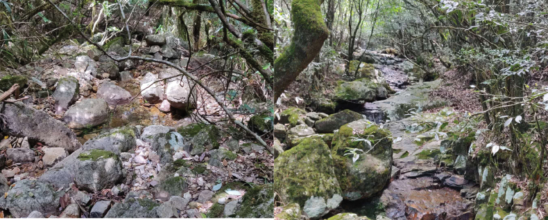 Figure 4
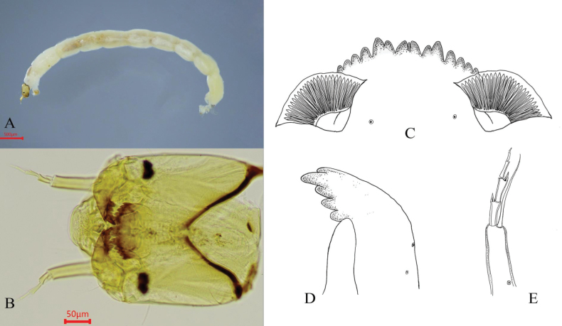 Figure 5
 Figure 6
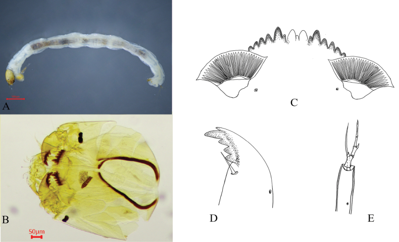 Figure 7
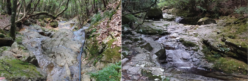 Figure 8
|
|
|
| |
|---|---|---|---|
|
| 2 | 3 | 3 |
| B.l. /mm | 7.1–7.8, 7.5 | 2.1–3.7, 2.8 | 2.9–9.6, 5.0 |
| H. w. | 450–460, 455 | 287.5–300, 292.5 | 287.5–620.0, 480.6 |
|
| 200–202.5, 201.25 | 150–157.5, 153.3 | 125–228, 190.8 |
|
| 105–100, 102.5 | 67.5–77.5, 72.1 | 71–128, 109.8 |
| Ant1 | 102.5–112.5, 107.5 | 100–102.5, 101.2 | 42.5–117.5, 75.8 |
| Ant2 | 25–27.5, 26.3 | 27–28, 27.5 | 15.5–25, 22.6 |
| Ant3 | 27.0 | 23–26.3, 24.8 | 17.5–27.5, 21.9 |
| Ant4 | 25.0 | 16–21.3, 19.1 | 15–18, 17.1 |
| Ant5 | 17–17.5, 17.3 | 10–11.3, 10.4 | 11.0–15.0, 12.9 |
| Ant6 | 10.0 | 7.5–8.8, 7.9 | 5.0–8.0, 6.1 |
|
| 1.0–1.1, 1.0 | 1.1–1.2, 1.1 | 0.8–1.3, 0.9 |
| AIR | 3.0–3.2, 3.1 | 3.7–4.4,4.0 | 1.4–3.2, 2.5 |
|
| 0.2–0.3, 0.27 | 0.2–0.2, 0.2 | 0.3–0.7, 0.4 |
| Bl | 106–125, 115.5 | 53–72, 59.67 | 87.5–137.5, 114.4 |
|
| 1.0–1.2, 1.1 | 0.6–0.8, 0.7 | 1.3–1.6, 1.4 |
| Pmd | 97.5–120, 108.8 | 62.5–65, 63.8 | 60–110, 90 |
|
| 168–195, 181.5 | 125–132.5, 129.8 | 115–200, 174.4 |
| M. w | 152.5–152.5, 152.5 | 105–110, 108 | 92.5–185, 151.9 |
|
| 37.5 | 23–25, 24.3 | 20.5–42.5, 33.3 |
| AS | 520–611, 565.5 | 197.4–445, 327.5 | 347–602.3, 492.5 |
|
| 125–132.5, 128.8 | 83–122.5, 96.8 | 75–150, 120 |
|
| 75–80, 77.5 | 37.5–57.5, 49.3 | 48–87.5, 72.3 |
|
| 83–90, 86.5 | 59–85, 68.8 | 51–125, 105.9 |
|
| 35–40, 37.5 | 22–29, 26 | 28–43, 38.3 |
|
| 8.0–10.0, 9.0 | 8.5–9.5, 9.0 | 8.5–12.5, 10.3 |
| 1 | Mental plate lacking distinct median teeth; anterior margin straight. Median teeth subequal in size; all teeth on the mental plate dark brown. Distributed in Afrotropical and Oriental regions |
|
| – | Mental plate with three characteristic median teeth, equal to or slightly lower than second lateral teeth; bases of 1st and 2nd lateral teeth fused |
|
| 2 | Median teeth distinct, always paler than lateral teeth and equal/subequal in size (if equal at base, median teeth slightly smaller); pecten epipharyngis single, with 3–4 large central teeth and 2–3 smaller lateral teeth; premandible with 5 teeth |
|
| – | Median teeth trifid, central median tooth often reduced/indistinct; all three median teeth paler or concolorous with lateral teeth; premandible with 3 teeth |
|
| 3 | Central median tooth slightly wider than or equal to outer median teeth; blade reaching apex of fourth antennal segment; labral lamella with 12–14 small teeth; pecten epipharyngis comprising 3–4 large and 3–5 small teeth |
|
| – | Central median tooth smaller than outer lateral teeth |
|
| 4 | Pecten epipharyngis bearing three broad apical marginal teeth; labral lamella with 20–22 small teeth; blade significantly longer than flagellum |
|
| – | Pecten epipharyngis with 9–11 teeth (mixed large and small); labral lamella with 22–24 small teeth; blade slightly shorter than or equal to flagellum |
|
| 5 | Median teeth dark brown, concolorous with lateral teeth |
|
| – | Median teeth distinctly paler than other teeth; pecten epipharyngis often trilobed |
|
| 6 | Labral lamella with 14–20 fine teeth |
|
| – | Labral lamella with 12–16 teeth |
|
| 7 | Pecten epipharyngis with three broad marginal teeth apically; premandible with two apical teeth (length 60.0–110.0 μm, mean 90.0); labral lamella with 16–17 teeth; cephalic capsule light brown, mentum dark brown with pale median teeth |
|
| – | Pecten epipharyngis not with three broad marginal teeth apically; premandible with three teeth |
|
| 8 | Pecten epipharyngis with three broad marginal teeth apically; premandible with two apical teeth (length 97.5–120.0 μm, mean 108.8); labral lamella with 15 teeth |
|
| – | Pecten epipharyngis not with three broad marginal teeth; premandible not bifid; striae: 28–32 (mean 30); head width: 360–410 μm (mean 380) |
|
| 9 | Premandible bifid (length 62.5–65.0 μm, mean 63.8); pecten epipharyngis with 8 teeth; total length: 2.1–3.7 mm (mean 2.8) |
|
| – | Premandible not bifid; pecten epipharyngis not with 8 teeth; total length: 5.5–9.4 mm (mean 6.9) |
|
| 10 | Labral lamella with 14–18 teeth (mode 16) |
|
| – | Labral lamella with 19–22 teeth (mode 20) |
|
- —National Natural Science Foundation of China 501100001809 https://ror.org/01h0zpd94 http://doi.org/10.13039/501100001809
Peer Reviews
No public reviews on file for this paper yet. If you reviewed it on a platform where reviews are public (OpenReview, ICLR, NeurIPS, ICML), you can paste yours below so the community can read it here.
Videos
No videos yet. Explain this paper in a talk, walkthrough, or lecture? Add one.
Taxonomy
TopicsAquatic Invertebrate Ecology and Behavior · Freshwater macroinvertebrate diversity and ecology · Environmental DNA in Biodiversity Studies
Introduction
The genus Microtendipes Kieffer was established by Kieffer in 1915 and currently comprises over 60 described species (Freeman and Cranston 1980; Cranston and Martin 1989; Ashe and Cranston 1990; Oliver et al. 1990; Zorina 2001; Qi and Wang 2006; Qi et al. 2014; Yamamoto and Yamamoto 2014; Hazra et al. 2016; Tang and Hiromi 2017; Song et al. 2023). In China, 18 recorded species have been described (Qi and Wang 2006; Qi et al. 2012, 2014; Song et al. 2023). Taxonomic research on Microtendipes has primarily focused on adult morphology, and larval descriptions are scarce. This gap is largely due to the high morphological homogeneity of larvae and the lack of reliable diagnostic traits, particularly distinct autapomorphies. These limitations collectively hinder species differentiation, precise taxonomic classification, and the establishment of a robust larval classification system. Furthermore, significant technical challenges persist in specimen collection and analysis, requiring specialized apparatus and microscopic techniques, and database limitations hinder reliable larva–adult matching. Despite these challenges, DNA barcoding has emerged as an effective identification tool (Carew et al. 2005, 2007; Gadawski et al. 2022).
DNA barcodes are included in most new species descriptions as a standard method for delimiting non-biting midges (Song et al. 2016, 2022; Yan et al. 2017; Lin et al. 2019). DNA barcoding has also proven effective for associating specimens across different life stages (Song et al. 2018). However, the larval stages of these species have remained undiscovered. To address this gap in the literature, we conducted extensive larval collections and performed DNA barcode analyses. Consequently, we successfully collected and identified the larvae of three Microtendipes species: M. baishanzuensis Song & Qi, 2023, M. robustus Song & Qi, 2023, and M. tuberosus Qi & Wang, 2006, providing the first descriptions of their larval stages.
Material and methods
Larval specimens were collected from multiple rivers and streams using D-shaped nets. All specimens were preserved in 75% ethanol and stored at −20 °C in the laboratory until subsequent morphological and molecular analyses. For morphological examination, specimens were slide-mounted in Euparal and examined under a microscope. Measurements followed standardized protocols, and morphological terminology and abbreviations were adopted from Sæther (1980) and Maschwitz and Cook (2000). Values are reported as ranges accompanied by mean values, with the number of observed specimens (N) indicated in parentheses. All specimens are deposited in the College of Life Sciences, Taizhou University.
Genomic DNA was extracted using the protocol outlined by Song et al. (2018). The standard barcode region of the 5′ portion of the mitochondrial gene cytochrome c oxidase I (COI-5P) was amplified using the universal primers LCO1490 and HCO2198 (Folmer et al. 1994). PCR amplifications were performed as described by Song et al. (2018). PCR products were electrophoresed on a 1.0% agarose gel, purified, and sequenced using an ABI 3730XL capillary sequencer (Beijing Genomics Institute Co., Ltd., Hangzhou, China). Raw sequences were edited using BioEdit v. 7.2.5 (Hall 1999).
Publicly available Microtendipes sequences (≥500 bp in length) were retrieved from the Barcode of Life Data System (BOLD; http://www.boldsystems.org/) on June 20, 2024 (see Suppl. material 1 for details). Sequence alignment was conducted in MEGA v. 12 (Kumar et al. 2024) using the ClustalW algorithm. Pairwise genetic distances were calculated using the K2P model in MEGA v. 12. A neighbor-joining (NJ) tree was constructed based on the Kimura 2-Parameter (K2P) substitution model with 1000 bootstrap replicates, using the “pairwise deletion” option to handle missing data. A maximum-likelihood (ML) tree was generated using IQ-TREE v. 2.1.3 (Nguyen et al. 2015), with node support assessed through ultrafast bootstrapping with 1000 replicates. Bayesian inference (BI) analysis was conducted using MrBayes v. 3.2.7 (Ronquist et al. 2012), employing Markov chain Monte Carlo (MCMC) randomization for 10 million generations, with the first 25% of trees discarded as burn-in. Trace files from the BI analysis were examined using Tracer v. 1.7 (Rambaut et al. 2018), and the final phylogenetic tree was visualized in FigTree v. 1.4.2.
Abbreviations
Morphological abbreviations of larval characters are used as follow: Ant 1–6, length of antennal segments 1–6 in µm; A1R, first antennal segment ratio, length of segment 1: width of segment 1 through the ring organ; AR, antennal ratio, length of basal segment: combined lengths of segments 2 to apex; B. l., total body length in mm; Bl, antennal blade length; BlR, blade ratio, length of blade: total length of Ant 2–6; Pmd, premandible length; H. l., head capsule length, length from anterior labrum to posterior margin of capsule; L, larva; Md, mandible length; M. w, mentum width; Mmw, median mental tooth width; PM, postmentum length; ROR, ring organ ratio, distance from basal to location of ring organ: length of basal antennal segment; SAS, length of supraanal seta; SSm–SSm: distance between setae submenti; V. w, ventromentum width; V. l, ventromentum length; IPD, inter-ventromentum plates distance; Str, striae (Sæther 1980; Maschwitz and Cook 2000).
Results and discussion
DNA barcodes analysis
This study analyzed a total of 161 sequences, including eight newly obtained larval sequences. A clear barcode gap of 4–6% was observed (Fig. 1), confirming the effectiveness of DNA barcoding for species delimitation within this genus. Based on three known male specimens, the maximum intraspecific genetic distance was 0.7% (mean: 0.5%) for M. baishanzuensis, 5.8% (mean: 2.9%) for M. robustus, and 3.2% (mean: 1.4%) for M. tuberosus. After including larval DNA sequences, these values changed slightly: M. baishanzuensis exhibited a maximum of 0.9% (mean: 0.5%), M. robustus a maximum of 5.8% (mean: 2.5%), and M. tuberosus a maximum of 6.1% (mean: 2.6%) (Suppl. material 2). Notably, the maximum intraspecific distance observed in M. tuberosus (6.1%) remained below the minimum interspecific distance (8.0%) between M. baishanzuensis and M. robustus.
Histogram of pairwise K2P distances between morphological species of Microtendipes. The horizontal axis represents the pairwise K2P distance, the vertical axis represents the number of pairwise sequence comparisons.
Phylogenetic analyses (Neighbor-joining, maximum likelihood, and Bayesian inference) revealed strong concordance between larval and adult sequences (Fig. 2). For M. tuberosus, although minor topological variations occurred across trees, all three methods provided robust support for the same clade structure. In M. robustus, consistent results across all three methods (NJ/ML/BI) confirmed larval–adult conspecificity via DNA barcoding. For M. baishanzuensis, all three phylogenetic reconstructions yielded well-resolved, congruent clades. This high consistency in branching patterns, combined with the low maximum pairwise genetic distance (0.9%), conclusively identifies the larvae as M. baishanzuensis using DNA barcoding.
Phylogeny tree for Microtendipes based on DNA barcode sequences. The tree was based on partial COI sequences and the generalized time-reversible substitution model. Omisus caledonicus (Edwards) was used as an outgroup. The phylogenetic analysis incorporated data from maximum-likelihood (ML), Bayesian inference (BI), and neighbor-joining (NJ) methods for all three identified species; the data are presented as ML/BI/NJ analyses. Only nodes with Ultra-BS (ML) > 80%, PP > 0.80, and BS(NJ) > 0.75 are shown; different colors within each area represent larvae.
Taxonomy
Microtendipes
baishanzuensis
Taxon classificationAnimaliaDipteraChironomidae
Song & Qi, 2023
CA90D76E-C0CE-514E-AF3A-E16EF1C09F9C
Microtendipes baishanzuensis Song & Qi, 2023: 8.
Material examined
(N = 2): 2 larvae, China, Zhejiang Province, Lishui City, Qingyuan County, Baishanzu Town, 1600 m a.s.l., 27.750°N, 119.198°E, 14.VIII.2020, C. Song.
Diagnosis
(Fig. 3B, C). Mentum dark brown, with median teeth distinctly paler than lateral teeth; cephalic capsule brown. Total length 7.1–7.8, 7.5 mm; head capsule length 490.0–520.0, 505.0 μm; head capsule width 450.0–460.0, 455.0 μm.
Larva of Microtendipes baishanzuensis Song & Qi, 2023. A. Habitus of larva; B. Head capsule; C. Mentum; D. Mandible; E. Antenna.
Mentum (Fig. 3C). Width 152.5 μm; median tooth width 37.5 μm; ventromental plate width 125.0–132.5, 128.8 μm; distance between ventromental plates 83.0–90.0, 86.5 μm; striae count 35.0–40.0, 37.5.
Mandible (Fig. 3D). Length 168.0–195.0, 181.5 μm.
Antenna (Fig. 3E). Segment lengths, 102.5–112.5, 107.5 μm; 25.0–27.5, 26.3 μm; 27.0 μm; 25.0 μm; 17.0–17.5,17.3 μm; 10.0 μm. Antennal ratio (AR) 1.0–1.1, 1.0. Ring organ width 34.0–35.0, 34.5 μm; ring organ positioned at 0.2–0.3, 0.3 of basal segment length, 22.5–35.0, 28.8 μm from segment base; antennal blade 106.0–125.0, 115.5 μm in length, width 2.0–18.0 μm.
Labrum. Premandible with two apical teeth; length 97.5–120.0, 108.8 μm. Pecten epipharyngis with three broad marginal teeth apically; labral lamella composed of 15 small teeth.
Thorax. Anterior claws pale with a little golden posterior claws pale golden, both simple and dense.
Abdomen. 8 tail hairs of anal seta, 520.0–611.0, 565.5 μm (Table 1).
Table 1.: Mensural features of the larvae of Microtendipes baishanzuensis, Microtendipes tuberosus and Microtendipes robustus. Abbreviations follow as in the text.
Ecological environment
(Fig. 4). A forest stream, winding through wooded areas, exhibiting high-clarity water flowing over a predominantly rocky substrate.
Habitat of Microtendipes baishanzuensis larvae.
Remarks.
A comparative analysis was conducted between our data and the larval morphological measurements reported by Tang (2006). The larvae of M. baishanzuensis resemble Microtendipes pedellus (De Geer, 1929) and Microtendipes chloris (Meigen, 1933) in the shape of the mentum. However, M. baishanzuensis has more striae than M. pedellus (mean 37.5 vs. 30.0). The total length of the postmentum in M. baishanzuensis is shorter than in M. pedellus (mean 201.3 μm vs. 223.0 μm). The ring organ in M. baishanzuensis is located at 0.2–0.3 (mean 0.267) of the basal segment, compared to approximately 0.33. Compared to M. chloris, M. baishanzuensis has a distinctly shorter blade (mean 115.5 μm vs. 145.0 μm), a longer postmentum (mean 201.3 μm vs. 215.0 μm), and more striae (mean 37.5 vs. 28.0).
Microtendipes
tuberosus
Taxon classificationAnimaliaDipteraChironomidae
Qi & Wang, 2006
80E993F8-5CDF-5499-A18D-925216EB36C6
Microtendipes tuberosus Qi & Wang, 2006: 43.
Material examined
(N = 3): 1 larva, China, Zhejiang Province, Lishui City, Suichang County, Jiulong Mountain National Nature Reserve, 370 m a.s.l., 28.408°N, 118.811°E, 29.VIII. 2020, C. Song; 2 larvae, China, Zhejiang Province, Lishui City, Suichang County, Jiulong Mountain National Nature Reserve, 400 m a.s.l., 28.380°N, 118.799°E, 29.VIII. 2020, C. Song.
Diagnosis
(Fig. 5B, C). Mentum dark brown, with median teeth concolorous with lateral teeth; median tooth slightly shorter than the second lateral tooth. The cephalic capsule is light brown. Total length 2.1–3.7, 2.8 mm; head capsule length 375.0–400.0, 387.5 μm; head capsule width 287.5–300.0, 292.5 μm.
Larva of Microtendipes tuberosus Qi & Wang, 2006. A. Habitus of larva; B. Head capsule; C. Mentum; D. Mandible; E. Antenna.
Mentum (Fig. 5C). Width 105.0–110.0, 108.0 μm; median tooth width 23.0–25.0, 24.3 μm; ventromental plate width 83.0–122.5, 96.8 μm; distance between ventromental plates 59.0–85.0, 68.8 μm; striae count 22–29, 26.
Mandible (Fig. 5D). Length 125.0–132.5, 129.8 μm.
Antenna (Fig. 5E). Segment lengths: 100.0–102.5, 101.2 μm; 27.0–28.0, 27.5 μm; 23.0–26.3, 24.8 μm; 16.0–21.3, 19.1 μm; 10.0–11.3, 10.4 μm; 7.5–8.8, 7.9 μm).
Antennal ratio (AR) 1.1–1.2, 1.1. Ring organ width 25.5 μm; ring organ positioned at 0.15–0.22, 0.20 of basal segment length, 15.0–22.5, 20.0 μm from segment base; antennal blade 53.0–72.0, 59.7 μm in length.
Labrum. Premandible bifid; length 62.5–65.0, 63.8 μm. Pecten epipharyngis is composed of 8 teeth.
Thorax. Anterior claws pale with a faint golden tint; posterior claws pale golden. Procercus and apical setae are yellowish brown.
Abdomen. Anal setae comprising 8 hairs, length 197.4–445.0, 327.5 μm (Table 1).
Ecological environment
(Fig. 6). The stream flows through forests and villages, featuring a streambed composed of bedrock and sediment, with excellent water clarity.
Habitat of Microtendipes tuberosus larvae.
Remarks.
A comparative analysis was conducted between our data and the larval morphological measurements reported by Tang (2006). The larvae of M. tuberosus resemble those of Microtendipes britteni (Edwards, 1983) in mentum morphology. However, M. tuberosus is noticeably smaller in body size (mean 2.8 mm vs. 6.9 mm in M. britteni) and has a shorter mandibular pecten (mean 153.3 μm vs. 195.0 μm in M. britteni). Among congeners, M. tuberosus is notably smaller overall. Both its median and lateral teeth are uniformly dark brown.
Microtendipes
robustus
Taxon classificationAnimaliaDipteraChironomidae
Song & Qi, 2023
A6D2358A-FD95-56BB-BD27-18BC432B3B56
Microtendipes robustus Song & Qi, 2023:16.
Material examined
(N = 3): 1 larva, China, Zhejiang Province, Lishui City, Qingyuan County, Baishanzu Town, 1370 m, 27.754°N, 119.186°E, 14.VIII.2020, C. Song; 1 larva, China, Zhejiang Province, Lishui City, Qingyuan County, Baishanzu Town, 1650 m a.s.l., 27.751°N, 119.199°E, 14.VIII.2020, C. Song; 1 larva, China, Zhejiang Province, Wenzhou City, Taishun County, Wuyanling National Nature Reserve, 1060 m a.s.l., 27.424°N, 119.404°E, 25.IX.2020, C. Song; 1 larva, China, Zhejiang Province, Wenzhou City, Taishun County, Wuyanling National Nature Reserve, 660 m a.s.l., 27.423°N, 119.404°E, 25.IX.2020, C. Song.
Diagnosis
(Fig. 7B, C). The larvae possess a light brown cephalic capsule and dark brown mentum, with the pale median teeth forming a distinct contrast against the darker lateral teeth.
Larva of Microtendipes robustus Song & Qi, 2023. A. Habitus of larva; B. Head capsule; C. Mentum; D. Mandible; E. Antenna.
Total length. 2.9–9.6 mm, 5.0 mm; head capsule length 325.0–650.0, 521.3 μm; head capsule width 287.5–620.0, 480.6 μm.
Mentum (Fig. 7C). Width 92.5–185.0, 151.9 μm; median tooth width 20.5–42.5, 33.3 μm; ventromental plate width 75.0–150.0, 120.0 μm; distance between ventromental plates 51.0–125.0, 105.9 μm; number of striae 28–43, 38.3.
Mandible (Fig. 7D). Length 115.0–200.0, 174.4 μm.
Antenna (Fig. 7E). Segment lengths, 42.5–117.5, 75.8 μm; 15.5–25.0, 22.6 μm; 17.5–27.5, 21.9 μm; 15.0–18.0, 17.1 μm; 11.0–15.0, 12.9 μm; 5.0–8.0, 6.1 μm. Antennal ratio (AR) 0.8–1.3, 0.9. Ring organ width 20.0–37.0, 30.5 μm; ring organ positioned at 0.3–0.6, 0.4 of basal segment, 22.5–32.5, 26.3 μm from segment base.
Labrum. Premandible with two apical teeth; length 60.0–110.0, 90.0 μm. Pecten epipharyngis with three broad marginal teeth apically; labral lamella composed of 16–17 small teeth.
Thorax. Anterior claws pale with golden hints; posterior claws darker, pale brown. Procercus and apical setae are yellowish brown.
Abdomen. 8 tail hairs of anal seta, 347.0–602.3, 492.5 μm (Table 1).
Ecological environment
(Fig. 8). A mountain stream, fed by natural springs, meandering through forested terrain with a bedrock-dominated streambed and pristine water.
Habitat of Microtendipes robustus larvae.
Remarks.
A comparative analysis was conducted between our data and the larval morphological measurements reported in Tang (2006). The larvae of M. robustus are similar to those of M. pedellus in mentum shape. However, M. robustus has a shorter overall body length (mean 5.0 mm vs. 8.8 mm in M. pedellus), shorter postmentum (mean 190.8 μm vs. 223.0 μm in M. pedellus), as well as a shorter mentum (mean 174.4 μm vs. 210.0 μm in M. pedellus) compared to M. pedellus.
Conclusions
Microtendipes baishanzuensis resembles M. pedellus and M. chloris in mentum shape but differs by having more striae and a longer postmentum than M. pedellus, as well as a larger body size and more striae than M. chloris. Microtendipes tuberosus is generally similar to congeners but is notably smaller in overall size and has a shorter postmentum than M. britteni. Microtendipes robustus is challenging to identify because of its morphological overlap with M. pedellus, but key distinguishing characteristics include a shorter ventromental plate length and a greater plate distance. These specific morphometric differences, combined with molecular evidence, provide reliable criteria for species identification within this taxonomically complex genus.
Key to known larvae of Microtendipes Kieffer, modified from Tang (2006)
**: **
Supplementary Material
XML Treatment for Microtendipes baishanzuensis
XML Treatment for Microtendipes tuberosus
XML Treatment for Microtendipes robustus
The reference list from the paper itself. Each links out to its DOI / PubMed record.
- 1Ashe P Cranston PS (1990) Family Chironomidae. In: SoósÁPapp L (Eds) Catalogue of Palaearctic Diptera, Vol.2. Akadémiai Kiadó, Budapest, 113–499.
- 2Carew ME Pettigrove V Hoffmann AA (2005) The utility of DNA markers in classical taxonomy: Using Cytochrome Oxidase I markers to differentiate Australian Cladopelma (Diptera: Chironomidae) midges. Annals of the Entomological Society of America 98(4): 587–594. 10.1603/0013-8746(2005)098[0587:TUODMI]2.0.CO;2 · doi ↗
- 3Carew ME Pettigrove V Cox RL Hoffmann AA (2007) DNA identification of urban Tanytarsini chironomids (Diptera: Chironomidae).Journal of the North American Benthological Society 26(2): 587–600. 10.1899/06-120.1 · doi ↗
- 4Cranston PS Martin J (1989) Family Chironomidae. In: Evenhuis NL (Ed.) Catalog of the Diptera of the Australasian and Oceanian Regions.Bishop Museum Press, Honolulu and Brill, E.J., Leiden, 252–274.
- 5Folmer O Black M Hoeh W Lutz R Vrijenhoek R (1994) DNA primers for amplification of mitochondrial cytochrome c oxidase subunit I from diverse metazoan invertebrates.Molecular Marine Biology and Biotechnology 3: 294–299.7881515 · pubmed ↗
- 6Freeman P Cranston PS (1980) Family Chironomidae. In: Crosskey RW (Ed.) Catalogue of the Diptera of the Afrotropical region.British Museum (Natural History), London, 175–202.
- 7Gadawski P Montagna M Rossaro B Gilka W Pesic V Grabowski M Magoga G (2022) DNA barcoding of Chironomidae from the Lake Skadar region: Reference library and a comparative analysis of the European fauna.Diversity & Distributions 28(12): 2838–2857. 10.1111/ddi.13504 · doi ↗
- 8Hall TA (1999) Bio Edit: A user-friendly biological sequence alignment editor and analysis program for Windows 95/98/NT.Nucleic Acids Symposium Series 41: 95–98. 10.1021/bk-1999-0734.ch 008 · doi ↗