Spinal Leclercia adecarboxylata infection in an immunocompetent patient: case report
Mohannad M. Mallat, Abdullah Alatar, Duaa Alhumoudi, Ali Alhijji, Abdulaziz Alsubaie, Ali Somily, Sherif Elwatidy

TL;DR
A rare spinal infection caused by Leclercia adecarboxylata in a healthy patient is reported, highlighting the need for better understanding and treatment of this uncommon pathogen.
Contribution
This is the first documented case of a spinal disc infection caused by Leclercia adecarboxylata in an immunocompetent individual.
Findings
L. adecarboxylata was isolated from a spinal disc and showed resistance to multiple antibiotics.
A literature review identified 227 global cases of L. adecarboxylata infections, with high recovery rates but significant mortality in some cases.
The patient initially responded to ciprofloxacin treatment but was lost to follow-up.
Abstract
Leclercia adecarboxylata (Family: Enterobacteriaceae) is a gram-negative bacillus that is found in diverse environments but has rarely been isolated from the human microbiota. Notably, no patient with a spinal disc infection caused by L. adecarboxylata has been documented to date. A 60-year-old male presented with chronic lower back pain and decreased extensor hallucis longus muscle power upon examination. Magnetic resonance imaging of the spine revealed canal stenosis at L3-4 and L4-5, along with migrating L4-5 disc material compressing the left lateral recess and foramina. The patient underwent L4-5 decompression and fixation surgery. Culturing of the excised disc material revealed the presence of lactose-fermenting oxidase-negative L. adecarboxylata. This isolate was found to be resistant to ampicillin; first-, second-, and third-generation cephalosporins; and…
Genes, proteins, chemicals, diseases, species, mutations and cell lines named across the full text — each resolved to its canonical identifier and authoritative record.
Click any figure to enlarge with its caption.
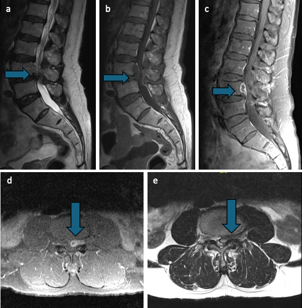 Fig 1
Fig 1| Drug | Result | CLSI MIC breakpoints for Enterobacterales | ||
|---|---|---|---|---|
| S | I | R | ||
| Ampicillin | R | ≤8 | 16 | ≥32 |
| Cefepime | S | ≤2 | – | ≥16 |
| Cefotaxime | R | ≤1 | 2 | ≥4 |
| Ciprofloxacin | S | ≤0.25 | 0.5 | ≥1 |
| Ertapenem | S | ≤0.5 | 1 | ≥2 |
| Gentamicin | S | ≤2 | 4 | ≥8 |
| Imipenem | S | ≤1 | 2 | ≥4 |
| Meropenem | S | ≤1 | 2 | ≥4 |
| Trimethoprim–sulfamethoxazole | R | ≤2/38 | – | ≥4/76 |
| Authors | No. of pts | Year | Sex | Age | Country | Clinical presentation | Isolation source | Risk factors | Immunity status | Outcome | Polymicrobial infection |
|---|---|---|---|---|---|---|---|---|---|---|---|
| Otani and Bruckner ( | 1 | 1991 | M | 8 m | USA | Bacteremia | Blood | Gastroschisis, intestinal atresia | Unk | Recovery | − |
| Cai et al. ( | 3 | 1992 | Unk | Unk | China | Diarrhea | Blood, feces, gallbladder | Previous cardiac surgery | Unk | Unk | − |
| Daza et al. ( | 1 | 1993 | M | 45 | Spain | Bacteremia | Liver | Hepatic cirrhosis | Compromised | Death | − |
| Dudkiewicz and Szewczyk ( | Uk | 1997 | Unk | Unk | Poland | Endocarditis | Cardiac valve | Unk | Unk | Unk | Unk |
| Temesgen et al. ( | 5 | 1997 | F | 35 | USA | Neutropenia | Blood | Leukemia | Compromised | Death | − |
| M | 54 | Pneumonia | Sputum | Esophagitis, degenerative arthritis, Still’s disease | Compromised | Recovery | + | ||||
| M | 23 | Soft tissue infection | Wound | None | Competent | Recovery | + | ||||
| M | 35 | Soft tissue infection | Wound | None | Competent | Recovery | + | ||||
| F | 43 | Soft tissue infection | Wound | None | Competent | Recovery | + | ||||
| Martínez et al. ( | 1 | 1998 | M | 78 | Spain | Foot ulcer | Wound | DM | Compromised | Unk | − |
| Hwang et al. ( | 1 | 1998 | M | 60 | South Korea | Peritonitis | Peritoneal fluid | PD, ESRD | Compromised | Unk | − |
| Lee et al. ( | 1 | 1999 | F | 69 | South Korea | Bacteremia | Blood, catheter tip | Chemotherapy | Compromised | Unk | + |
| de la Obra et al. ( | 1 | 1999 | F | 42 | Spain | Bacteremia | Blood | MM | Compromised | Recovery | − |
| Fattal and Deville ( | 1 | 2000 | M | 5 | USA | Peritonitis | Peritoneal fluid | PD, ESRD | Compromised | Recovery | − |
| de Baère et al. ( | 2 | 2001 | F | 78 | Belgium | Cholecystitis | Gallbladder | Compromised | Recovery | + | |
| F | 80 | Bacteremia | Blood | Compromised | Recovery | + | |||||
| Greco et al. ( | 3 | 2001 | M | 38 | Argentina | Bacteremia | Blood | Renal transplant, dialysis | Compromised | Unk | − |
| M | 77 | Soft tissue infection | Wound | Colon tumor | Compromised | Unk | + | ||||
| M | 75 | Soft tissue infection | Wound | None | Unk | Unk | + | ||||
| Rodríguez et al. ( | 1 | 2001 | M | 74 | Spain | Peritonitis | Peritoneal fluid | PD | Compromised | Unk | + |
| Longhurst and West ( | 1 | 2001 | F | 11 m | USA | Bacteremia | Blood | ALL | Compromised | Recovery | + |
| Pérez-Moreno et al. ( | 1 | 2003 | M | 60 | Spain | Septic arthritis | Synovial fluid | None | Competent | Recovery | − |
| Mazzariol et al. ( | 1 | 2003 | M | 58 | Italy | Bacteremia | Blood | AML | Compromised | Recovery | − |
| Beltrán et al. ( | 1 | 2004 | F | 61 | Spain | Soft tissue infection | Wound | DM, SVI | Compromised | Recovery | + |
| Sawamura et al. ( | 1 | 2005 | M | 59 | Japan | Pyelonephritis | Urine | Bladder cancer | Compromised | Recovery | + |
| Kim et al. ( | 1 | 2008 | M | 71 | South Korea | Peritonitis | Peritoneal fluid | HCC and cirrhosis | Compromised | Recovery | − |
| Jover-Sáenz et al. ( | 1 | 2008 | F | 81 | Spain | Cholecystitis | Bile | Metabolic syndrome, atrial fibrillation | Compromised | Recovery | − |
| Hess et al. ( | 1 | 2008 | F | 40 | USA | Soft tissue infection | Wound | None | Competent | Recovery | − |
| Dalamaga et al. ( | 1 | 2008 | M | 30 | Greece | Epididymo-orchitis | Blood, urine | T10 paraplegia | Compromised | Recovery | + |
| Lee et al. ( | 1 | 2009 | F | 48 | South Korea | Endocarditis, bacteremia | Blood | Endometrial cancer | Compromised | Recovery | − |
| Dalamaga et al. ( | 1 | 2009 | M | 53 | Greece | Soft tissue infection, bacteremia | Blood | Burn | Compromised | Recovery | − |
| Corti et al. ( | 1 | 2009 | M | 37 | Italy | Soft tissue infection | Wound | Trauma | Competent | Recovery | + |
| Fernández-Ruiz et al. ( | 2 | 2009 | M | 72 | Spain | Catheter-related bacteremia | Blood | Hemodialysis, transplant | Compromised | Recovery | + |
| M | 81 | Catheter-related bacteremia | Blood | DM, hemodialysis | Compromised | Recovery | − | ||||
| Marco Lattur et al. ( | 1 | 2010 | M | 22 | France | Bacteremia | Blood | Ewing sarcoma with metastasis, PE | Compromised | Recovery | − |
| Shah et al. ( | 1 | 2011 | M | 8 | USA | Soft tissue infection | Wound | ALL | Compromised | Recovery | − |
| Marina et al. ( | 1 | 2011 | M | 58 | USA | Catheter-related bacteremia | Blood | ESRD, DM | Compromised | Recovery | − |
| Tam and Nayak ( | 1 | 2012 | M | 81 | USA | Soft tissue infection | Wound | DM, hypertension | Compromised | Recovery | + |
| Shin et al. ( | 1 | 2012 | F | 47 | South Korea | Bacteremia | Blood | Breast cancer | Compromised | Recovery | − |
| Myers et al. ( | 1 | 2012 | F | 7 d | Canada | Sepsis | Blood | Prematurity | Compromised | Recovery | − |
| Forrester et al. ( | 1 | 2012 | M | 55 | USA | Bacteremia, septic shock | Blood | Trauma | Competent | Recovery | + |
| Michael et al. ( | 1 | 2013 | M | 25 | USA | Soft tissue infection | Wound | Trauma, foreign body, explosives | Competent | Recovery | − |
| Nelson et al. ( | 1 | 2013 | F | 1 m | USA | Sepsis, gastric perforation | Blood | Prematurity, RDS | Compromised | Death | − |
| Eiland et al. ( | 1 | 2013 | F | 55 | USA | Acute respiratory and renal failure | Bronchial wash | Leukopenia | Compromised | Death | − |
| De Mauri et al. ( | 1 | 2013 | M | 81 | Italy | Catheter-related bacteremia | Blood | Hemodialysis | Compromised | Recovery | − |
| Bali et al. ( | 1 | 2013 | M | 32 | India | Peritonsillar and pharyngeal space abscess + SIRS) | Oral cavity | None | Competent | Recovery | − |
| Sethi et al. ( | 1 | 2014 | M | 5 | USA | Sepsis | Blood | Intestinal inertia, central line | Compromised | Death | − |
| Sanchez Porto et al. ( | 1 | 2014 | F | 56 | Spain | Soft tissue infection, bacteremia | Blood | Metabolic syndrome | Compromised | Recovery | − |
| Keren et al. ( | 1 | 2014 | M | 46 | Israel | Soft tissue infection | Wound | None | Competent | Recovery | + |
| Kashani et al. ( | 1 | 2014 | F | 43 | USA | Ulcerative colitis, enteropathic arthritis | Colon | IBD | Compromised | Recovery | − |
| García-Fulgueiras et al. ( | 1 | 2014 | M | 59 | Uruguay | Necrotizing lesion, osteomyelitis | Bone | DM, Liver disease | Compromised | Unk | − |
| Haji et al. ( | 1 | 2014 | M | 70 | Japan | Septic arthritis, Bacteremia | Blood | None | Competent | Recovery | − |
| Chao et al. ( | 1 | 2014 | F | 48 | Taiwan | Peritonitis | Peritoneal fluid | PD | Compromised | Recovery | − |
| Anuradha ( | 2 | 2014 | F | 31 | India | Vaginitis | Vaginal swab | None | Competent | Recovery | − |
| M | 50 | Soft tissue infection (gluteal abscess) | Pus discharge | None | Competent | Recovery | − | ||||
| Prakash et al. ( | 3 | 2015 | M | 41 | India | Pneumonia | Tracheal aspirate | Head trauma | Competent | Recovery | − |
| F | 32 | Pneumonia | Tracheal aspirate | - | Competent | Recovery | − | ||||
| M | 32 | Pneumonia | Tracheal aspirate | HIV, spinal TB | Compromised | Death | − | ||||
| Hurley et al. ( | 1 | 2015 | M | 2 | USA | Soft tissue infection | Wound | None | Competent | Recovery | − |
| Grantham et al. ( | 1 | 2015 | F | 9 | USA | Soft tissue infection | Wound | None | Competent | Recovery | + |
| Allawh and Camp ( | 1 | 2015 | F | 26 | USA | Soft tissue infection | Wound | None | Competent | Recovery | + |
| Papacharalampous et al. ( | 1 | 2015 | M | 57 | Greece | Infective aortic aneurysm | Blood, abscess | COPD | Competent | Recovery | − |
| Jean et al. ( | 1 | 2016 | M | 66 | Taiwan | Bacteremia | Blood | Long term use of NSAIDs | Compromised | Recovery | − |
| Voulalas et al. ( | 1 | 2016 | M | 55 | Greece | Mycotic aneurysm | Bone | None | Compromised | Recovery | − |
| Zamora ( | 1 | 2016 | F | 34 | USA | Soft tissue infection, bacteremia | Blood | ESRD, RHD, SLE, liver cirrhosis, venous ulcers | Compromised | Death | + |
| Riazzo et al. ( | 1 | 2017 | M | 30 | Spain | Soft tissue infection | Deep tissue | Fracture and crush injury | Competent | Recovery | + |
| Atas et al. ( | 1 | 2017 | F | 72 | Turkey | Peritonitis | Peritoneal fluid | PD | Compromised | Recovery | + |
| Matsuura and Sugiyama ( | 1 | 2018 | M | 81 | Japan | Pneumonia, sepsis | Blood | Pneumonia, stroke, prostate cancer, spinal cord injury | Compromised | Recovery | − |
| Capretta et al. ( | 1 | 2018 | M | 9 | USA | Soft tissue infection | Wound | None | Competent | Recovery | − |
| Botero-García et al. ( | 1 | 2018 | M | 69 | Columbia | Soft tissue infection | Wound | DM | Competent | Recovery | − |
| Spiegelhauer et al. ( | 1 | 2018 | F | 61 | Denmark | Pneumonia, urinary tract infection, diarrhea | Tracheal aspirate, urine, feces | Neurofibromatosis type I, bilateral lung transplant | Compromised | Death | − |
| Sánchez-Códez et al. ( | 1 | 2019 | M | 11 | Spain | Catheter-related bacteremia | Blood | Central line, Lennox-Gastaut syndrome, intestinal pseudo-obstruction | Compromised | Recovery | + |
| Adapa et al. ( | 1 | 2019 | F | 48 | USA | Peritonitis | Peritoneal fluid | ESRD, DM | Compromised | Recovery | − |
| Mayfield et al. ( | 1 | 2019 | F | 65 | USA | Postoperative infection | Wound | Fracture repair | Competent | Recovery | − |
| Merza et al. ( | 1 | 2019 | F | 51 | USA | Cholecystitis, septic shock | Bile | None | Competent | Death | − |
| Broderick et al. ( | 1 | 2019 | M | 12 | USA | Folliculitis | Skin | None | Competent | Recovery | − |
| Gupta et al. ( | 1 | 2019 | M | 13 m | India | Pneumonia | Blood | None | Competent | Recovery | − |
| Quan et al. ( | 1 | 2019 | M | 55 | USA | Soft tissue infection | Tissue | Trauma | Competent | Recovery | + |
| Keyes et al. ( | 2 | 2020 | M | 11 | USA | Soft tissue infection | Wound | Penetrating trauma | Competent | Recovery | − |
| M | 16 | Urinary tract infection | Urine | Stage 4 CKD, solitary left kidney, neurogenic bladder | Compromised | Recovery | − | ||||
| Alosaimi and Kaaki ( | 1 | 2020 | F | 50 | Saudi Arabia | Catheter-related bacteremia | Blood | ESRD, DM, hemodialysis | Compromised | Recovery | − |
| Courtois et al. ( | 1 | 2020 | M | 8 | Argentina | Urinary tract infection | Urine | ALL, chemotherapy | Compromised | Recovery | − |
| Lonneman et al. ( | 1 | 2020 | F | 72 | USA | Soft tissue infection | Wound | None | Competent | Recovery | − |
| Kaushik et al. ( | 1 | 2020 | F | 22 | USA | Soft tissue infection | Pus, necrotic tissue | None | Competent | Unk | + |
| Gómez-Arroyo et al. ( | 1 | 2020 | F | 72 | Spain | Soft tissue infection | Wound | Prosthetic limb | Compromised | Recovery | + |
| Hassan et al. ( | 1 | 2020 | M | 7 | India | Peritonitis | Blood | Nephrotic syndrome | Compromised | Recovery | − |
| Fadeyi et al. ( | 1 | 2020 | M | 63 | USA | Left hip fracture | Blood | ESRD, dialysis, cirrhosis | Compromised | Death | + |
| Sng et al. ( | 6 | 2021 | 6 M | 66 | Singapore | Five with bacteremia, one with soft tissue infection | Blood | DM, hypertension, HCC, metastatic lung cancer | Unk | 4 recoveries, | Unk |
| Zayet et al. ( | 6 | 2021 | F | 71 | France | Peritoneal dialysis peritonitis | Dialysis fluid | Kidney transplantation | Compromised | Recovery | − |
| F | 19 | Corneal abscess with superficial punctate keratitis | Contact lens fluid | None | Competent | Recovery | + | ||||
| M | 68 | Ventilator-associated pneumonia/ARDS | Bronchial aspiration | Mechanical ventilation | Compromised | Death | + | ||||
| M | 74 | Catheter-associated male urinary tract infection | Urine | Urinary catheter, BPH | Compromised | Recovery | - | ||||
| M | 81 | Vascular prosthetic graft infection | Iliofemoral prosthetic vascular graft | Prosthetic graft | Compromised | Recovery | + | ||||
| M | 84 | Catheter-associated male urinary tract infection | Urine | Urinary catheter, BPH | Compromised | Recovery | + | ||||
| Garza-González et al. ( | 25 | 2021 | 16M, 9F | 6 | Mexico | Bacteremia | Blood | Contaminated TPN formula (outbreak) | Compromised | 24 Recoveries, | Unk |
| Malik et al. ( | 1 | 2021 | M | 62 | Georgia | Endocarditis | Blood | Cardiac disease, pacemaker, hypertension, colostomy | Compromised | Recovery | − |
| Shaikhain et al. ( | 1 | 2021 | F | 74 | Saudi Arabia | Pneumonia | Blood | None | Competent | Death | − |
| Li et al. ( | 1 | 2021 | M | 93 | USA | Urinary tract infection | Urine | Radiation cystitis, Foley catheter | Competent | Recovery | + |
| Aarab et al. ( | 1 | 2021 | F | 9d | Morocco | Intestinal obstruction | Blood, Cerebrospinal fluid | Hirschsprung disease | Compromised | Recovery | − |
| Gamon et al. ( | 1 | 2021 | M | 55 | Germany | Septic shock | Bronchial aspiration | COVID-19 pneumonia | Competent | Recovery | + |
| King et al. ( | 1 | 2021 | M | 21 | USA | Osteomyelitis, septic arthritis | Bone, synovial fluid | Trauma | Competent | Recovery | + |
| Benachinmardi et al. ( | 85 | 2022 | Unk | Unk | India | Unk | Blood, tracheal aspirate, ventriculoperitoneal shunt tip, pus, wound, urine | Hospital outbreak | Unk | 85 Recoveries | Unk |
| Arasu et al. ( | 2 | 2022 | Unk | 7 | Australia | Septic arthritis | Synovial fluid | Trauma | Competent | Recovery | − |
| Unk | 3 | Septic arthritis | Synovial fluid | Trauma | Competent | Recovery | − | ||||
| Harper et al. ( | 1 | 2022 | M | 34 | USA | Sepsis | Blood | Chemotherapy, central venous catheter | Compromised | Unk | − |
| Anaut et al. ( | 1 | 2022 | Unk | 24 wk | Spain | Nosocomial sepsis | Blood | Prematurity | Compromised | Death | − |
| Householder et al. ( | 1 | 2022 | M | 54 | USA | Septic arthritis | Synovial fluid | Knee arthroscopy | Competent | Unk | − |
| Tan et al. ( | 1 | 2022 | M | 51 | China | Endocarditis | Blood | Cardiac disease | Competent | Recovery | − |
| Sahu et al. ( | 1 | 2022 | M | 10 m | India | Meningoencephalitis | CSF | None | Competent | Recovery | − |
| Al Shuhoumi et al. ( | 1 | 2022 | F | 63 | Oman | Cerebral hemorrhage | Rectum | DM, CKD | Competent | Death | + |
| Dotis et al. ( | 1 | 2022 | M | 14.5 | Greece | Peritonitis | Peritoneal fluid | Nephrotic syndrome, PD | Competent | Recovery | − |
| Colangelo et al. ( | 1 | 2022 | F | 38 | Italy | Catheter-related bacteremia | Blood | DLBCL | Compromised | Recovery | − |
| Meng et al. ( | 1 | 2023 | F | 5 d | China | Unk | Feces | None | Competent | Unk | − |
| Present case | 1 | 2023 | M | 60 | Saudi Arabia | Back pain and radiculopathy | Spinal disc | None | Competent | Recovery | − |
Peer Reviews
No public reviews on file for this paper yet. If you reviewed it on a platform where reviews are public (OpenReview, ICLR, NeurIPS, ICML), you can paste yours below so the community can read it here.
Videos
No videos yet. Explain this paper in a talk, walkthrough, or lecture? Add one.
Taxonomy
TopicsEnterobacteriaceae and Cronobacter Research · Parasitic Infections and Diagnostics · Amoebic Infections and Treatments
INTRODUCTION
Leclercia adecarboxylata, a gram-negative bacillus in the Enterobacterales family, was initially described by H. Leclerc as Escherichia adecarboxylata in 1962 but subsequently reclassified to its current name in 1986 (1). Despite its rarity, this bacterium has been historically isolated from diverse environmental reservoirs, food, and animal guts (2). Furthermore, it was found to be a constituent of the human gastrointestinal microbiota (3, 4).
The first-known case of a patient infected with L. adecarboxylata was reported in the United States in 1991 by Otani and Bruckner (5), and subsequent reports of other cases have been emerging globally, albeit without a discernible pattern. Furthermore, the disease reportedly manifested as bacteremia, pneumonia, infective endocarditis, peritonitis, septic arthritis, and soft tissue infections (2). In Saudi Arabia, only two L. adecarboxylata-infected patients have been reported, one presenting with catheter-related bacteremia and the other with pneumonia (6, 7). Herein, we report a 60-year-old immunocompetent patient with L. adecarboxylata infection of the spine and present a literature review on reported cases of infection by this bacterium. Notably, our extensive literature search did not uncover any reports of L. adecarboxylata infection of the spinal disc, making this case report noteworthy.
CASE PRESENTATION
A 60-year-old male, working as a general practitioner, was referred to our neurosurgical clinic at King Saud University Medical City (Riyadh, Saudi Arabia) with an 8-year history of chronic lower back pain radiating to the posterior aspect of the thighs bilaterally. His pain, which had increased in severity over the past 9 months, was aggravated by walking and standing and relieved by sitting and bending forward. Neurogenic claudication was noted at 5–10 m. He also complained of numbness, mainly in the soles of both feet. There was no associated weakness, sphincter dysfunction, or upper motor signs. Any history of trauma was denied. His past medical history was remarkable for hepatitis C that was treated 10 years ago, with no history of intravenous drug abuse. The sequela of the patient’s hepatitis C infection was not fully investigated in our institution. His past surgical history was significant for an appendectomy and a cholecystectomy, which were performed more than 10 years prior. On physical examination, his vitals were stable, and his BMI was 28 kg/m^2^. Neurological assessment revealed a power of 5/5 throughout the limbs, except for decreased power in the extensor hallucis longus muscle (3/5 on the left side and 4/5 on the right). No sensory deficit was found on examination, and both the patellar and Achilles reflexes were +1. The straight leg raise and clonus tests were negative.
His laboratory results revealed anemia 125 gm/L (130–180 gm/L) but normal white blood cell (WBC) and C-reactive protein (CRP). The patient underwent electromyography and nerve conduction study, both of which showed evidence of subacute to chronic L5-S1 radiculopathy on the left side. Contrast magnetic resonance imaging of the spine showed canal stenosis at the L3-4 and L4-5 vertebrae, with migrating L4-5 disc material behind the L4 vertebral body compressing the left lateral recess and L4 foramina and with peripheral contrast enhancement (Fig. 1). Computed tomography imaging revealed disc degeneration (more pronounced in the L4-5 level), resulting in severe canal and foraminal stenosis.
Preoperative magnetic resonance images of sagittal T2 (a), sagittal T1 (b), and sagittal T1 with contrast sequences (c) showing canal stenosis at the level L3-4 and L4-5 vertebrae, with migrating L4-5 disc material behind the L4 vertebral body compressing the left lateral recess and with peripheral contrast enhancement, and axial T1 with contrast (d) and axial T2 sequences (e) showing significant canal stenosis with left L4 foraminal stenosis.
The patient was admitted and scheduled for L4-5 decompression and fixation surgery. Intraoperatively, laminectomy and discectomy were performed with pedicle screws and rod fixation at L4-5. Because the excised disc material was abnormally yellowish in color and consistency, it was sent for microscopic and histopathological examination. The patient was discharged on day 4 after surgery in good clinical condition, with satisfactory postoperative X-ray results. The excised disc material was inoculated onto 5% sheep blood agar, MacConkey agar plates, and Todd Hewitt broth and incubated at 36 ± 1°C. Two days later, lactose-fermenting oxidase-negative gram-negative bacilli had grown on the solid and liquid media. Bacterial strain identification and antimicrobial susceptibility tests were performed using MicroScan WalkAway Plus (Becton Dickinson, Heidelberg, Germany) according to the manufacturer’s instructions. The antimicrobial susceptibility test results were interpreted according to breakpoints defined by the Clinical and Laboratory Standards Institute (CLSI) (8). The bacterial cells were identified as L. adecarboxylata with 100% probability. The isolate was susceptible to aminoglycosides, piperacillin–tazobactam, carbapenems, cefepime, ciprofloxacin, and tigecycline but resistant to ampicillin, first-, second-, and third-generation cephalosporins and trimethoprim–sulfamethoxazole (Table 1). Subsequently, the patient was started on ciprofloxacin 500 mg twice daily for 6 weeks. Upon initial follow-up in the clinic after 2 weeks, the condition of the patient was favorable. However, because follow-up was lost thereafter, further clinical, laboratory, and radiological assessments to determine his full antibiotic response could not be performed.
DISCUSSION
L. adecarboxylata has previously been isolated from various environments, such as water, soil, the surface of chicken eggshells, and the oral cavities of sharks (2). Clinically, the bacterium has been isolated from a diverse array of human biological specimens, namely, blood, urine, wound swabs, deep tissue, gallbladder, peritoneal fluid, cerebrospinal fluid, synovial fluid, respiratory tract, bone, and abscess aspirate (2).
Colonies of this facultative anaerobic bacterium typically appear gray and smooth in texture on blood agar, with some strains producing a yellow to light-yellow pigment, which can be a distinguishing feature. On MacConkey agar, the organism is typically identified as a lactose fermenter, producing pink colonies due to lactose fermentation and subsequent acid production (9, 10).
Although the clinical characteristics of L. adecarboxylata remain unclear, its low pathogenicity and high antibiotic susceptibility suggest that it is primarily non-lethal (6). Several hypotheses regarding its possible routes of infection have been proposed: for example, bacterial translocation from the genitourinary tract, direct host access through catheters and wounds, and bacterial translocation across mucosal barriers of the gastrointestinal tract (2, 3, 11). L. adecarboxylata tends to be part of a polymicrobial infection in immunocompetent patients (12, 13). However, it can present as a monomicrobial infection in immunocompromised patients, particularly those with predisposing factors such as peritoneal dialysis, central lines, wounds, and other immunosuppression factors (2, 12, 14, 15). Interestingly, although our patient was immunocompetent, he presented with a monomicrobial intervertebral disc infection.
L. adecarboxylata is likely underreported in the literature (2), particularly in countries with minimally equipped laboratories that do not have access to automated identification systems, such as matrix-assisted laser desorption/ionization time-of-flight mass spectrometry and 16S rRNA or whole-genome sequencing, and are instead using Analytical Profile Index for Enterobacterales and Vitek 2 software that have yet to be updated to accommodate new bacterial strains (2, 16, 17). L. adecarboxylata can potentially be misidentified when using conventional methods because it exhibits very similar yields and properties to those of other species from the same family of Enterobacterales, such as Escherichia coli (7, 17).
The breakpoints of antibiotics are specific for each bacterial species, and some laboratories hold off susceptibility testing until the identity of the isolate is confirmed. Given the rarity of L. adecarboxylata, the challenge posed in its identification may lead to a delay in susceptibility testing and in devising the optimal antibiotic regimen. Additionally, despite its excellent antibiotic susceptibility profile, strains resistant to antibiotics such as fosfomycin and ampicillin are emerging (18, 19). In our patient, the strain was sensitive to ciprofloxacin, meropenem, imipenem, and gentamicin and resistant to ampicillin, cefotaxime, and trimethoprim–sulfamethoxazole. Our antibiotic of choice was ciprofloxacin 500 mg twice daily for 6 weeks. Unfortunately, the patient was lost to long-term follow-up but was responding well to treatment at his postoperative 2-week assessment.
We conducted a literature review of all reported human cases of L. adecarboxylata infection using three databases: PubMed, Scopus, and Google Scholar. English and non-English (translated) articles were retrieved using the search terms “Leclercia adecarboxylata,” “L. adecarboxylata,” and “Escherichia adecarboxylata.” Each publication was separately reviewed by two authors. The most recent search was conducted on 7 March 2024. In total, 96 publications covering 227 patients infected with L. adecarboxylata were identified (Table 2).
The infections occurred across different continents without a discernible pattern: Asia (120 patients), North America (38 patients), Europe (35 patients), South America (31 patients), Australia (2 patients), and Africa (one patient). Of the 227 patients, 88 were males, 48 were females, and 91 were of unreported sex. The patients ranged from 5 days to 93 years of age (median age: 47 years) as follows: eight infants (≤12 months), 15 children (≤12 years), 53 adolescents and adults (12–60 years), 33 older adults (≥60 years), and the rest unknown.
Bacteremia was the main clinical manifestation in 50 patients (22% of all patients), 35 of whom likely acquired the infection from catheter use, with 25 of this subset (catheter-related bacteremia) reported in an outbreak in Mexico caused by the contamination of total parenteral nutrition formula. Furthermore, 28 patients presented with soft tissue or wound infections (12.3% of all patients). Of nine patients who developed peritonitis, seven were on peritoneal dialysis. Furthermore, polymicrobial infection was reported in 31 patients (13.7%), was absent in 73 patients (32%), and was not specified in 123 patients (54%). Regarding outcomes, 196 patients (86%) achieved recovery, whereas 16 patients (7%) died. The outcomes for the remaining 15 patients were unreported.
We identified several key risk factors associated with L. adecarboxylata infection. Chronic conditions such as diabetes mellitus and renal failure (end-stage renal disease) were prevalent, and cancer, hepatic cirrhosis, and metabolic syndrome were common. Patients with a history of organ transplant, chemotherapy, or long-term use of medical devices such as central lines and catheters were also at higher risk, whereas a small percentage of patients had no precipitating risk factor to begin with. Overall, these risk factors underscore the complex and multifactorial nature of L. adecarboxylata in vulnerable patient populations.
The bacterium exhibited resistance to beta-lactams, specifically ampicillin (as observed in all patients in a report of 85 patients), cephalosporins (e.g., cefazolin, ceftriaxone, and cefuroxime), and aztreonam. It also demonstrated resistance to the aminoglycosides gentamicin and tobramycin, as well as to trimethoprim–sulfamethoxazole (cotrimoxazole), a combination frequently used in treating urinary tract and respiratory infections. The presence of extended-spectrum beta-lactamases (one patient) and carbapenemase (one patient) in some isolates has further exacerbated the difficulty in managing infections, as these enzymes confer resistance to a wide range of beta-lactam antibiotics. Additionally, resistance to amoxicillin–clavulanate, fosfomycin, and piperacillin–tazobactam has been documented.
The main limitation of this literature review was data inconsistency, as different studies reported variable case information that complicated classification and comparison. Additionally, we did not include results on antibiotic sensitivity, as the lack of a standardized antibiotic selection approach and the use of different regimens across patients made comprehensive insights on treatment outcomes difficult to achieve. Furthermore, the decision to continue antibiotics for 6 weeks was based on follow-up imaging and clinical assessment to reassess the need for further treatment. However, the patient was lost to follow-up after the initial treatment, preventing assessment of long-term outcomes or potential complications.
In conclusion, we have presented both a unique case of a patient with spinal L. adecarboxylata infection and an extensive related literature review. The microbiological diagnosis of L. adecarboxylata presents a challenge as it can be easily misidentified as other species of Enterobacterales. Moreover, although the bacterium has a good susceptibility profile, resistance is emerging. Importantly, large gaps in the literature about the route of infection, risk factors, and best therapeutic regimen remain.
The reference list from the paper itself. Each links out to its DOI / PubMed record.
- 1Tamura K, Sakazaki R, Kosako Y, Yoshizaki E. 1986. Leclercia adecarboxylata gen. nov., comb. nov., formerly known as Escherichia adecarboxylata. Curr Microbiol 13:179–184. doi:10.1007/BF 01568943 · doi ↗
- 2Spiegelhauer MR, Andersen PF, Frandsen TH, Nordestgaard RLM, Andersen LP. 2019. Leclercia adecarboxylata: a case report and literature review of 74 cases demonstrating its pathogenicity in immunocompromised patients. Infect Dis (Lond) 51:179–188. doi:10.1080/23744235.2018.153683030488747 · doi ↗ · pubmed ↗
- 3Sethi K, Barker EM, Metlay LA, Caserta MT, Daugherty LE. 2014. Leclercia adecarboxylata sepsis and cerebral herniation. J Pediatric Infect Dis Soc 3:e 1–e 3. doi:10.1093/jpids/pis 13126624912 · doi ↗ · pubmed ↗
- 4Kashani A, Chitsazan M, Che K, Garrison RC. 2014. Leclercia adecarboxylata bacteremia in a patient with ulcerative colitis. Case Rep Gastrointest Med 2014:457687. doi:10.1155/2014/45768725405041 PMC 4227368 · doi ↗ · pubmed ↗
- 5Otani E, Bruckner DA. 1991. Leclercia adecarboxylata isolated from a blood culture. Clin Microbiol Newsl 13:157–158. doi:10.1016/0196-4399(91)90067-6 · doi ↗
- 6Alosaimi RS, Muhmmed Kaaki M. 2020. Catheter-related esbl-producing Leclercia adecarboxylata septicemia in hemodialysis patient: an emerging pathogen? Case Rep Infect Dis 2020:7403152. doi:10.1155/2020/740315232089912 PMC 6996699 · doi ↗ · pubmed ↗
- 7Shaikhain T, Al-Husayni F, Al-Fawaz S, Alghamdi EM, Al-Amri A, Alfares M. 2021. Leclercia adecarboxylata bacteremia without a focus in a non-immunosuppressed patient. Am J Case Rep 22:e 929537–1. doi:10.12659/AJCR.92953733782375 PMC 8019838 · doi ↗ · pubmed ↗
- 8Humphries R, Bobenchik AM, Hindler JA, Schuetz AN. 2021. Overview of changes to the Clinical and Laboratory Standards Institute Performance Standards for Antimicrobial Susceptibility Testing, M 100, 31st edition. J Clin Microbiol 59:e 0021321. doi:10.1128/JCM.00213-2134550809 PMC 8601225 · doi ↗ · pubmed ↗