Solobacterium moorei sepsis secondary to flexor tenosynovitis: a case report and review of literature
Katarina Popovic, Satya Sai Venkata Lakshmi Arepalli, Sachin Saju, Douglas Drevets

TL;DR
A rare case of Solobacterium moorei sepsis is reported, highlighting its identification and treatment in a patient with a skin infection.
Contribution
This case report adds to the limited literature on S. moorei sepsis and highlights the use of MALDI-TOF for identification.
Findings
S. moorei was identified using MALDI-TOF instead of traditional 16s RNA sequencing.
The patient recovered after surgical drainage and antibiotic treatment with aminopenicillin/beta-lactamase inhibitor.
Literature review shows S. moorei is generally susceptible to penicillins and carbapenems but may resist metronidazole and others.
Abstract
Solobacterium moorei is a gram-positive, non-sporulating, strict anaerobic bacillus and an uncommon human pathogen typically found in skin and soft tissue infections. Additionally, S. moorei is a rare cause of severe infections associated with bacteremia. We report a case of a 56-year-old African American man with S. moorei bacteremia, likely due to a bite wound, and review 25 previously reported cases. The patient recovered after incision and drainage of flexor tenosynovitis and treatment with 15 days of aminopenicillin/beta-lactamase inhibitor antibiotics. S. moorei was identified with matrix-assisted laser desorption/ionization time-of-flight mass spectrometry, whereas most other reports used 16s RNA sequencing. Literature review indicates isolates are typically susceptible to penicillins, beta-lactam/beta-lactamase inhibitors, carbapenems, and 3rd/4th-generation cephalosporins but…
Genes, proteins, chemicals, diseases, species, mutations and cell lines named across the full text — each resolved to its canonical identifier and authoritative record.
Click any figure to enlarge with its caption.
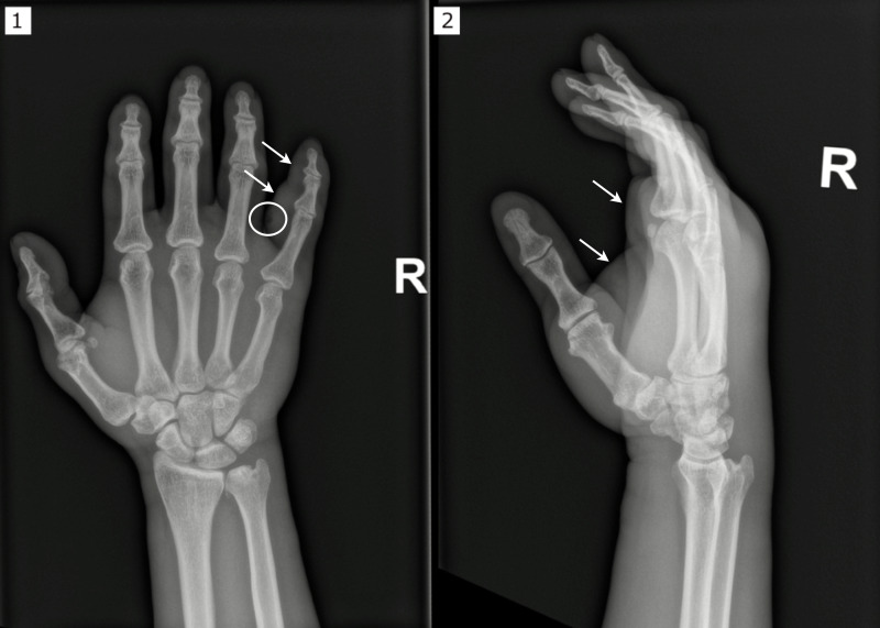 Fig 1
Fig 1| No. | Age/sex | Infection | Comorbidities | ID | Other isolated bacteria | Treatment | Treatment duration | Yr (ref.) |
|---|---|---|---|---|---|---|---|---|
| 1 | 67 M | Sepsis; multiple dentoalveolar abscesses | Multiple myeloma; h/o autologous bone marrow graft | 16s rRNA PCR | None | FEP; source control: dental extraction | 15 days | 2006 |
| 2 | 43 F | Acute proctitis post-radiotherapy | Cervical cancer | 16s rRNA PCR | None | TZP | 14 days | 2006 |
| 3 | 37 M | Septic pulmonary embolism; femoral vein | Intravenous drug abuse | 16s rRNA PCR |
| PCNG + FLU → | Unknown | 2007 |
| 4 | 43 M | Dento-alveolar abscess; fever, anemia, diarrhea | Lymphoma; h/o kidney transplant | 16s rRNA PCR | None | PCNV + MTZ → PCNV + | Unknown | 2011 |
| 5 | 66 F | Sepsis; pulmonary abscess | Lung cancer; meningeal carcinomatosis | 16s rRNA PCR | None | CXM + GEN → MEM + | Unknown | 2011 |
| 6 | 64 M | Sepsis | Colon cancer; h/o complicated abdominal surgery | 16s rRNA PCR | None | CXM + MTZ; source control: acute exploratory laparotomy (revealed colon cancer relapse) | 4 weeks | 2011 |
| 7 | 33 F | Femoral vein thrombosis | Intravenous drug abuse; chronic hepatitis B | 16s rRNA PCR |
| CXM → PEN + MTZ | 5 weeks | 2011 |
| 8 | 77 M | Pneumonia; toothache | Prostate cancer; chronic heart disease | 16s rRNA PCR |
| PCNG → PCNV | 4 weeks | 2011 |
| 9 | 56 M | Fournier gangrene | Unknown | Unknown | Unknown | Unknown | Unknown | 2017 |
| 10 | 58 M | Sepsis; bronchiolitis; | Lung cancer; COPD; encephalopathy; neuromuscular | Unknown | None | Declined treatment | Unknown | 2016 |
| 11 | 36 M | Acute cholangitis | Recurrent chronic pancreatitis | 16s rRNA PCR and MALDI-TOF MS | None | CTX → MEM → TZP → unspecified | 25 days | 2020 |
| 12 | 61 M | Sepsis; thrombotic | Hypertension; hyperlipidemia; type 2 diabetes mellitus; rectal cancer | 16s rRNA PCR | CXM + MXF → minocycline + rifampicin for brucellosis → VAN + MEM | Unknown | 2019 | |
| 13 | 70 M | Sepsis; pneumonia; HSV-1 esophagitis | HIV infection; periodontitis | MALDI-TOF MS |
| AMC | Unknown | 2019 |
| 14 | 76 M | Sepsis; peritonitis | Colon cancer; diabetes mellitus | 16s rRNA PCR |
| Unknown | Unknown | 2021 |
| 15 | 85 M | Unknown | Unknown | 16s rRNA PCR |
| Unknown | Unknown | 2021 |
| 16 | 65 M | Diabetic foot infection | Diabetes mellitus | 16s rRNA PCR | Unknown | Unknown | 2021 | |
| 17 | 52 M | Diabetic foot infection | Diabetes mellitus | 16s rRNA PCR |
| Unknown | Unknown | 2021 |
| 18 | 37 M | Peritonsillar phlegmon | Unknown | 16s rRNA PCR | Unknown | Unknown | 2021 | |
| 19 | 93 M | Severe sepsis; diarrhea | Unknown | 16s rRNA PCR | None | Unknown | Unknown | 2021 |
| 20 | 42 F | Decompensated alcoholic cirrhosis with ascites | Unknown | 16s rRNA PCR | None | Unknown | Unknown | 2021 |
| 21 | 88F | Unknown | Unknown | 16s rRNA PCR | None | Unknown | Unknown | 2021 |
| 22 | 70M | Unknown | Unknown | 16s rRNA PCR | None | Unknown | Unknown | 2021 |
| 23 | 36M | Unknown | Unknown | 16s rRNA PCR | None | Unknown | Unknown | 2021 |
| 24 | 75 F | Appendicular peritonitis; bowel obstruction | Unknown | 16s rRNA PCR | None | Unknown | Unknown | 2021 |
| 25 | 19 F | Sepsis; eft maxillary sinusitis; subperiosteal abscess | None | MALDI-TOF MS | None; intraoperative culture: mixed aerobic and anaerobic flora, including | AMC + MTZ → AMC; source control: endoscopic sinonasal surgery | 18 days | 2023 |
| 26 | 56 M | Sepsis; flexor tenosynovitis | Schizoaffective disorder; polysubstance abuse disorder; ADHD | MALDI-TOF MS | None; intraoperative culture: | TZP → SAM → AMC; source control: I&D x3 | 15 days | 2024 (current) |
| Antibiotic/antibiotic group | No. of isolates tested ( | Susceptibility (%) |
|---|---|---|
| Penicillin | 11 | 100 |
| Aminopenicillins | 2 | 100 |
| Aminopenicillins/BLI | 6 | 100 |
| Piperacillin/tazobactam | 8 | 100 |
| 3rd-generation cephalosporins | 2 | 100 |
| Cefepime | 1 | 100 |
| Carbapenems | 8 | 100 |
| Vancomycin | 9 | 100 |
| Clindamycin | 8 | 100 |
| Clarithromycin | 1 | 100 |
| Moxifloxacin | 5 | 100 |
| Tigecycline | 5 | 100 |
| Metronidazole | 11 | 91 |
| Rifampin | 1 | 0 |
| Trimethoprim/sulfamethoxazole | 1 | 0 |
| Linezolid | 1 | 0 |
Peer Reviews
No public reviews on file for this paper yet. If you reviewed it on a platform where reviews are public (OpenReview, ICLR, NeurIPS, ICML), you can paste yours below so the community can read it here.
Videos
No videos yet. Explain this paper in a talk, walkthrough, or lecture? Add one.
Taxonomy
TopicsMycobacterium research and diagnosis · Otolaryngology and Infectious Diseases · Infections and bacterial resistance
INTRODUCTION
Solobacterium moorei is the only known species of the Solobacterium genus. It is a gram-positive, non-sporulating, strict anaerobic bacillus that is part of the oral and intestinal microbiota. It is a rare human pathogen predominantly causing skin and soft tissue infections. It is a very rare cause of severe infections associated with bacteremia, with only 25 previous cases reported identified by literature search. Here, we report a case of S. moorei bacteremia with sepsis secondary to flexor tenosynovitis.
CASE PRESENTATION
A 56-year-old African American man experiencing homelessness with underlying schizoaffective disorder, polysubstance abuse disorder, and attention deficit hyperactivity disorder (ADHD) presented to the emergency department with severe pain, swelling, and decreased range of motion in his right hand.
He reported biting off a callus on the palmar side of the proximal phalanx of the right fifth finger about 2 weeks prior to presentation. The following day, he noted swelling and pain in his right hand. He developed fevers, chills, and dizziness a few days prior to seeking medical care. Physical examination revealed significant swelling and erythema of the entire right hand and forearm, most prominent on the fifth digit with a greatly reduced range of motion of each finger on that hand. He was febrile (38.4°C), tachycardic (90–120 bpm), and hypertensive (180–200/100 mmHg), meeting the systemic inflammatory response syndrome criteria for sepsis. Notable laboratory tests included a white blood cell count of 21.31 × 10^3^/µL (normal 4.0–11.0 × 10^3^/µL) with 81.9% neutrophils, lactate of 2.7 mmol/L (normal 0.5–1.9 mmol/L), C-reactive protein 162.2 g/L (normal < 5.0 mg/L), and creatinine of 1.4 mg/dL (normal 0.78–1.34 mg/dL). The urine drug screen was positive for methamphetamine, cocaine, and cannabinoids, whereas human immunodeficiency virus (HIV), hepatitis B, and hepatitis C serologies were non-reactive. An X-ray of his right hand showed soft tissue swelling and subcutaneous emphysema on the palmar aspect of the fifth proximal phalanx (Fig. 1). He was treated with fluid resuscitation, cefepime 2 g every 8 h, and vancomycin (25 mg/kg loading dose with maintenance dosing adjusted by vancomycin trough levels) and underwent emergent incision and drainage (I&D) on admission and on hospital days two and four. During the initial I&D, significant purulence was encountered upon deep dissection of the A1 pulley of the right small finger. Aerobic and anaerobic cultures were collected from the tissue.
X-ray of the patient’s right hand in (1) posteroanterior and (2) lateral projections with soft tissue swelling and subcutaneous emphysema on the palmar aspect of the fifth proximal phalanx. The circle demonstrates where the subcutaneous emphysema is located, while the arrows indicate soft tissue swelling.
Due to a shortage of blood culture bottles, a single blood culture sample was collected using BACTEC Plus Aerobic/F bottle. The instrument bottle was incubated using the BC BACTEC Automated Blood Culture System and subcultured to Columbia blood agar and CDC anaerobic blood agar per the microbiology laboratory standard protocol. All subcultures were incubated at 35–37°C in CO_2_. Bacterial growth was strictly anaerobic with pinpoint colonies. Time to positivity of the blood culture from the moment it was subcultured was roughly 24 h. The Gram stain was initially reported as gram-negative bacilli; however, the Genmark ePlex Blood Culture Nucleic Acid Test (GenMark, Roche Diagnostics USA, Indianapolis, Indiana, USA) for gram-negative bacteria was negative. Matrix-assisted laser desorption/ionization time of flight mass spectrometry (MALDI-TOF MS) (BD Bruker MALDI-TOF MS Microflex, software version flexAnalysis 3.4.79.0) was performed on growth from a subculture and revealed S. moorei with an ID/confidence score of 2.49, indicating high confidence. Susceptibility testing was performed using the E-test method (ETEST, bioMérieux, Hazelwood, Missouri, USA) under anaerobic conditions at 35°C for 48 h. The medium used was pre-reduced Brucella blood with vitamin K and Hemin agar and the inoculum in brain heart infusion broth. E-test results were interpreted using the 35th edition Clinical & Laboratory Standards Institute (CLSI) M100 Performance Standards for Antimicrobial Susceptibility Testing for anaerobes (1). Susceptibility testing revealed resistance to metronidazole with a minimal inhibitory concentration (MIC) > 256 µg/mL and susceptibility to piperacillin-tazobactam (MIC 0.064 µg/mL), ampicillin-sulbactam (MIC 0.032 µg/mL), and clindamycin (MIC 0.125 µg/mL). Intraoperative cultures from the initial I&D grew Streptococcus constellatus, Prevotella species, and Fusobacterium species. A second blood culture obtained 3 days after admission was sterile. After susceptibility testing was completed, antibiotics were deescalated on day seven to intravenous ampicillin-sulbactam 3 g every 6 h, and oral amoxicillin-clavulanic acid 875–125 mg every 12 h was prescribed on discharge to complete a 15-day course of antibiotics from source control. Clinical improvement was noted by resolution of fever by day two with decreased swelling and restored range of motion in his fingers.
DISCUSSION
S. moorei is a gram-positive, non-sporulating, strict anaerobe, rod-shaped bacteria and the only species in the genus Solobacterium. It was first described in 2000 by Kageyama et al. after isolation from human feces and also found in oral (particularly tongue) and intestinal microbiota. Despite phenotypic resemblance to Eubacterium species, phylogenetic analysis with 16s rDNA sequencing revealed it was distinct from Eubacterium, Erysipelothrix rhusiopathiae, and Holdemania filiformis, leading to a new genus: Solobacterium (2). It is implicated in halitosis (3, 4) and periodontal and endodontic diseases (5) and may play a role in colorectal carcinogenesis along with other intestinal anaerobes (6).
S. moorei is considered an opportunistic pathogen and rarely causes severe infections. Including the current case, there are only 26 reported cases of S. moorei bacteremia. These typically arise after damage to mucosal or cutaneous barriers (7). Most patients have serious underlying illnesses, with the most common being malignancies (Table 1) (8–12). Two cases have been reported in patients with intravenous drug abuse (10, 13). One case was associated with acute cholangitis in the setting of chronic pancreatitis and bile duct stricture (14) and one with HIV infection and herpes simplex virus 1 (HSV-1) esophagitis (15). Interestingly, Alejo-Cancho et al. reported a case in a 19 year-old with no significant medical history in the setting of left maxillary sinusitis with subperiosteal abscess (16). In the present case, the patient had a history of polysubstance abuse disorder but denied using drugs intravenously.
S. moorei is also associated with dental (4), skin and soft tissue, and wound infections (18). It can cause osteoarticular infections, otitis media, abdominal infections, perirectal abscesses, and even intracranial infections (5, 12, 18). S. moorei is usually found in polymicrobial tissue infections with mixed aerobic and anaerobic microorganisms, most commonly Fusobacterium species, Streptococcus constellatus, Bacteroides species, Actinomyces species, Prevotella species, Parvimonas micra, Staphylococcus aureus, Porphyromonas species, Escherichia coli, and Atopobium species (5, 9–12, 15, 18). However, in blood cultures, it is typically the only pathogen isolated (Table 1). This aligns with the microbiological findings in our patient. S. moorei was not isolated from the wound culture, even though this was the likely source of infection. This is likely due to difficult cultivation and slow growth of S. moorei compared to the other pathogens isolated.
Because S. moorei exhibits slow growth, has few specific positive biochemical reactions, and displays phenotypic variation, there are no commercially available identification kits (18). MALDI-TOF MS and 16s rRNA polymerase chain reaction (PCR) are the primary diagnostic tools for S. moorei identification. Most cases have been identified with 16s rRNA PCR (Table 1), which is considered the gold standard due to slightly higher precision (19). However, MALDI-TOF MS is a reliable alternative with a faster turnaround time and lower cost than 16s rRNA PCR (20). The available models include Bruker Biotyper (Bruker), Vitek MS (bioMérieux), Vitek MS Prime (bioMérieux), EX2600 (Zybio), and MicroIDSys (ASTA) (21, 22). The present case used the Bruker Biotyper Microflex with flexAnalysis 3.4.79.0 software. MALDI-TOF MS requires a pure culture, while 16s rRNA PCR can be done with mixed samples (23). However, this is rarely done routinely due to cost, complexity, and potential decrease in specificity as more microorganisms are identified (24). A laboratory’s ability to identify this microorganism depends on their routine identification procedures. It is easily missed, as it is not included in all identification cards and is not in all MALDI-TOF MS databases.
S. moorei bacteremia has been treated with a wide variety of antibiotics (9–11, 13–16). Our review indicates it is reliably susceptible to beta-lactams and clindamycin. Lee et al. reported a case with resistance to both trimethoprim/sulfamethoxazole and rifampin (Table 2) (14). In the present case, E-test using the latest CLSI-M100 standards showed resistance to metronidazole, which contrasts with reports of other blood isolates. A common challenge for anaerobic susceptibility testing is the limited availability of CLSI-approved methods. Other challenges include the need for a stable level of anaerobiosis during incubation, longer turnaround time compared to susceptibility testing for aerobes, the need for extensive staff experience and quality control, and breakpoint differences between CLSI and other used standards (25). Most patients survive S. moorei bacteremia, as did the patient described here. There were two reported deaths: one patient with sepsis, bronchiolitis, pulmonary abscess, pyothorax, and lung cancer who declined treatment (12); and another with S. moorei bacteremia, pneumonia, and brucellosis who ultimately died of thrombotic thrombocytopenic purpura (11).
There are no specific guidelines for the management of anaerobic bacteremia, specifically for treatment duration and whether echocardiogram is necessary. Previous cases reported a treatment duration of 2 to 5 weeks (Table 1). In the present case, the patient was treated for 15 days from the final I&D.
Conclusion
S. moorei sepsis with bacteremia is a very rare infection, with only 26 cases (including this case) reported. It is usually associated with underlying diseases, such as malignancy, often with compromised integrity of protective skin and mucosal barriers. The literature review indicates isolates are typically susceptible to penicillins, beta-lactam/beta-lactamase inhibitors, carbapenems, and 3rd/4th-generation cephalosporins but may be resistant to metronidazole, levofloxacin, and rifampin. Improvements in diagnostic methods and expansion of databases, such as for MALDI-TOF MS, may yield more frequent diagnosis of this infection and distinguish it from other types of anaerobic bacteremia.
The reference list from the paper itself. Each links out to its DOI / PubMed record.
- 1Lewis IIJSM, Amy J, Bobenchik AM, Bryson AL, Campeau S, Cullen SK, Dingle T, Esparza G, Humphries Romney M, Kirn TJ, Lutgring J, Narayanan N, Palavecino E, Pierce VM, Schuetz AN, Sharp S, Simner Patricia, Tamma PD, Weinstein MP. 2025. CLSI M 100, Performance Standards for Antimicrobial Susceptibility Testing. 35th Edition. Clinical and Laboratory Standards Institure.
- 2Kageyama A, Benno Y. 2000. Phylogenic and phenotypic characterization of some Eubacterium-like isolates from human feces: description of Solobacterium moorei gen. nov., sp. nov. Microbiol Immunol 44:223–227. doi:10.1111/j.1348-0421.2000.tb 02487.x 10832964 · doi ↗ · pubmed ↗
- 3Kazor CE, Mitchell PM, Lee AM, Stokes LN, Loesche WJ, Dewhirst FE, Paster BJ. 2003. Diversity of bacterial populations on the tongue dorsa of patients with halitosis and healthy patients. J Clin Microbiol 41:558–563. doi:10.1128/JCM.41.2.558-563.200312574246 PMC 149706 · doi ↗ · pubmed ↗
- 4Haraszthy VI, Zambon JJ, Sreenivasan PK, Zambon MM, Gerber D, Rego R, Parker C. 2007. Identification of oral bacterial species associated with halitosis. J Am Dent Assoc 138:1113–1120. doi:10.14219/jada.archive.2007.032517670880 · doi ↗ · pubmed ↗
- 5Alauzet C, Aujoulat F, Lozniewski A, Ben Brahim S, Domenjod C, Enault C, Lavigne J-P, Marchandin H. 2021. A new look at the genus Solobacterium: a retrospective analysis of twenty-seven cases of infection involving S. moorei and a review of sequence databases and the literature. Microorganisms 9:1229. doi:10.3390/microorganisms 906122934198943 PMC 8229177 · doi ↗ · pubmed ↗
- 6Yu J, Feng Q, Wong SH, Zhang D, Liang QY, Qin Y, Tang L, Zhao H, Stenvang J, Li Y, et al.. 2017. Metagenomic analysis of faecal microbiome as a tool towards targeted non-invasive biomarkers for colorectal cancer. Gut 66:70–78. doi:10.1136/gutjnl-2015-30980026408641 · doi ↗ · pubmed ↗
- 7Nagy E, Boyanova L, Justesen US. 2018. How to isolate, identify and determine antimicrobial susceptibility of anaerobic bacteria in routine laboratories. Clin Microbiol Infect 24:1139–1148. doi:10.1016/j.cmi.2018.02.00829458156 · doi ↗ · pubmed ↗
- 8Detry G, Pierard D, Vandoorslaer K, Wauters G, Avesani V, Glupczynski Y. 2006. Septicemia due to Solobacterium moorei in a patient with multiple myeloma. Anaerobe 12:160–162. doi:10.1016/j.anaerobe.2006.04.00216723262 · doi ↗ · pubmed ↗