Deep Learning-Based Assessment of Brainstem Volume Changes in Spinocerebellar Ataxia Type 2 (SCA2): A Study on Patients and Preclinical Subjects
Robin Cabeza-Ruiz, Luis Velázquez-Pérez, Evelio González-Dalmau, Alejandro Linares-Barranco, Roberto Pérez-Rodríguez

TL;DR
A deep learning model accurately segments brainstem regions in SCA2 patients and preclinical subjects, revealing volume differences that could help track disease progression.
Contribution
A modified U-Net with attention and inception modules achieves superior brainstem segmentation and reveals atrophy patterns in SCA2.
Findings
The modified U-Net outperforms existing methods in brainstem segmentation with higher Dice scores across all substructures.
Controls have significantly larger brainstem volumes compared to preclinical and patient groups, indicating atrophy linked to disease progression.
Pons volume is reduced by nearly 50% in SCA2 patients and preclinical carriers compared to controls.
Abstract
What are the main findings? Superior Segmentation Performance: The proposed modified U-Net architecture (with attention-enhanced skip connections and inception modules) significantly outperforms three comparative approaches in brainstem parcellation, achieving higher scores across all substructures (medulla, pons, and mesencephalon) and the whole brainstem.Volume Differences Across Groups: Automated segmentation reveals distinct volumetric patterns, with controls exhibiting larger volumes (whole brainstem: 1.62) compared to preclinical (1.49) and patient groups (1.12), suggesting potential atrophy linked to disease progression. Superior Segmentation Performance: The proposed modified U-Net architecture (with attention-enhanced skip connections and inception modules) significantly outperforms three comparative approaches in brainstem parcellation, achieving higher scores across all…
Genes, proteins, chemicals, diseases, species, mutations and cell lines named across the full text — each resolved to its canonical identifier and authoritative record.
Click any figure to enlarge with its caption.
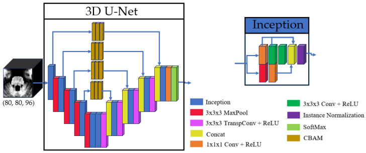 Figure 1
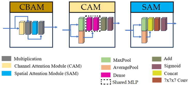 Figure 2
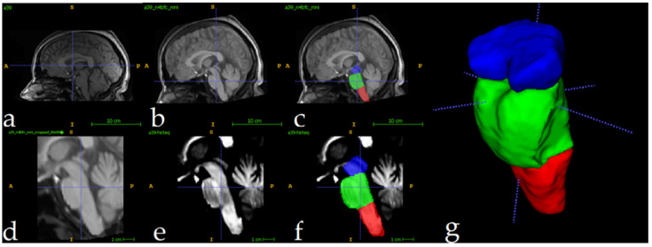 Figure 3
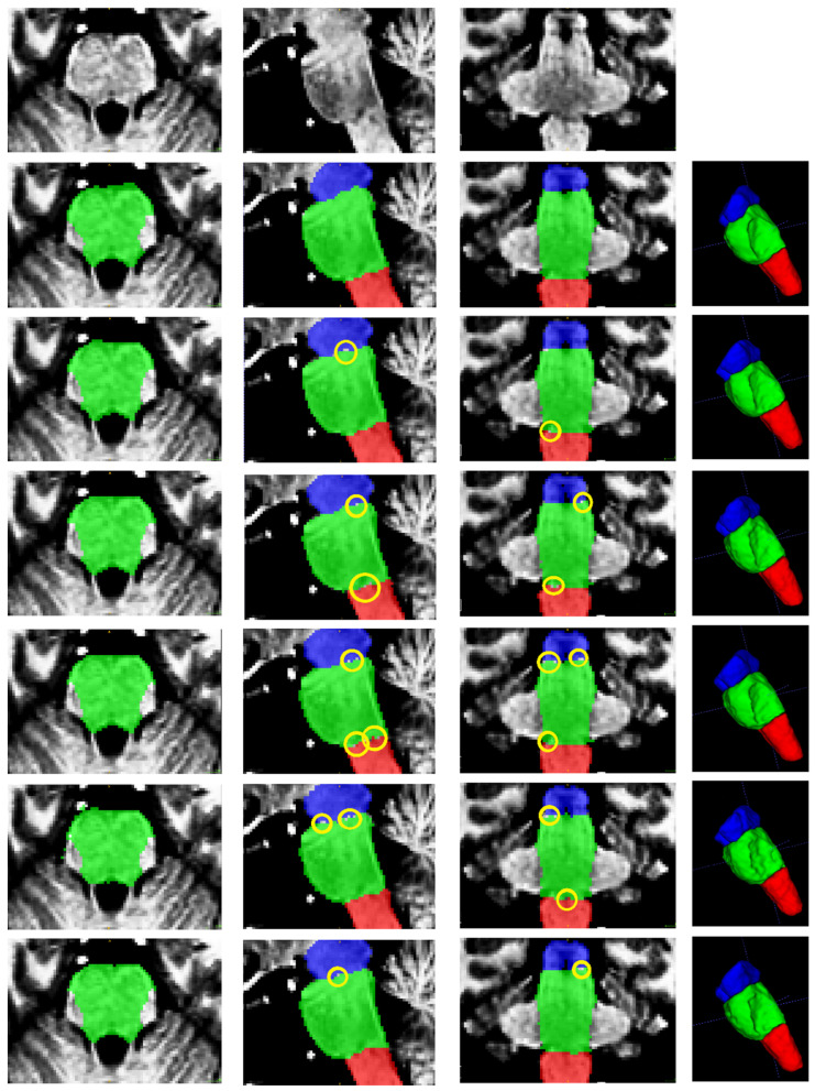 Figure 4
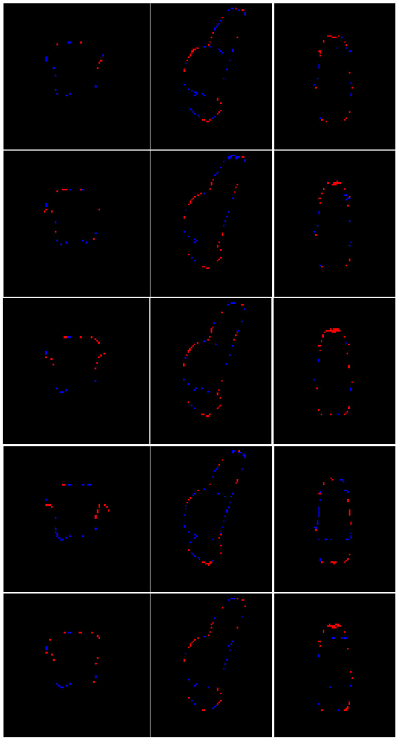 Figure 5
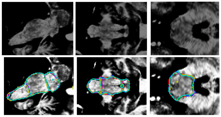 Figure 6
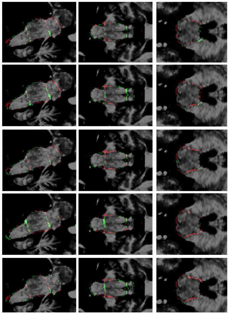 Figure 7
 Figure 8
Peer Reviews
No public reviews on file for this paper yet. If you reviewed it on a platform where reviews are public (OpenReview, ICLR, NeurIPS, ICML), you can paste yours below so the community can read it here.
Videos
No videos yet. Explain this paper in a talk, walkthrough, or lecture? Add one.
Taxonomy
TopicsGenetic Neurodegenerative Diseases · Advanced Neuroimaging Techniques and Applications · Parkinson's Disease Mechanisms and Treatments