Evaluation of [11C]-Methionine Positron Emission Tomography and Cerebral Blood Volume Imaging in the Diagnosis of Non-Contrast-Enhanced Gliomas
Naoya Imai, Hirohito Yano, Yuka Ikegame, Shoji Yasuda, Ryo Morishima, Soko Ikuta, Noriyuki Nakayama, Takashi Maruyama, Naoyuki Ohe, Morio Kumagai, Yoshihiro Muragaki, Jun Shinoda, Tsuyoshi Izumo

TL;DR
This study compares [11C]-Methionine PET and cerebral blood volume imaging for diagnosing non-contrast-enhanced brain tumors called gliomas.
Contribution
The study evaluates how cerebral blood volume imaging performs compared to PET in diagnosing different glioma subtypes.
Findings
Relative cerebral blood volume and MET imaging were significantly correlated in glioma assessment.
Cerebral blood volume showed comparable diagnostic accuracy to MET for differentiating glioma subtypes.
Cerebral blood volume was more reliable for identifying MET-positive regions in some glioma subtypes than others.
Abstract
Background/Objectives: Methionine (MET) positron emission tomography (PET) and cerebral blood volume (CBV) imaging provide complementary glioma assessment. This study compared MET and CBV across glioma subtypes defined by the 2021 World Health Organization Classification. Methods: This retrospective study enrolled 106 patients (mean age 41.9 ± 12.4 years; 57 males) with MRI non-contrast-enhanced gliomas: 21 glioblastoma, isocitrate dehydrogenase (IDH)-wildtype (G); 50 astrocytoma, IDH-mutant (A); and 35 oligodendrogliomas, IDH-mutant, and 1p/19q-codeleted (O). Relative CBVs (rCBVs) were measured in VOI-T2 and VOI-MET, and the MET tumor-to-normal (T/N) ratio was calculated. Results: MET and rCBV were significantly correlated (r = 0.5, p < 0.001); rCBV was higher in MET-positive tumors and predicted MET accumulation (area under the curve [AUC] = 0.72, cutoff = 2.99). In VOI-T2, rCBV and…
Genes, proteins, chemicals, diseases, species, mutations and cell lines named across the full text — each resolved to its canonical identifier and authoritative record.
Click any figure to enlarge with its caption.
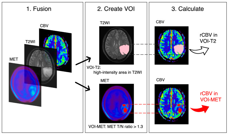 Figure 1
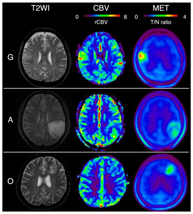 Figure 2
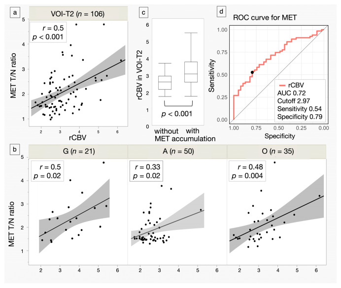 Figure 3
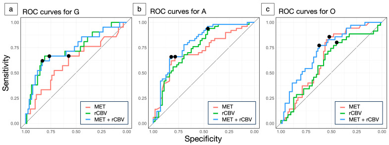 Figure 4
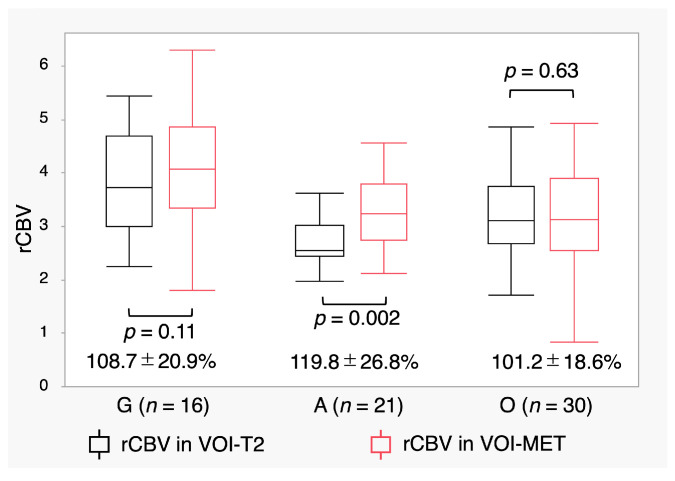 Figure 5
Peer Reviews
No public reviews on file for this paper yet. If you reviewed it on a platform where reviews are public (OpenReview, ICLR, NeurIPS, ICML), you can paste yours below so the community can read it here.
Videos
No videos yet. Explain this paper in a talk, walkthrough, or lecture? Add one.
Taxonomy
TopicsGlioma Diagnosis and Treatment · Radiomics and Machine Learning in Medical Imaging · MRI in cancer diagnosis