Correlation Between Volumetric Soft Tissue Asymmetry and Postero-Anterior Cephalometric Measurements in Patients with Skeletal Facial Asymmetry: A Cross-Sectional Pilot Study
Saki Tanaka, Yudai Shimpo, Hiromi Sato, Toshiko Sekiya, Shotaro Ueda, Chihiro Kariya, Takashi Oikawa, Hiroshi Tomonari

TL;DR
This study finds that lower facial soft tissue asymmetry is strongly linked to mandibular skeletal deviations, while midface asymmetry has weaker skeletal correlations.
Contribution
This pilot study is the first to correlate 3D volumetric soft tissue asymmetry with PA cephalometric measurements in skeletal facial asymmetry.
Findings
Menton deviation from the midline shows the strongest correlation with whole facial asymmetry (R2 = 0.630).
Midfacial asymmetry has only moderate skeletal correlations (maximum R2 = 0.186).
Maxillary occlusal cant parameters do not significantly correlate with soft tissue asymmetry.
Abstract
Background/Objectives: While skeletal facial asymmetry is commonly assessed using posteroanterior (PA) cephalometric radiographs, the association between skeletal measurements and volumetric soft tissue asymmetry remains unclear. This study aimed to identify which skeletal parameters are most strongly correlated with soft tissue asymmetry measured using three-dimensional (3D) imaging. Methods: Thirty-three Japanese patients (8 males and 25 females; mean age: 26.85 ± 12.13 years) undergoing orthodontic–orthognathic treatment were included. Three-dimensional facial surface data were acquired using the VECTRA® H1 imaging system. Soft tissue asymmetry was quantified by calculating the volumetric difference between the original and mirrored 3D facial images, divided into three regions: whole face, midface, and lower face. PA cephalometric radiographs were traced, and 28 skeletal variables…
Genes, proteins, chemicals, diseases, species, mutations and cell lines named across the full text — each resolved to its canonical identifier and authoritative record.
Click any figure to enlarge with its caption.
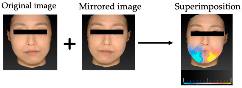 Figure 1
 Figure 2
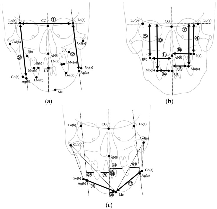 Figure 3
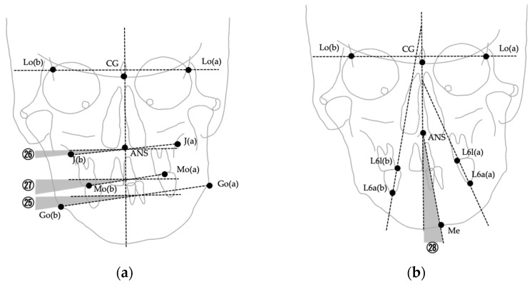 Figure 4
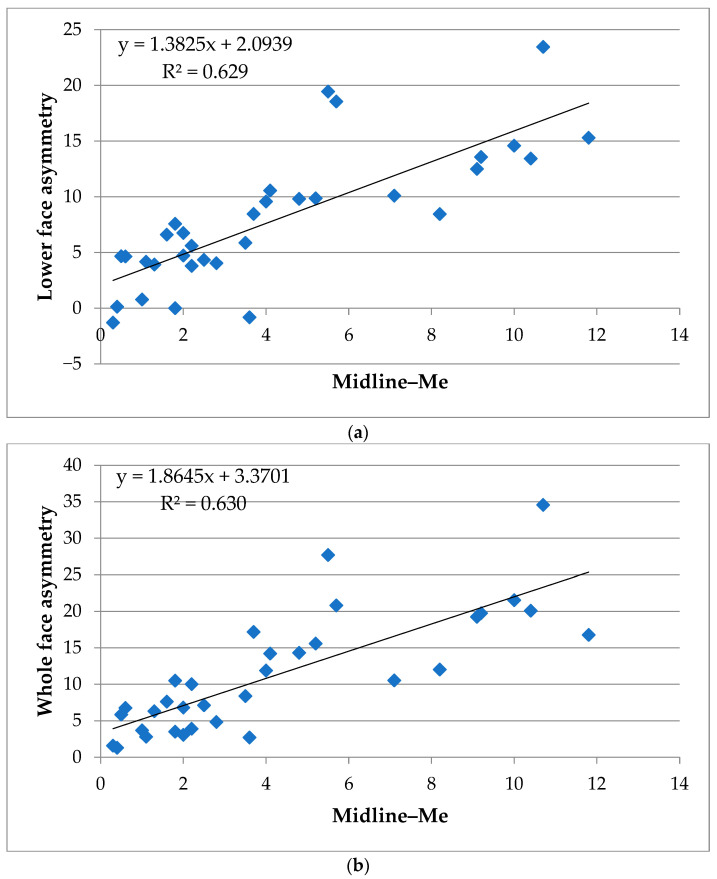 Figure 5
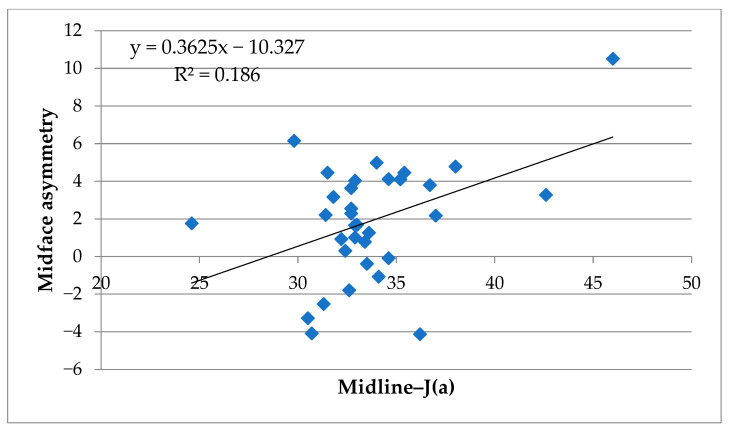 Figure 6
Peer Reviews
No public reviews on file for this paper yet. If you reviewed it on a platform where reviews are public (OpenReview, ICLR, NeurIPS, ICML), you can paste yours below so the community can read it here.
Videos
No videos yet. Explain this paper in a talk, walkthrough, or lecture? Add one.
Taxonomy
TopicsOrthodontics and Dentofacial Orthopedics · Temporomandibular Joint Disorders · Dental Radiography and Imaging