Morphological Characterization of Plasma-Derived Nanoparticles Isolated by High-Speed Ultracentrifugation: A Scanning Electron Microscopy Study
Lubov A. Kungurova, Alexander A. Artamonov, Evgeniy A. Grigoryev, Aleksei Yu. Aronov, Olga S. Vezo, Ruslan I. Glushakov, Kirill A. Kondratov

TL;DR
This study uses low-voltage scanning electron microscopy to examine the size and structure of plasma-derived exomeres and supermeres, which are small nanoparticles involved in cell communication.
Contribution
The study demonstrates the feasibility of low-voltage scanning electron microscopy for characterizing exomeres and supermeres in plasma.
Findings
Plasma-derived exomeres and supermeres measured 10–50 nm in diameter, as observed using low-voltage scanning electron microscopy.
Dynamic light scattering detected particles as small as 10–18 nm in the isolated fractions.
Low-voltage scanning electron microscopy can detect the size of these nanoparticles but cannot distinguish them from other particles without immunological methods.
Abstract
Extracellular vesicles are critical mediators of intercellular signaling. Recent studies have revealed that, in addition to vesicular structures, smaller non-vesicular nanoparticles—termed exomeres and supermeres—also participate in intercellular communication. Detailed characterization of these nanoscale entities within biological systems is essential for elucidating their structural and functional roles. Due to their sub-50 nm dimensions, high-resolution imaging modalities such as atomic force microscopy and electron microscopy are currently the primary techniques available for their visualization. In the present study, we employed low-voltage scanning electron microscopy to investigate the size of exomeres and supermeres isolated from human blood plasma via high-speed ultracentrifugation. Platelet-poor plasma was obtained from the blood of six healthy donors (two women and four men,…
Genes, proteins, chemicals, diseases, species, mutations and cell lines named across the full text — each resolved to its canonical identifier and authoritative record.
Click any figure to enlarge with its caption.
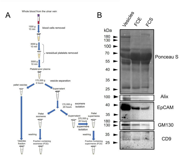 Figure 1
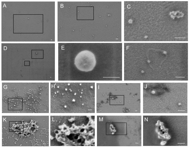 Figure 2
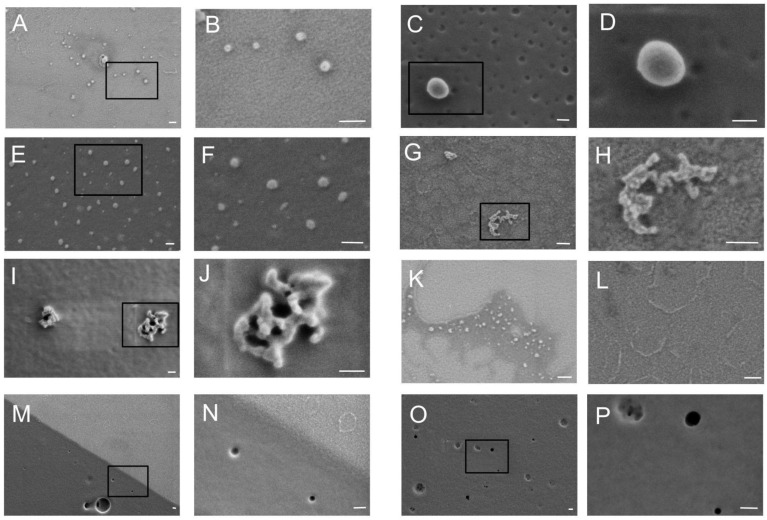 Figure 3
 Figure 4
Peer Reviews
No public reviews on file for this paper yet. If you reviewed it on a platform where reviews are public (OpenReview, ICLR, NeurIPS, ICML), you can paste yours below so the community can read it here.
Videos
No videos yet. Explain this paper in a talk, walkthrough, or lecture? Add one.
Taxonomy
TopicsExtracellular vesicles in disease · Nanopore and Nanochannel Transport Studies · Inhalation and Respiratory Drug Delivery